Brachial Plexus Diagram Generator: Roots to Branches
Generate a brachial plexus diagram with the wiring right: five roots from C5 to T1, upper, middle and lower trunks, six divisions, lateral, posterior and medial cords, and the musculocutaneous, axillary, radial, median and ulnar nerves, with the side branches where they really leave. Choose a flat schematic, an anatomical view around the axillary artery, the nerves of the arm, or the classic injuries, then a labeled diagram or a blank worksheet with its answer key.
Roots, trunks, divisions, cords, branchesMedian nerve from two cordsLabeled or blank worksheet
Checked against NCBI StatPearls anatomy chapters
Cords named by the axillary artery
Worksheet + answer key pairs
Print-ready PNG
AI Brachial Plexus Diagram Generator
Describe the plexus figure you need, or pick a view, a level, and labels from the menus.
820 / 2000 characters
Generate a publication-ready PNG from your description.
Preview
Brachial Plexus Diagram Example Previews
Target aspect ratio: 16:9
Sample previews. Describe your figure to generate your own brachial plexus diagram.
Labeled and Blank Brachial Plexus Diagram Examples
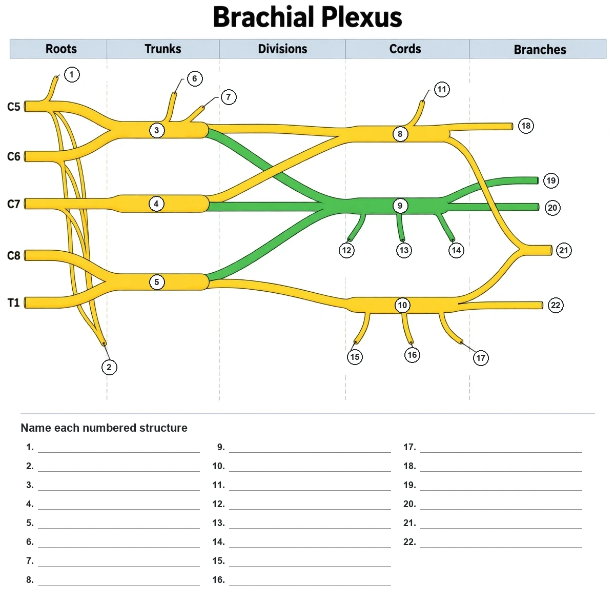
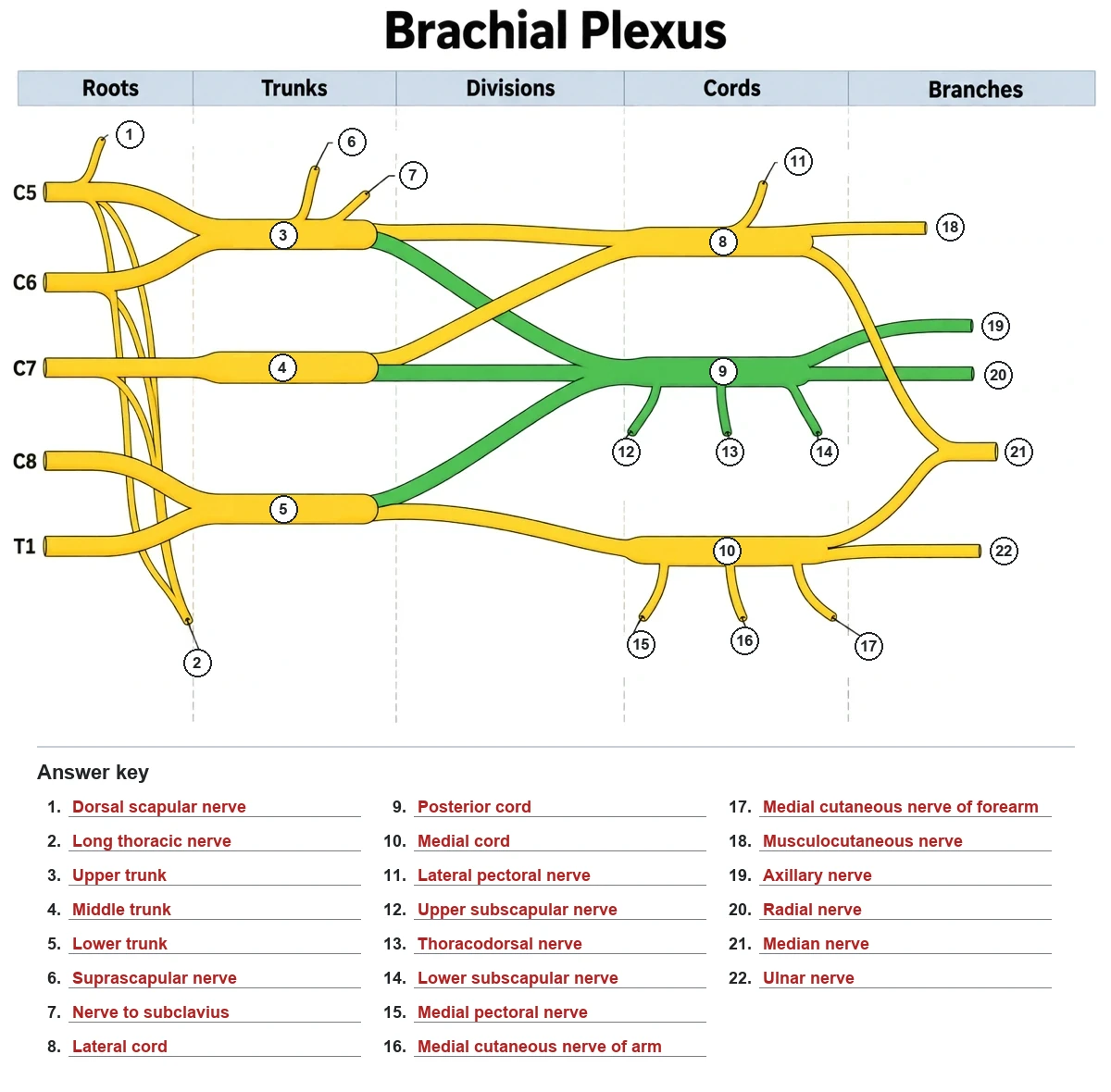
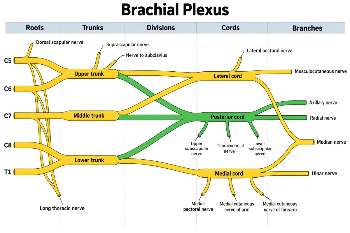
A fully labeled brachial plexus schematic with every side branch at its origin, and a 22-label blank worksheet with its matching answer key. Load one into the generator, then switch the view, level, or labels to make your own.
Prompt
Create a labeled brachial plexus diagram titled "Brachial Plexus": a flat brachial plexus schematic on a white background, read left to right under five column headers "Roots", "Trunks", "Divisions", "Cords", "Branches": the five roots C5, C6, C7, C8 and T1 labeled on the left; C5 and C6 joining as the upper trunk, C7 continuing alone as the middle trunk, C8 and T1 joining as the lower trunk; each trunk splitting into an anterior division drawn in yellow and a posterior division drawn in green; the three posterior divisions joining as the green posterior cord in the middle, the anterior divisions of the upper and middle trunks joining as the yellow lateral cord above it, and the anterior division of the lower trunk continuing as the yellow medial cord below it; the lateral cord ending as the musculocutaneous nerve and a root that curves down to meet a root from the medial cord, forming the median nerve; the posterior cord ending as the axillary and radial nerves; the medial cord ending as the ulnar nerve; and every named side branch drawn as a short stub where it leaves its root, trunk or cord. Label every structure in dark sans-serif text: the trunks and cords inside the nerve, the side branches at the end of short leader lines: dorsal scapular nerve from C5, long thoracic nerve from C5, C6 and C7, suprascapular nerve and nerve to subclavius from the upper trunk, lateral pectoral nerve from the lateral cord, upper subscapular, thoracodorsal and lower subscapular nerves from the posterior cord, medial pectoral nerve, medial cutaneous nerve of arm and medial cutaneous nerve of forearm from the medial cord, and the five terminal nerves on the right: musculocutaneous, axillary, radial, median and ulnar.
Use this example
Prompt
Create a blank brachial plexus worksheet titled "Brachial Plexus". Draw a flat brachial plexus schematic on a white background, read left to right under five column headers "Roots", "Trunks", "Divisions", "Cords", "Branches": the five roots C5, C6, C7, C8 and T1 labeled on the left; C5 and C6 joining as the upper trunk, C7 continuing alone as the middle trunk, C8 and T1 joining as the lower trunk; each trunk splitting into an anterior division drawn in yellow and a posterior division drawn in green; the three posterior divisions joining as the green posterior cord in the middle, the anterior divisions of the upper and middle trunks joining as the yellow lateral cord above it, and the anterior division of the lower trunk continuing as the yellow medial cord below it; the lateral cord ending as the musculocutaneous nerve and a root that curves down to meet a root from the medial cord, forming the median nerve; the posterior cord ending as the axillary and radial nerves; the medial cord ending as the ulnar nerve; and every named side branch drawn as a short stub where it leaves its root, trunk or cord. Keep the root names C5 to T1 and the five column headers. Replace every other name with a small numbered circle at the same place, 22 in all, in this order: 1 = Dorsal scapular nerve, 2 = Long thoracic nerve, 3 = Upper trunk, 4 = Middle trunk, 5 = Lower trunk, 6 = Suprascapular nerve, 7 = Nerve to subclavius, 8 = Lateral cord, 9 = Posterior cord, 10 = Medial cord, 11 = Lateral pectoral nerve, 12 = Upper subscapular nerve, 13 = Thoracodorsal nerve, 14 = Lower subscapular nerve, 15 = Medial pectoral nerve, 16 = Medial cutaneous nerve of arm, 17 = Medial cutaneous nerve of forearm, 18 = Musculocutaneous nerve, 19 = Axillary nerve, 20 = Radial nerve, 21 = Median nerve, 22 = Ulnar nerve. Below the figure print an answer grid of 22 empty numbered lines. No names, no word bank and no key anywhere on the image.
Blank worksheet
Use this example
Prompt
Create the answer key for the blank brachial plexus worksheet. Draw a flat brachial plexus schematic on a white background, read left to right under five column headers "Roots", "Trunks", "Divisions", "Cords", "Branches": the five roots C5, C6, C7, C8 and T1 labeled on the left; C5 and C6 joining as the upper trunk, C7 continuing alone as the middle trunk, C8 and T1 joining as the lower trunk; each trunk splitting into an anterior division drawn in yellow and a posterior division drawn in green; the three posterior divisions joining as the green posterior cord in the middle, the anterior divisions of the upper and middle trunks joining as the yellow lateral cord above it, and the anterior division of the lower trunk continuing as the yellow medial cord below it; the lateral cord ending as the musculocutaneous nerve and a root that curves down to meet a root from the medial cord, forming the median nerve; the posterior cord ending as the axillary and radial nerves; the medial cord ending as the ulnar nerve; and every named side branch drawn as a short stub where it leaves its root, trunk or cord, with the same 22 numbered circles in the same places, and below it the answer grid with every line filled in: 1 = Dorsal scapular nerve, 2 = Long thoracic nerve, 3 = Upper trunk, 4 = Middle trunk, 5 = Lower trunk, 6 = Suprascapular nerve, 7 = Nerve to subclavius, 8 = Lateral cord, 9 = Posterior cord, 10 = Medial cord, 11 = Lateral pectoral nerve, 12 = Upper subscapular nerve, 13 = Thoracodorsal nerve, 14 = Lower subscapular nerve, 15 = Medial pectoral nerve, 16 = Medial cutaneous nerve of arm, 17 = Medial cutaneous nerve of forearm, 18 = Musculocutaneous nerve, 19 = Axillary nerve, 20 = Radial nerve, 21 = Median nerve, 22 = Ulnar nerve. Keep the numbering in this order so the key matches the worksheet line for line.
Use this example
Prompt
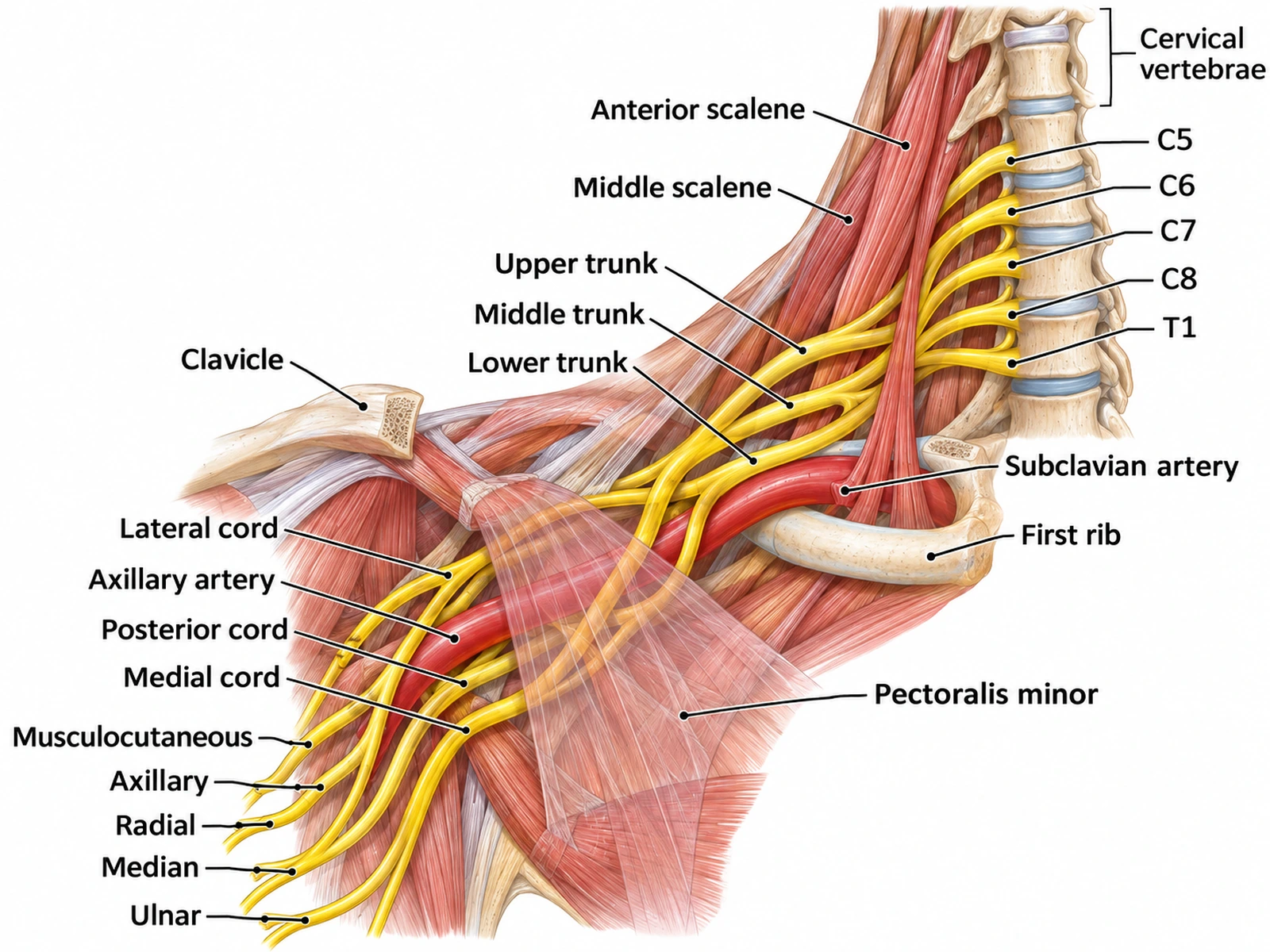
Create a labeled anatomical illustration of the right brachial plexus seen from the front, on a white background. Show the cervical vertebrae, the "Anterior scalene" and "Middle scalene" with the roots "C5", "C6", "C7", "C8" and "T1" emerging between them, the "Upper trunk", "Middle trunk" and "Lower trunk", the "Clavicle" cut away, the "First rib", the "Subclavian artery" becoming the "Axillary artery", the "Pectoralis minor" drawn transparent, the "Lateral cord", "Posterior cord" and "Medial cord" named by their position around the axillary artery, and the five terminal nerves "Musculocutaneous", "Axillary", "Radial", "Median" and "Ulnar".
Anatomical view
Use this example
Prompt
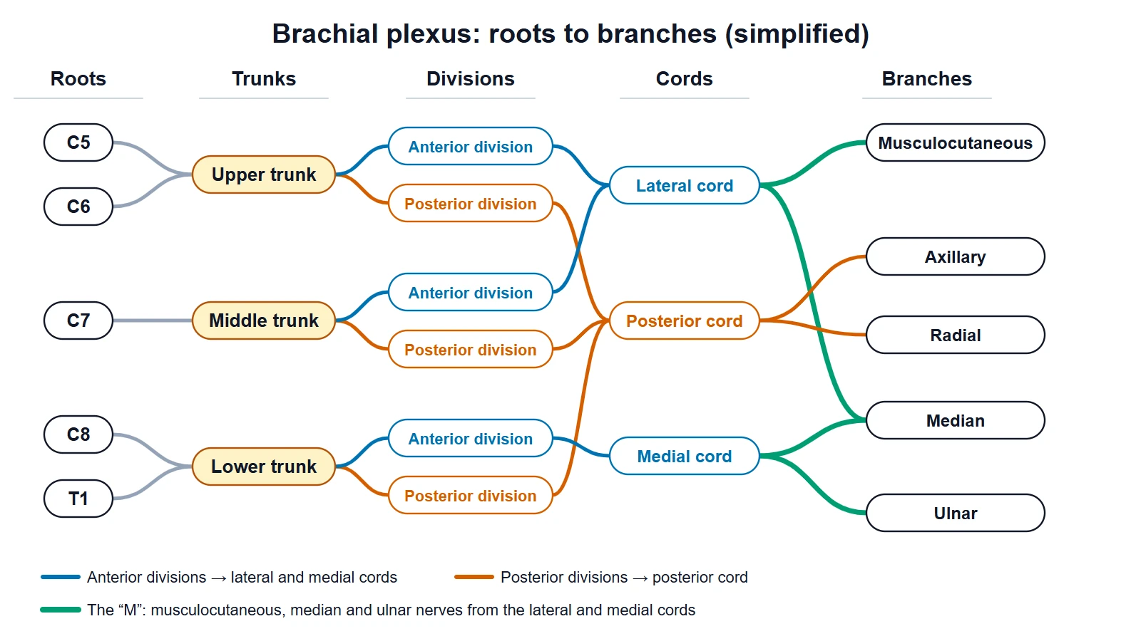
Create a simplified brachial plexus chart on a white background in five columns headed "Roots", "Trunks", "Divisions", "Cords" and "Branches". Roots C5 and C6 join the "Upper trunk", C7 continues as the "Middle trunk", C8 and T1 join the "Lower trunk". Each trunk splits into an "Anterior division" (blue) and a "Posterior division" (orange). The upper and middle anterior divisions form the "Lateral cord", the lower anterior division forms the "Medial cord", and all three posterior divisions form the "Posterior cord". Branches: "Musculocutaneous" from the lateral cord, "Axillary" and "Radial" from the posterior cord, "Median" from the lateral and medial cords, "Ulnar" from the medial cord. Highlight the "M" formed by the musculocutaneous, median and ulnar nerves with their cords, and add a short legend.
Simplified MIntro anatomy
Use this example
Prompt
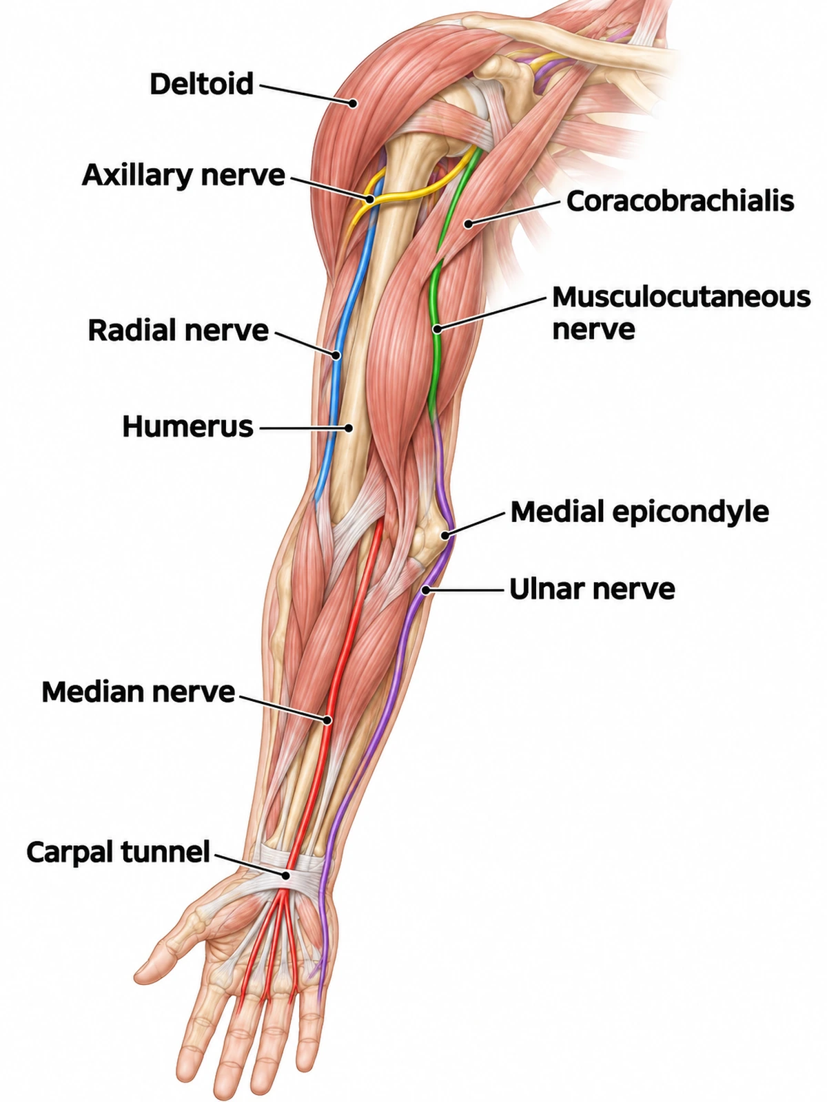
Create a labeled illustration of the nerves of the right upper limb seen from the front, muscles shown faintly, on a white background. Label the "Deltoid", "Humerus", "Coracobrachialis", "Medial epicondyle" and "Carpal tunnel", and draw each nerve in its own colour: the "Axillary nerve" wrapping the surgical neck of the humerus to the deltoid, the "Radial nerve" along the humerus, the "Musculocutaneous nerve" piercing coracobrachialis, the "Ulnar nerve" behind the medial epicondyle to the little-finger side of the hand, and the "Median nerve" down the middle of the forearm through the carpal tunnel to the thumb side.
Nerves of the armIntro anatomy
Use this example
Prompt
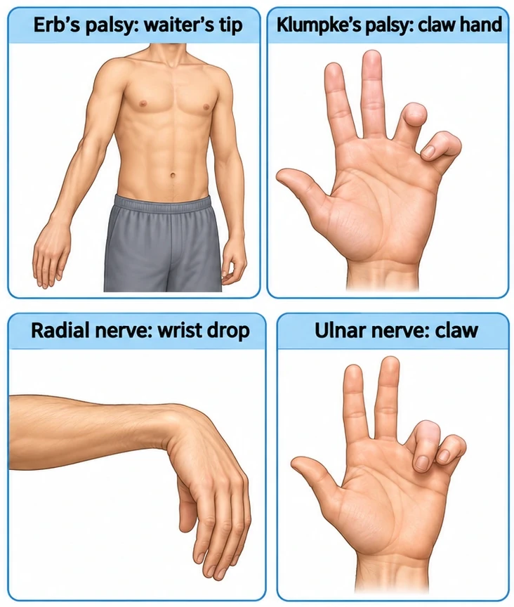
Create four labeled clinical panels on a white background in a 2 × 2 grid: "Erb’s palsy: waiter’s tip" (arm adducted and internally rotated, elbow extended, forearm pronated, wrist flexed), "Klumpke’s palsy: claw hand", "Radial nerve: wrist drop", and "Ulnar nerve: claw" with the ring and little fingers clawed. Clean medical illustration style, one pale header band per panel.
Injuries and lesionsClinical / root values
Use this example
How to make a brachial plexus diagram
1
Pick the view, level, and labels
A classic schematic, the simplified M, an anatomical view around the axillary artery, the nerves of the arm, or the injuries panel. Set the level from intro anatomy to clinical root values, then labeled, numbered, or a blank worksheet.
2
Generate the figure
The generator draws five roots, three trunks, six divisions and three cords, takes the median nerve from both the lateral and medial cords, and places each side branch on the root, trunk or cord it really leaves from.
3
Trace every path, then export
Follow C7 into the middle trunk, the posterior divisions into the posterior cord, and both roots into the median nerve. For a worksheet, make the answer key from the same result, then download the PNG.
The brachial plexus is the network of nerves that supplies the shoulder, arm and hand. It begins as five spinal nerve roots in the neck and ends, in the armpit, as the five nerves that run down the limb. In between, the fibres from each root are shuffled: they merge into trunks, split into divisions, regroup as cords, and leave in new combinations. That is why a single nerve such as the radial carries fibres from five spinal levels, and why one root injury weakens several muscles at once.
A brachial plexus diagram makes the shuffle readable. The classic version lays the plexus out flat, from the spine on the left to the arm on the right, so you can trace any fibre from its root to its nerve. Anatomical versions put the same wiring back in the body, between the neck muscles, behind the collarbone and around the axillary artery. Both answer the same questions: which roots feed which nerve, and where along the way each branch leaves.
Roots, trunks, divisions, cords and branches
The five sections always come in the same order, remembered as Randy Travis Drinks Cold Beer, and in the counts 5, 3, 6, 3, 5.
Roots (5): the anterior rami of C5, C6, C7, C8 and T1, emerging between the anterior and middle scalene muscles.
Trunks (3): upper (C5 + C6), middle (C7) and lower (C8 + T1), in the posterior triangle of the neck.
Divisions (6): each trunk splits into an anterior and a posterior division behind the clavicle.
Cords (3): lateral (anterior divisions of the upper and middle trunks), medial (anterior division of the lower trunk) and posterior (all three posterior divisions), in the axilla.
Terminal branches (5): musculocutaneous, axillary, radial, median and ulnar nerves, formed at the lower border of pectoralis minor.
How to draw the brachial plexus step by step
Write C5, C6, C7, C8 and T1 down the left side, evenly spaced.
Join C5 and C6 into the upper trunk, carry C7 straight across as the middle trunk, and join C8 and T1 into the lower trunk.
Split each trunk in two. Send the three posterior divisions to the middle and join them into the posterior cord; join the anterior divisions of the upper and middle trunks into the lateral cord above it; carry the anterior division of the lower trunk on as the medial cord below it.
From the lateral cord draw the musculocutaneous nerve and a branch angling down; from the medial cord draw the ulnar nerve and a branch angling up; let those two branches meet as the median nerve. You now have the M.
From the posterior cord draw the axillary nerve and the larger radial nerve.
Add the side branches last: dorsal scapular and long thoracic from the roots, suprascapular and the nerve to subclavius from the upper trunk, then the pectoral, cutaneous, subscapular and thoracodorsal nerves from the cords.
Why the cords are named lateral, posterior and medial
Students often expect the lateral cord to be at the top of the page because it is drawn there. The names come from the anatomy instead: in the axilla, the three cords lie around the second part of the axillary artery, deep to pectoralis minor, one on its lateral side, one behind it and one on its medial side.
A flat schematic keeps the order but loses the artery, so a good anatomical diagram restores it. Higher up, above the clavicle, the trunks lie above and to the outer side of the subclavian artery; lower down, after the cords split into terminal branches, the M of the musculocutaneous, median and ulnar nerves lies in front of the third part of the axillary artery. Showing the artery in red under the nerves is the quickest way to make the cord names make sense.
The side branches and where they leave
Most labeling errors are side branches put on the wrong part of the plexus. These are the standard named branches by origin, with their usual root values.
From the roots: dorsal scapular nerve (C5) to the rhomboids and levator scapulae; long thoracic nerve (C5–C7) to serratus anterior.
From the upper trunk: suprascapular nerve (C5–C6) to supraspinatus and infraspinatus; nerve to subclavius (C5–C6).
From the lateral cord: lateral pectoral nerve (C5–C7) to pectoralis major.
From the medial cord: medial pectoral nerve (C8–T1) to pectoralis minor and major; medial cutaneous nerve of arm; medial cutaneous nerve of forearm.
From the posterior cord: upper subscapular nerve to subscapularis; thoracodorsal nerve (C6–C8) to latissimus dorsi; lower subscapular nerve to subscapularis and teres major. Remembered, with the axillary and radial nerves, as STAR.
Nothing named leaves the middle trunk, the lower trunk or the divisions.
The M and the median nerve
The single most important check on any brachial plexus diagram is the median nerve. It has two roots: a lateral root from the lateral cord, carrying fibres from C5 or C6 to C7, and a medial root from the medial cord, carrying C8 and T1. They join in front of the axillary artery, and with the musculocutaneous nerve on one side and the ulnar nerve on the other they make the letter M. A median nerve drawn from one cord is the most common error in online images, and it breaks the logic of the plexus: the median nerve supplies both forearm muscles with upper-root fibres and hand muscles with C8–T1 fibres.
The terminal nerves in the arm
Musculocutaneous: pierces coracobrachialis, runs between biceps and brachialis and supplies all three, then continues as the lateral cutaneous nerve of the forearm.
Axillary: leaves the axilla through the quadrangular space with the posterior circumflex humeral artery, winds around the surgical neck of the humerus, and supplies deltoid and teres minor.
Radial: spirals behind the humerus in the radial groove, supplies triceps and the extensors on the back of the arm and forearm, and splits at the elbow into a superficial sensory branch and a deep motor branch.
Median: gives no branches in the arm, supplies most forearm flexors and the pronators, and passes through the carpal tunnel to the thenar muscles and the lateral two lumbricals.
Ulnar: gives no branches in the arm, passes behind the medial epicondyle (the "funny bone"), and supplies flexor carpi ulnaris, part of flexor digitorum profundus, and most of the small muscles of the hand.
Erb’s palsy and Klumpke’s palsy
Upper and lower plexus injuries are the classic clinical pair. Erb’s palsy damages the upper trunk (C5–C6), usually when the head and shoulder are forced apart, as in shoulder dystocia during birth or a fall onto the shoulder. Deltoid, the rotator cuff muscles from the suprascapular nerve, biceps and brachialis are weak, so the arm hangs adducted and internally rotated with the elbow extended, the forearm pronated and the wrist flexed: the waiter’s tip. Sensation is lost over the outer arm and forearm. StatPearls describes it as the most common neonatal brachial plexus injury, and most affected infants are treated with supportive care.
Klumpke’s palsy damages the lower trunk (C8–T1), usually when the arm is pulled upward, as in grabbing a branch while falling or a delivery with the arm above the head. The small muscles of the hand fail, giving a claw hand, and sensation is lost along the inner forearm and hand. A T1 root torn near the spinal cord also cuts the sympathetic supply to the face, causing Horner syndrome. On a diagram, highlight the upper trunk for Erb’s palsy and the lower trunk for Klumpke’s palsy.
Wrist drop, claw hand and other nerve lesions
Wrist drop: radial nerve, most often in a fracture of the shaft of the humerus at the radial groove.
Claw of the ring and little fingers: ulnar nerve, often at the elbow behind the medial epicondyle.
Hand of benediction: a high median nerve injury; when the patient tries to make a fist, the index and middle fingers stay straight.
Thenar wasting and numb thumb, index and middle fingers: median nerve compression in the carpal tunnel.
Flattened shoulder and weak abduction: axillary nerve, after a fractured surgical neck of the humerus or an anterior shoulder dislocation.
Winged scapula: long thoracic nerve, which supplies serratus anterior and is exposed on the side of the chest wall.
Dermatomes and reflexes of the upper limb
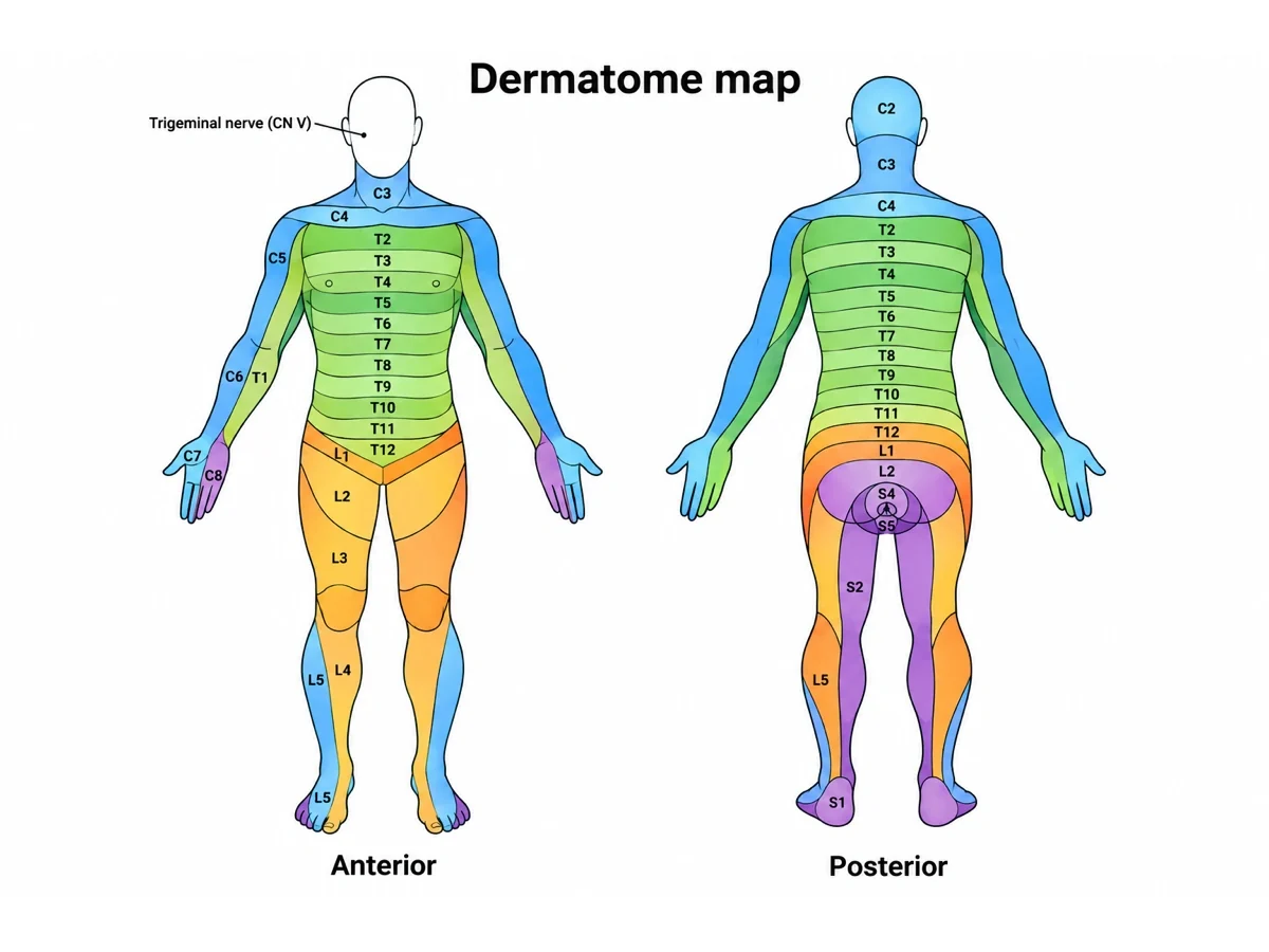
Root-level questions are answered with dermatomes and reflexes rather than with nerves, and a clinical brachial plexus figure often pairs the two.
C5: skin over the outer upper arm; biceps reflex (with C6).
C6: outer forearm and thumb; brachioradialis reflex.
C7: middle finger; triceps reflex.
C8: little finger and the inner side of the hand.
T1: inner side of the forearm.
For the full map of dermatomes from C2 to S5, use the dermatome chart generator; a dermatome follows a root, while a peripheral nerve’s skin area mixes several roots.
Brachial plexus variations
Real plexuses vary, and a research or surgical figure may need to show it. When C4 sends a sizeable branch, the plexus is called prefixed; when T2 contributes, it is postfixed. Communications between the median and musculocutaneous nerves are common. Root values given in textbooks are the usual pattern, not a law: the median nerve, for example, is often listed as C6–T1 but frequently receives C5 fibres, and the ulnar nerve often receives some from C7. Unless your figure is about a variant, draw the standard five-root plexus.
Brachial plexus blocks
Anaesthetists numb the arm by injecting local anaesthetic around the plexus, and the level of the block follows the sections of the diagram: interscalene blocks reach the roots and upper trunks and suit shoulder surgery, though they often spare the lower trunk and the ulnar nerve; supraclavicular blocks reach the trunks and divisions, where the plexus is most compact; infraclavicular blocks reach the cords around the axillary artery; and axillary blocks reach the terminal branches. A figure for a block protocol usually shows the anatomical view with the needle target marked at one of these levels.
Labeled, numbered and blank brachial plexus diagrams
A labeled brachial plexus diagram is the study reference. A numbered version with a key keeps a dense figure readable on a slide, and suits quizzing by covering the key. A blank brachial plexus worksheet keeps every nerve and leaves numbered answer lines; the answer-key action writes the names back onto the same drawing, so the two print as a matching pair. For exams, the 22-label worksheet on this page covers the trunks, cords, terminal nerves and every standard side branch; set the level to intro anatomy for a shorter version without the side branches.
Most common brachial plexus diagram mistakes
These are the errors that turn up most often in student drawings, online images, and AI output. Trace each one before the figure goes into a handout, a slide, or an exam.
MistakeCorrect
MistakeSix roots, or a C4 or T2 root on a standard plexus
CorrectFive roots: the anterior rami of C5, C6, C7, C8 and T1
MistakeC7 joining the upper or lower trunk
CorrectC7 alone forms the middle trunk; C5 + C6 the upper, C8 + T1 the lower
MistakeThe median nerve drawn from the medial cord only
CorrectThe median nerve has two roots: one from the lateral cord, one from the medial cord
MistakeThe posterior cord built from one trunk
CorrectThe posterior cord receives the posterior divisions of all three trunks
MistakeCords named by their place on the page
CorrectCords are named by their position around the axillary artery
MistakeThe ulnar nerve leaving the posterior cord
CorrectThe ulnar nerve comes from the medial cord; the radial and axillary from the posterior cord
MistakeThe lateral pectoral nerve on the medial cord
CorrectLateral pectoral from the lateral cord, medial pectoral from the medial cord
MistakeThe suprascapular nerve leaving a cord or a root
CorrectThe suprascapular nerve and the nerve to subclavius leave the upper trunk
The five terminal branches of the brachial plexus
Each terminal nerve, the cord it comes from, its usual root values, the main muscles it supplies, and the classic sign when it is injured.
Nerve
Cord and roots
Main muscles
Skin supplied
Classic injury sign
Musculocutaneous
Lateral cord, C5–C7
Coracobrachialis, biceps brachii, brachialis
Lateral forearm
Weak elbow flexion and supination
Axillary
Posterior cord, C5–C6
Deltoid, teres minor
Skin over the lower deltoid
Weak abduction after a fractured surgical neck of the humerus or a shoulder dislocation
Radial
Posterior cord, C5–T1
Triceps, brachioradialis, wrist and finger extensors
Back of the arm and forearm, back of the hand on the thumb side
Wrist drop after a mid-shaft humerus fracture
Median
Lateral + medial cords, C6–T1 (often C5)
Most forearm flexors, pronators, thenar muscles, lateral two lumbricals
Palm side of the thumb, index, middle and half the ring finger
Carpal tunnel syndrome; "hand of benediction" after a high injury
Ulnar
Medial cord, C8–T1 (often C7)
Flexor carpi ulnaris, half of flexor digitorum profundus, most small hand muscles
Little finger and half the ring finger
Claw of the ring and little fingers after an elbow injury
Brachial Plexus Diagram FAQ
It shows how five spinal nerve roots in the neck, the anterior rami of C5, C6, C7, C8 and T1, mix and regroup to become the nerves of the shoulder, arm and hand. Read from the spine outward, a brachial plexus diagram has five sections: roots, trunks, divisions, cords and terminal branches. The standard labeled version also names the side branches that leave along the way, from the dorsal scapular nerve at the C5 root to the thoracodorsal nerve on the posterior cord.
Generate nerve, muscle and bone diagrams, dermatome maps, blank labeling worksheets with matching answer keys, and print-ready anatomy figures with the same SciFigure AI workflow.