Digestive System Diagram AI

Digestive System Diagram Generator

Generate a labeled human digestive system diagram from mouth to anus, or a blank version for students to label. Describe the organs and the style you need, then download a clean figure for worksheets, revision notes, and slides.

Labeled and unlabeledMouth to anusAnatomical or flow chart

AI Digestive System Diagram Generator

Describe which digestive organs to label and the style you need.

Costs 5 credits358 / 2000 characters

Generate a publication-ready PNG from your description.

Preview

Digestive System Diagram Example Previews

Target aspect ratio: 3:4

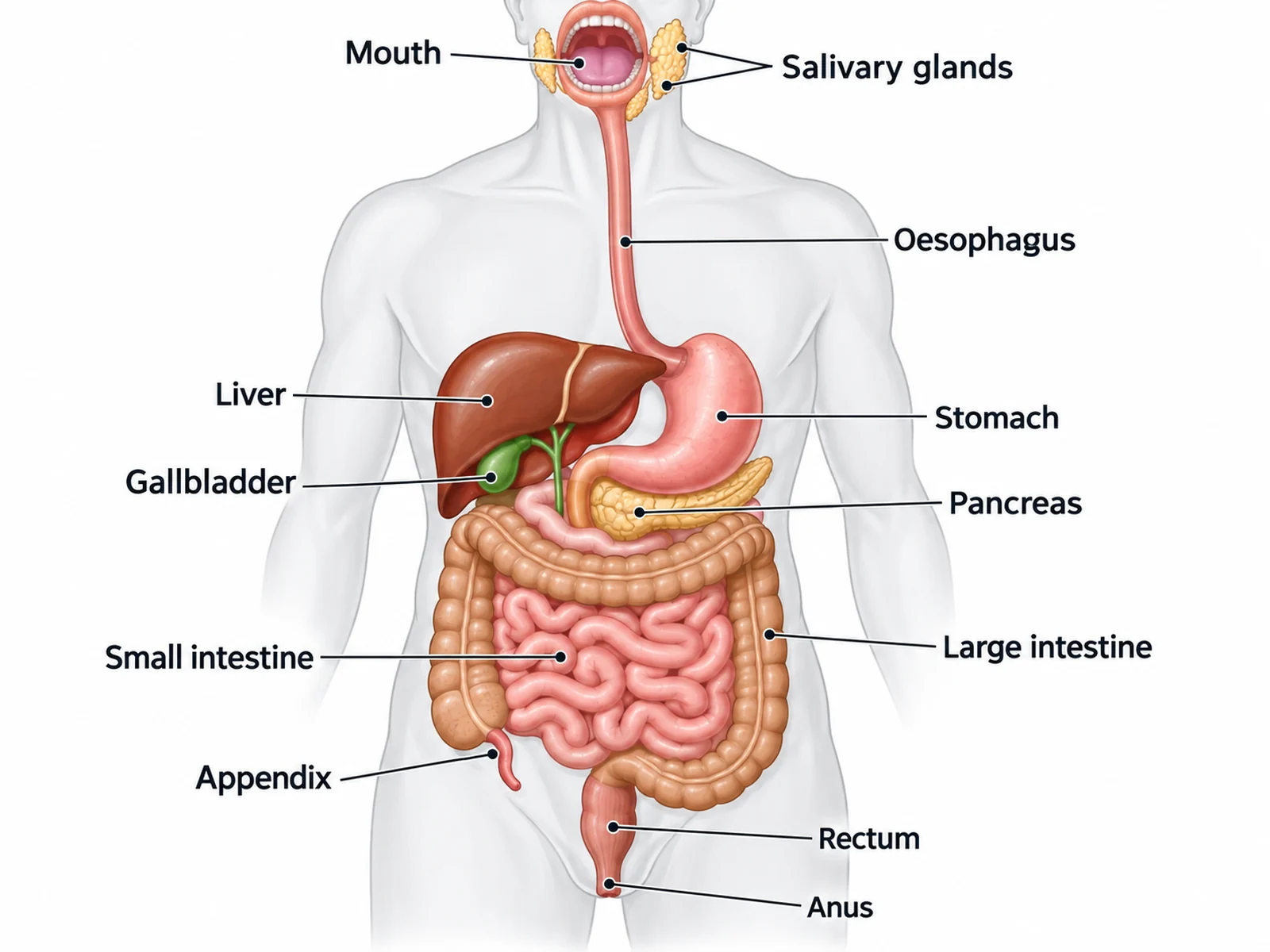
Labeled human digestive system diagram naming the mouth, salivary glands, oesophagus, stomach, liver, gallbladder, pancreas, small intestine, large intestine, appendix, rectum, and anus
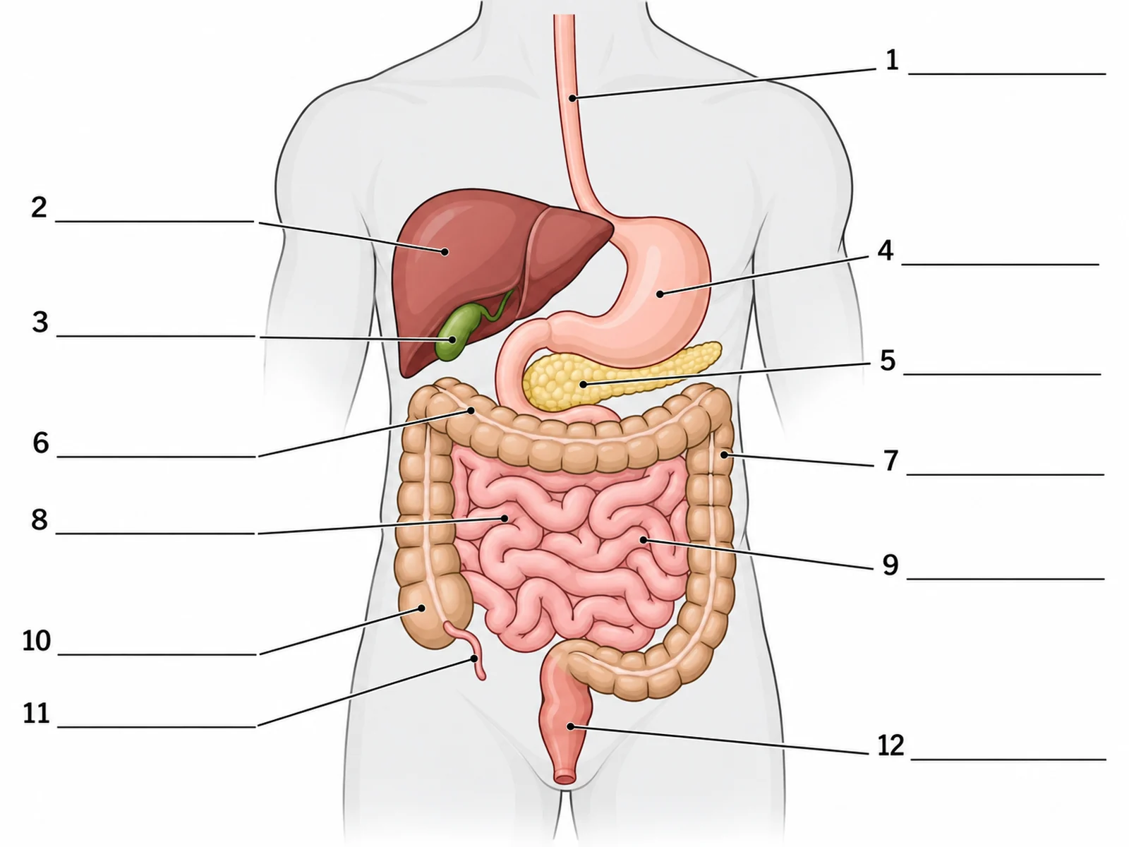
Blank unlabeled digestive system diagram with numbered empty label lines for students to complete
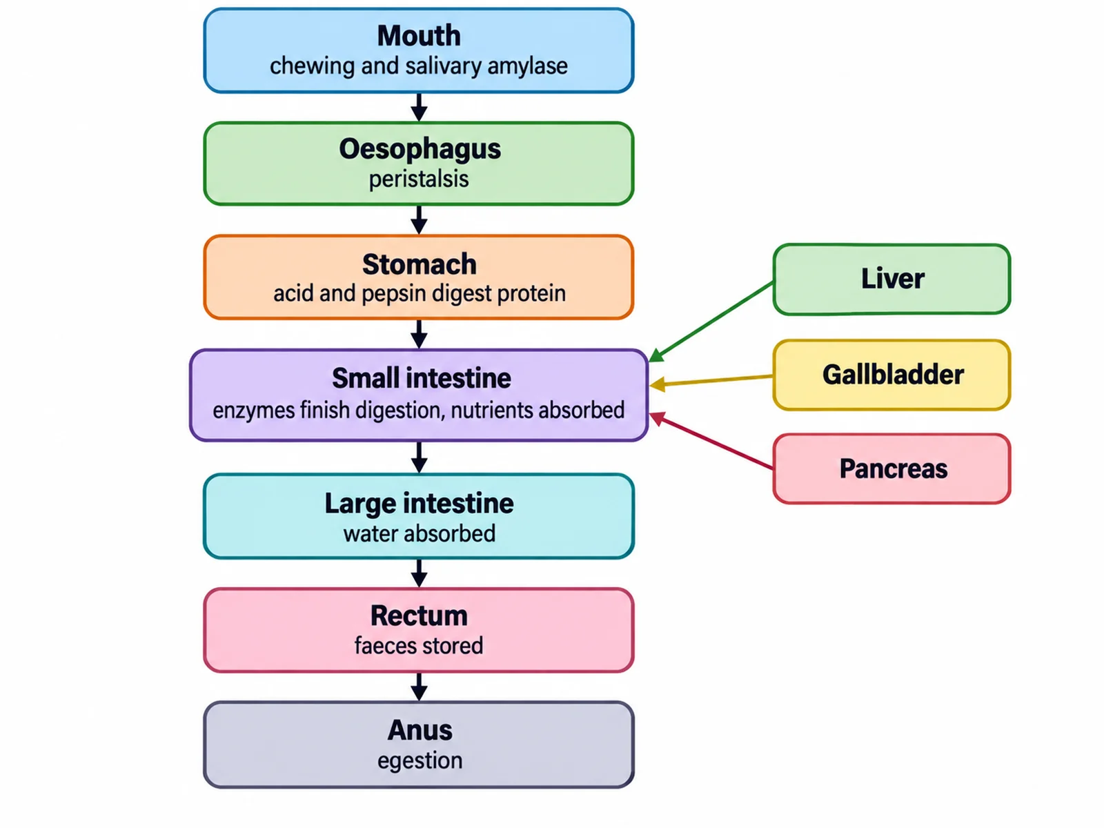
Flow chart of the human digestive system showing food passing from mouth to anus with the process at each stage

Sample previews. Describe your diagram to generate your own digestive figure.

Labeled Digestive System Diagram Examples

Use these as visual directions for labeled anatomy, blank worksheets, flow charts, simple versions for younger classes, and close-ups of the stomach and villi.

Labeled human digestive system diagram naming the mouth, salivary glands, oesophagus, stomach, liver, gallbladder, pancreas, small intestine, large intestine, appendix, rectum, and anus

Prompt

Create a fully labeled human digestive system diagram on a white background. Draw a simplified front-facing torso outline in pale grey with the digestive organs in soft flat colors inside it: mouth and salivary glands at the top, a tube-like oesophagus running down, a J-shaped stomach on the viewer’s right, a large brown liver on the viewer’s left with a small green gallbladder beneath it, a pale pancreas behind and below the stomach, a coiled pink small intestine in the centre, a thicker large intestine framing it on three sides, a small appendix branching off the caecum at the lower LEFT of the frame (the body’s right side), then rectum and anus. Label every organ with a thin leader line and readable dark sans-serif text.
Labeled Digestive System

Blank unlabeled digestive system diagram with numbered empty label lines for students to complete

Prompt

Create a blank digestive system worksheet. Draw the same torso and organ outlines in clean line art with pale flat fills, but leave every label empty: draw thin leader lines from each organ out to a short horizontal ruled blank line with no text on it. Number the blanks 1 to 12. Include generous white space and no answer key.

Blank Digestive System Worksheet

Flow chart of the human digestive system showing food passing from mouth to anus with the process at each stage

Prompt

Create a digestive system flow chart on a white background. Draw the alimentary canal as a vertical sequence of rounded boxes connected by downward arrows: Mouth, Oesophagus, Stomach, Small intestine, Large intestine, Rectum, Anus. Inside each box name the organ in bold and add a short process line beneath it, such as "chewing and salivary amylase", "peristalsis", "acid and pepsin digest protein", "enzymes finish digestion, nutrients absorbed", "water absorbed", "faeces stored", "egestion". To the right, draw three side boxes for the accessory organs Liver, Gallbladder, and Pancreas, each with a thin arrow pointing into the small intestine box.

Digestive System Flow Chart

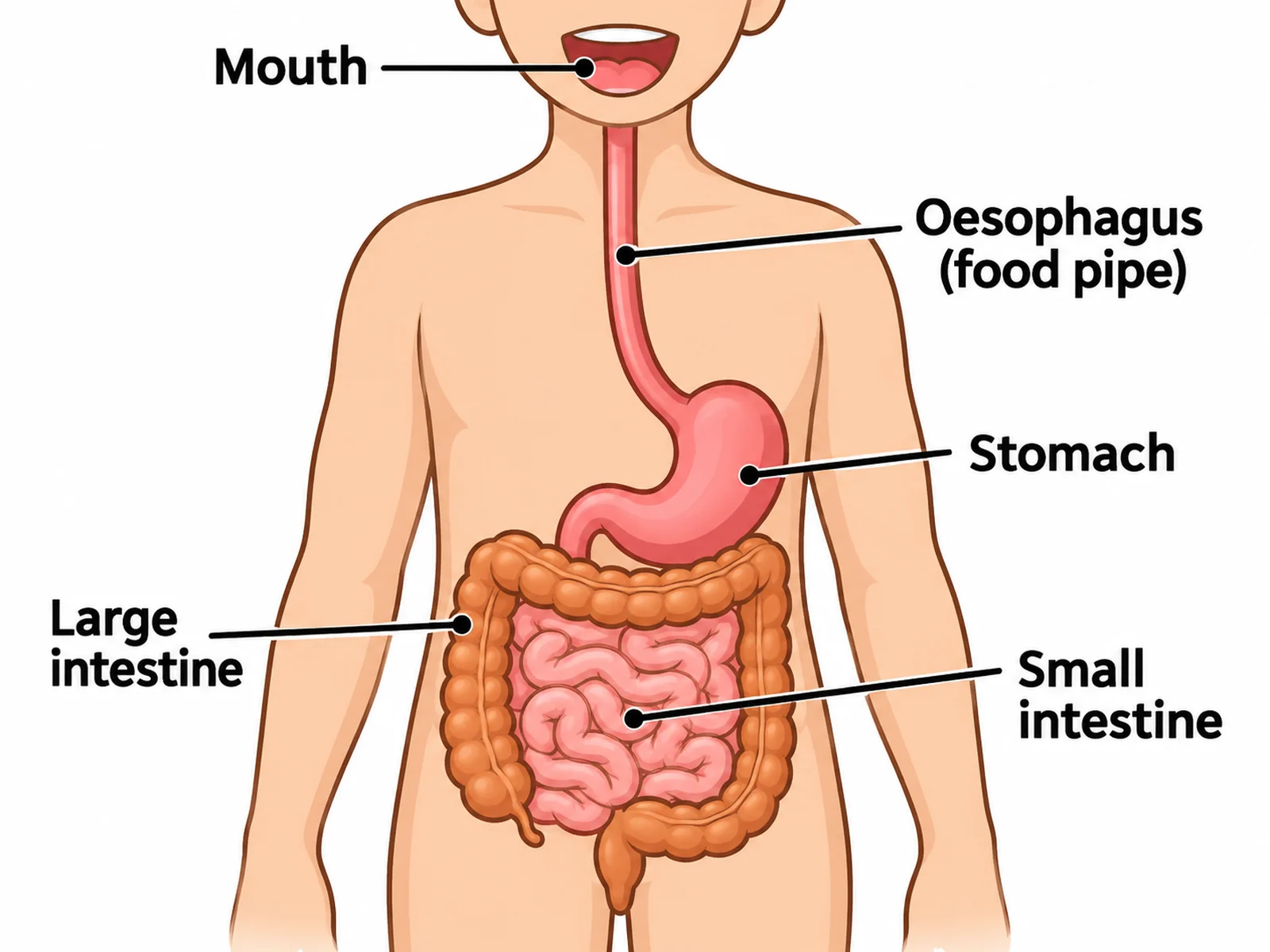
Simple digestive system diagram for younger students labeling only the mouth, oesophagus, stomach, small intestine, and large intestine

Prompt

Create a simple digestive system diagram for primary or lower-secondary students. Show only five labeled parts: mouth, oesophagus (food pipe), stomach, small intestine, and large intestine. Use bold flat colors, thick clean outlines, large readable text, and plenty of white space. Omit the liver, gallbladder, pancreas, and appendix.

Simple Digestive System

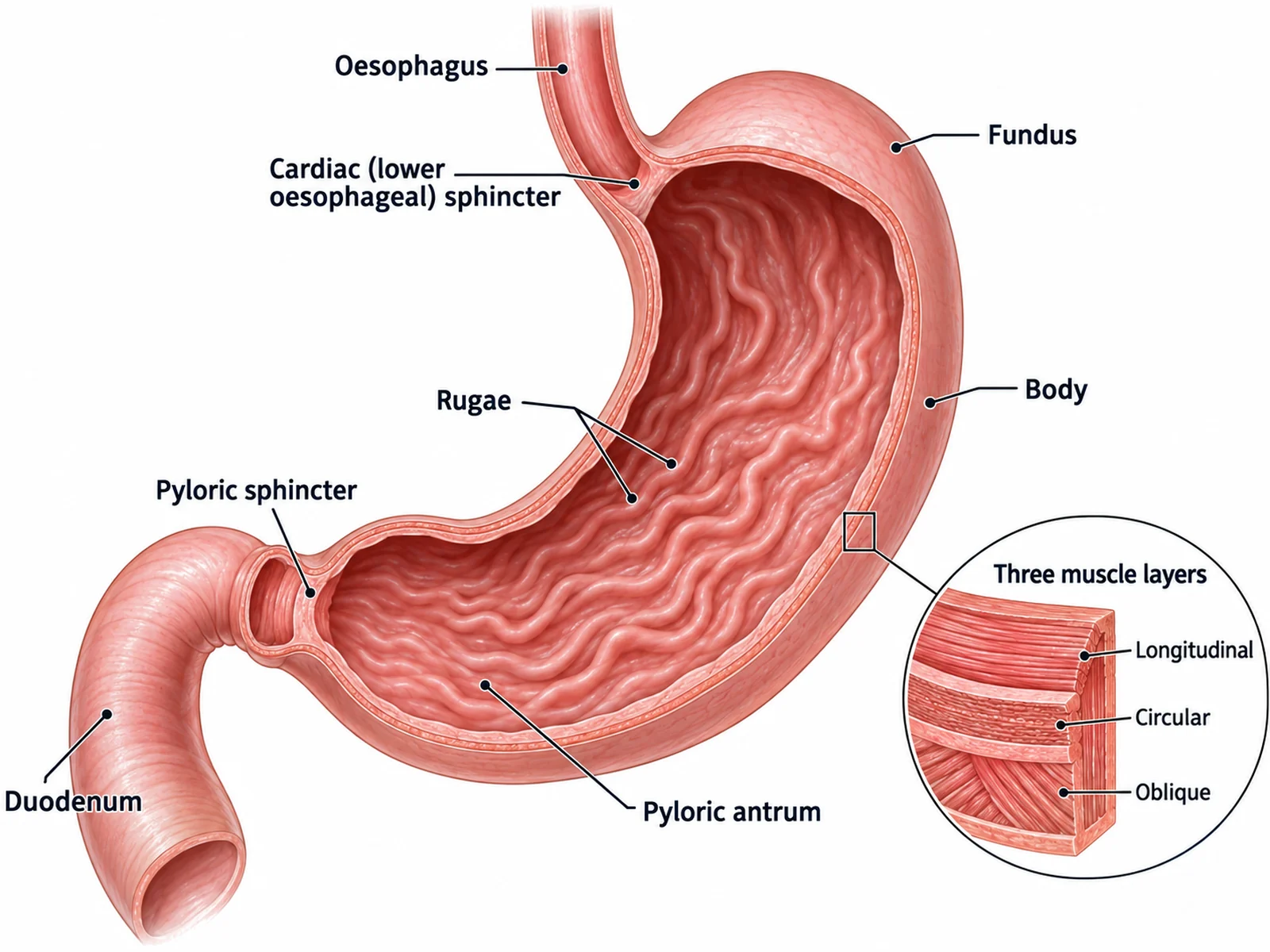
Labeled stomach diagram showing the cardiac sphincter, fundus, body, rugae, pyloric antrum, pyloric sphincter, and muscle layers

Prompt

Create a labeled stomach diagram on a white background, drawn as a cut-away so the interior is visible. Label the oesophagus entering at the top, the cardiac (lower oesophageal) sphincter, fundus, body, the folded rugae lining the inner wall, pyloric antrum, pyloric sphincter, and the duodenum leaving at the bottom. Add a small inset callout showing the three muscle layers of the stomach wall: longitudinal, circular, and oblique. Use soft anatomical pinks and reds with thin leader lines.

Stomach Anatomy

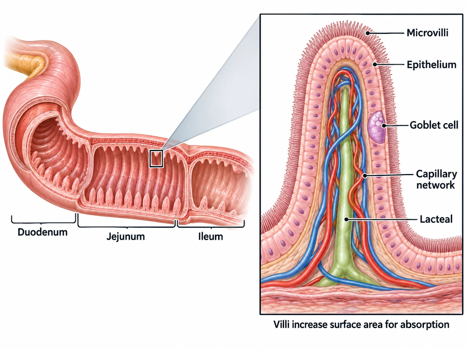
Labeled diagram of the small intestine with a magnified view of a villus showing epithelium, capillary network, and lacteal

Prompt

Create a labeled small intestine absorption diagram with two linked panels. On the left, a short section of small intestine drawn in cross-section with its folded inner lining, labeled duodenum, jejunum, and ileum along the organ. On the right, a magnified single villus connected to the left panel by a callout wedge, labeled epithelium, microvilli, capillary network, lacteal, and goblet cell. Add a short caption under the right panel reading "Villi increase surface area for absorption".

Small Intestine and Villi

How to make a labeled digestive system diagram

1

Choose the view and the organs

An anatomical torso view, a flow chart of the food route, or a close-up of one organ. List the organs your syllabus requires and say whether you want labels or blanks.

2

Generate the diagram

The generator places the organs anatomically and adds clean leader lines with readable text.

3

Check the anatomy, then export

Confirm the liver is on the body’s right, the stomach on the left, and each leader line points at the right organ, then download the PNG for a worksheet, slide, or revision sheet.

What is a digestive system diagram?

A digestive system diagram is a labeled map of the organs that break food down, absorb nutrients, and remove waste. It is usually drawn as a front-facing torso with the alimentary canal traced from mouth to anus and the accessory organs (liver, gallbladder, pancreas) placed alongside. The same content can also be drawn as a flow chart when the point is the sequence rather than the anatomy.

The organs and what each one does

  • Mouth and salivary glands: chewing increases surface area; salivary amylase begins starch digestion.
  • Oesophagus: a muscular tube that pushes food to the stomach by waves of peristalsis.
  • Stomach: hydrochloric acid kills bacteria and pepsin digests protein; the wall churns food into chyme.
  • Liver: produces bile, which emulsifies fats into smaller droplets.
  • Gallbladder: stores and concentrates bile, releasing it into the duodenum.
  • Pancreas: secretes amylase, protease, and lipase, plus bicarbonate to neutralise stomach acid.
  • Small intestine: duodenum, jejunum, and ileum; enzymes finish digestion and villi absorb nutrients.
  • Large intestine: caecum, colon, and rectum; absorbs water and mineral salts from the remaining material.
  • Anus: egests faeces, the undigested waste the body cannot absorb.

Common errors to check before you print

  • Liver and stomach swapped. In a front-facing diagram the liver appears on the viewer’s left and the stomach on the viewer’s right.
  • Pancreas drawn in front of the stomach. It lies behind and below it.
  • Appendix on the wrong side. It attaches to the caecum in the body’s right lower quadrant, which appears on the viewer’s left in a front-facing diagram.
  • Digestion and egestion confused. Egestion removes undigested food; excretion removes metabolic waste.

Digestive System Diagram Generator FAQ

The alimentary canal runs mouth, salivary glands, oesophagus, stomach, small intestine (duodenum, jejunum, ileum), large intestine (caecum, colon, rectum), and anus. The accessory organs that sit beside it are the liver, gallbladder, and pancreas. A complete diagram labels all of these, and most syllabuses also expect the appendix.

Need other anatomy figures?

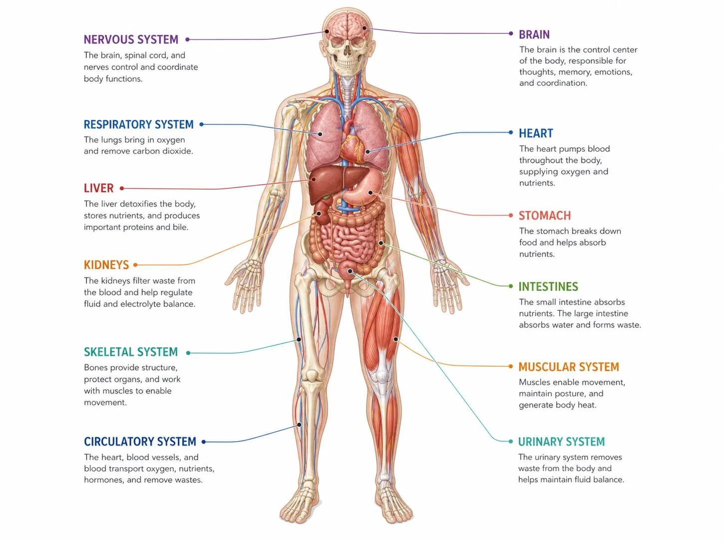
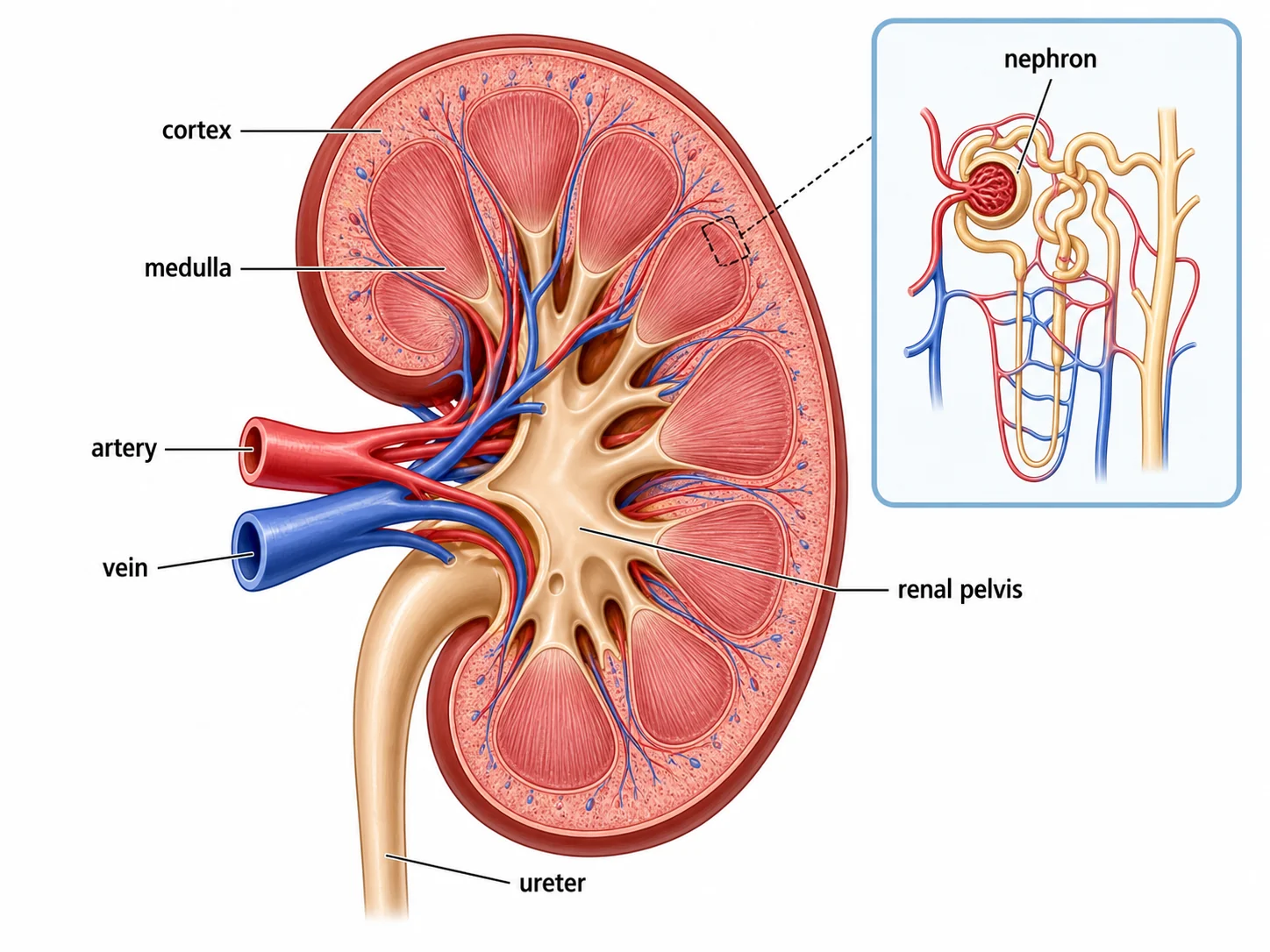
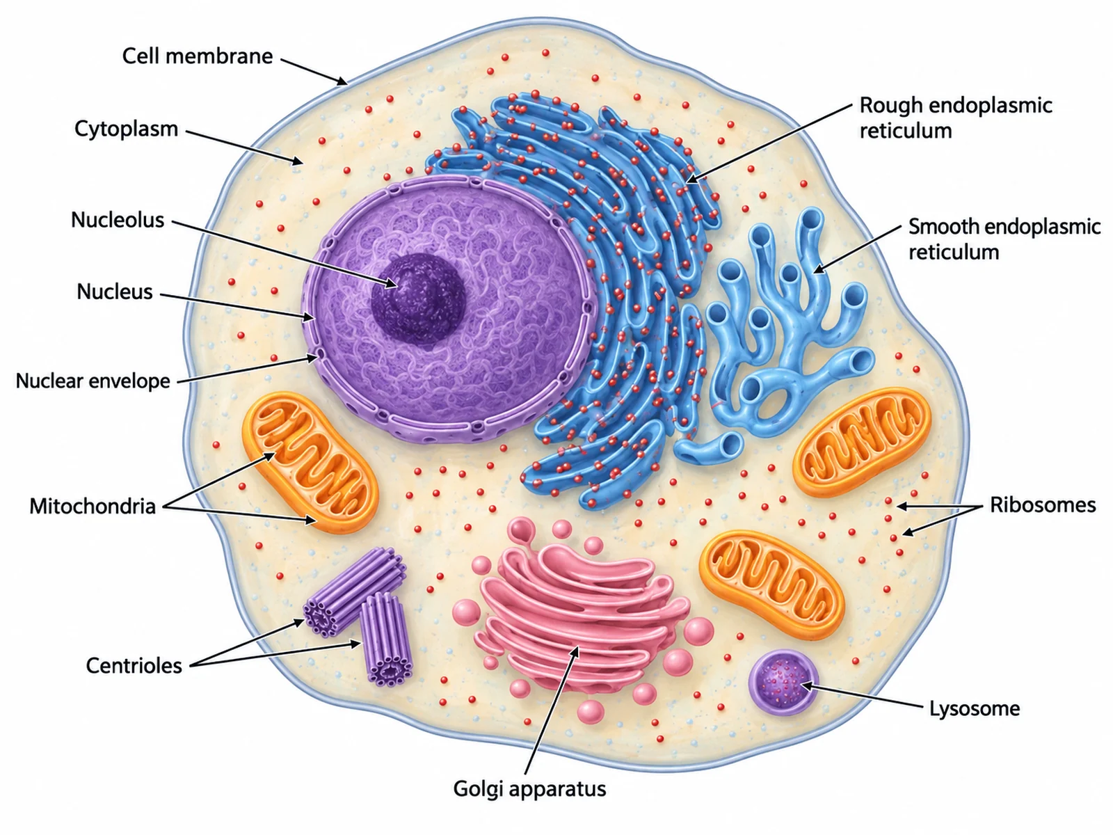
Generate body system diagrams, organ close-ups, cell diagrams, and worksheet-ready biology illustrations with the same SciFigure AI workflow.