Generate a labeled kidney diagram of a coronal section, from the renal capsule and cortex through the pyramids and calyces to the renal pelvis and ureter, or a blank kidney diagram with numbered lines and a matching answer key. Add the blood supply, the outside of the kidney, or the route from kidney to bladder.
Labeled, numbered or blankCortex to ureterMatching answer key
Checked against OpenStax A&P
Worksheet + answer key pairs
Print-ready PNG
AI Kidney Diagram Generator
Describe the section and the structures to label, or pick a blank worksheet from the Labels menu.
640 / 2000 characters
Generate a publication-ready PNG from your description.
Preview
Kidney Diagram Example Previews
Target aspect ratio: 4:3
Sample previews. Describe your diagram to generate your own kidney figure.
Labeled and Blank Kidney Diagram Examples
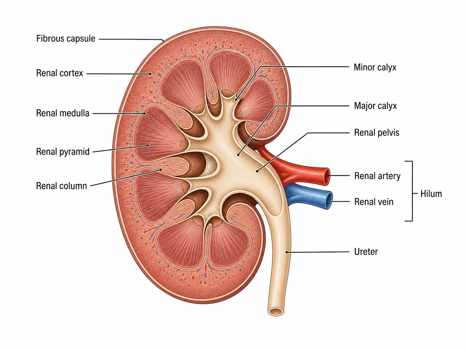
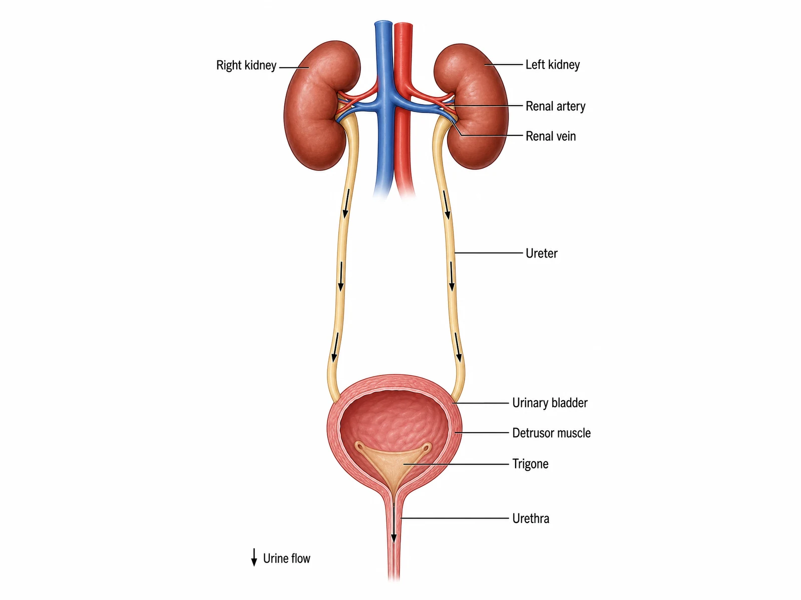
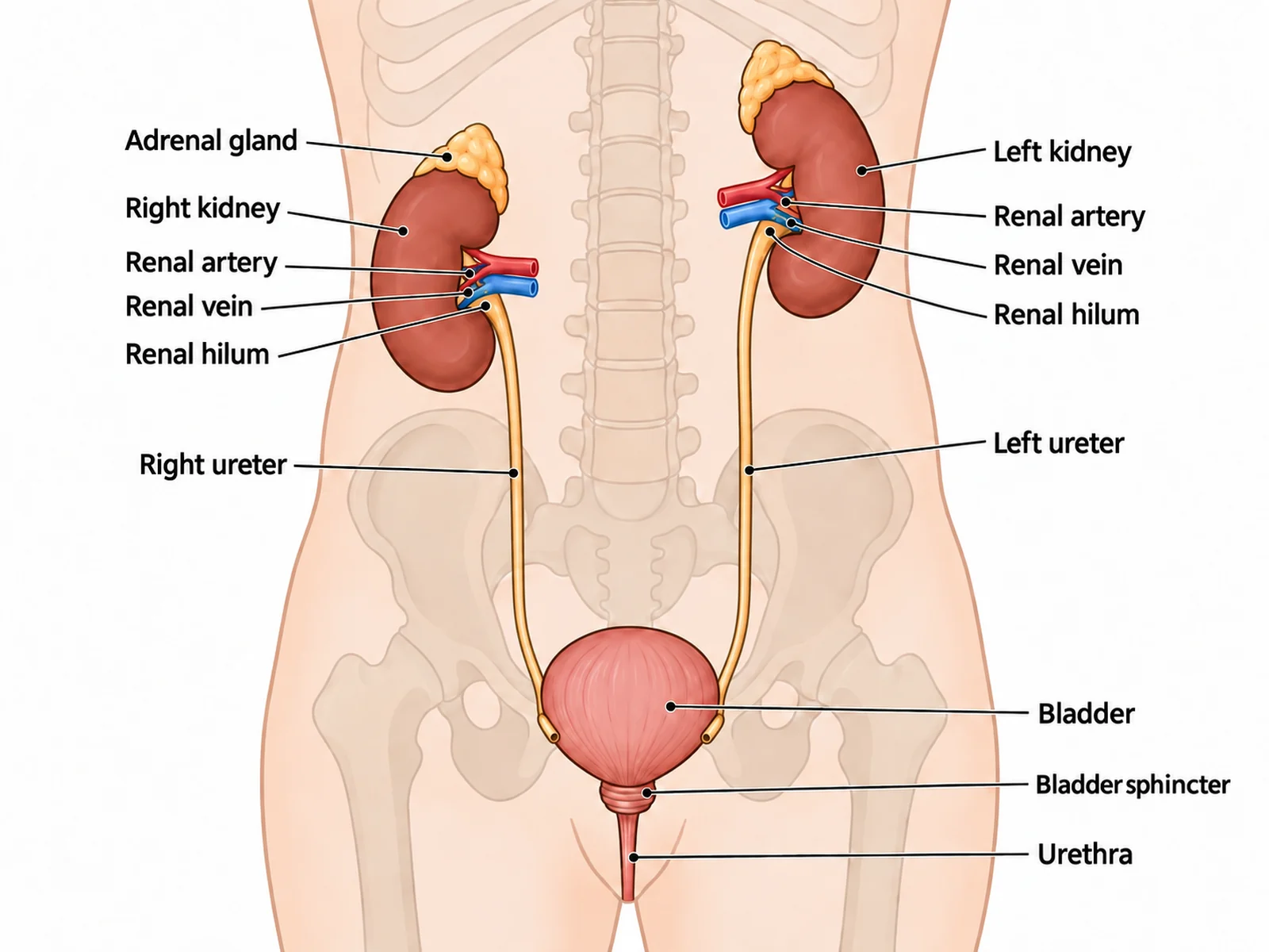
A labeled kidney cross section, a blank kidney diagram worksheet with its matching answer key, and the route of urine from the kidneys to the bladder.
Prompt
Create a labeled kidney diagram on a white background: one human kidney cut lengthwise in a coronal section, large, with the concave hilum facing the viewer’s right. Draw a thin fibrous capsule, a pale granular renal cortex, several striated cone-shaped renal pyramids with their tips pointing inward, renal columns of cortex between the pyramids, each papilla opening into a minor calyx, minor calyces joining into major calyces and a funnel-shaped renal pelvis, and the ureter running down from the hilum, with a red renal artery and a blue renal vein at the hilum. Label "Fibrous capsule", "Renal cortex", "Renal medulla", "Renal pyramid" and "Renal column" on the left, and "Minor calyx", "Major calyx", "Renal pelvis", "Renal artery", "Renal vein" and "Ureter" on the right, with a bracket labeled "Hilum" around the two vessels.
Use this example
Prompt
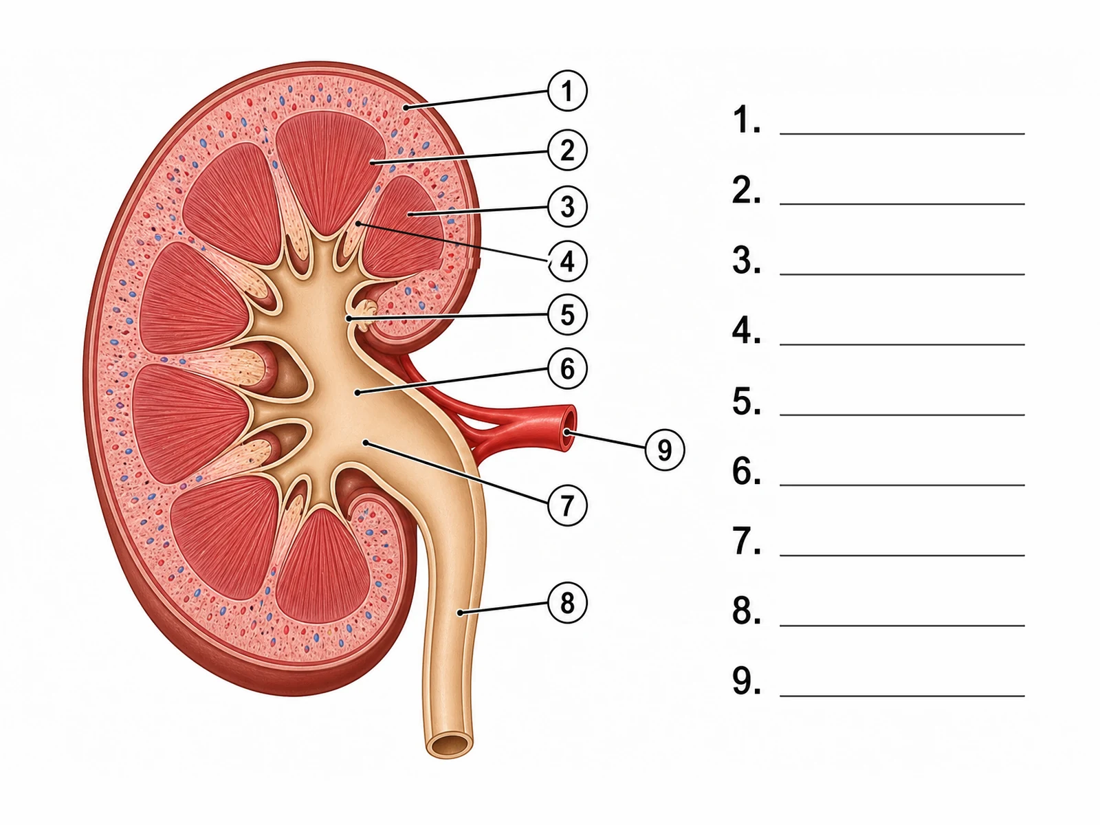
Create a blank kidney diagram worksheet for students to label. Draw one human kidney cut in a coronal section on a white background, its hilum facing the viewer’s right: a dark red rim for the renal capsule, a pale speckled renal cortex with small red and blue vessels, several dark red striated renal pyramids with their tips pointing inward, pale strips of renal column between neighboring pyramids, each papilla sitting in a cup-shaped minor calyx, the minor calyces joining into major calyces and a tan, funnel-shaped renal pelvis that narrows into the ureter running down from the hilum, and a red renal artery entering at the hilum. Draw nine thin leader lines out to the right, each ending in a small numbered circle, in this order: 1 = Renal cortex, 2 = Renal medulla, 3 = Renal pyramid, 4 = Renal column, 5 = Minor calyx, 6 = Major calyx, 7 = Renal pelvis, 8 = Ureter, 9 = Renal artery. To the right of the kidney add a column of numbered answer lines, 1 to 9, each an empty ruled line. Put no structure names anywhere on the image.
Blank worksheet
Use this example
Prompt
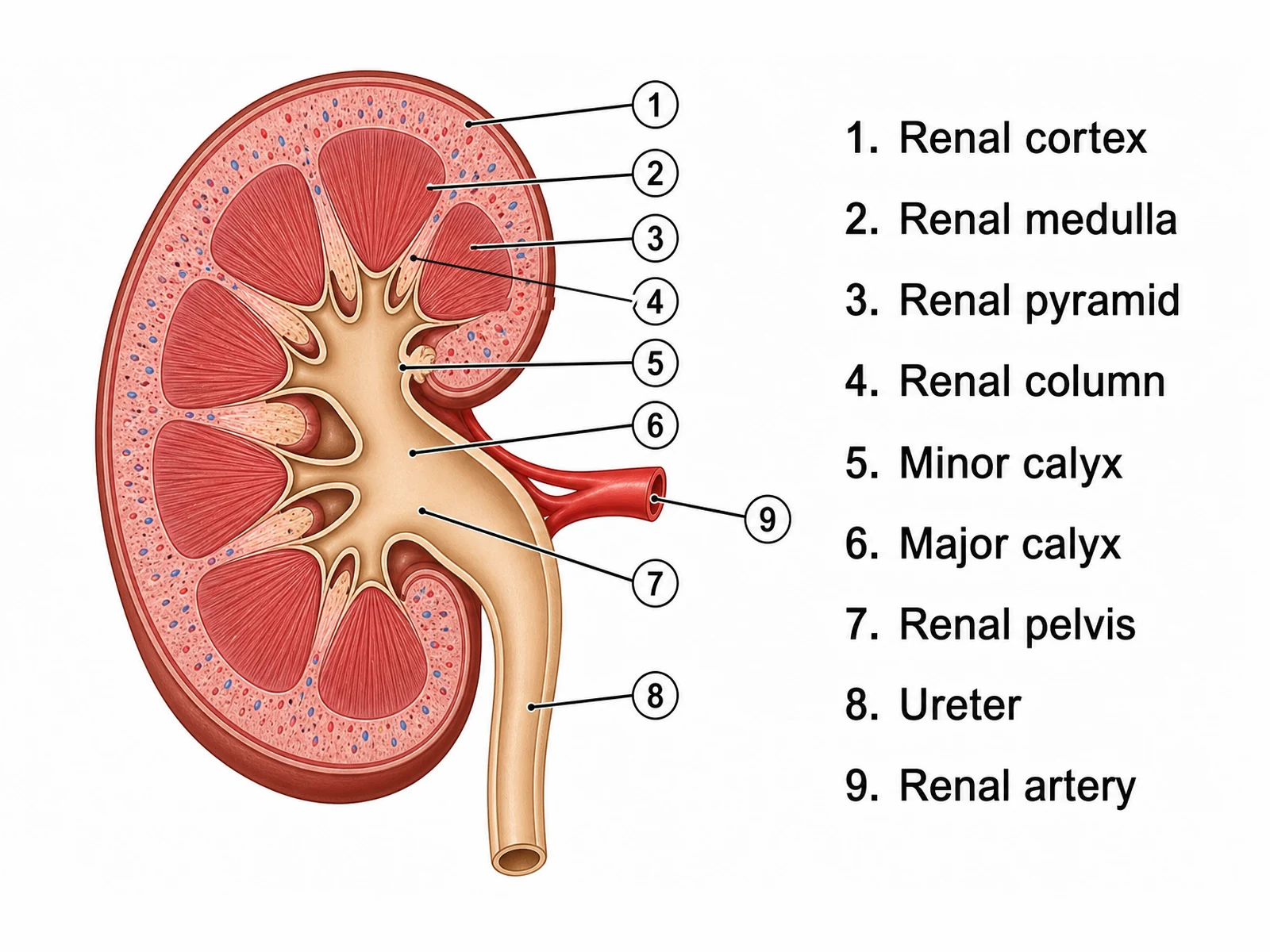
Create the answer key for a blank kidney worksheet. Draw one human kidney cut in a coronal section on a white background, its hilum facing the viewer’s right: a dark red rim for the renal capsule, a pale speckled renal cortex with small red and blue vessels, several dark red striated renal pyramids with their tips pointing inward, pale strips of renal column between neighboring pyramids, each papilla sitting in a cup-shaped minor calyx, the minor calyces joining into major calyces and a tan, funnel-shaped renal pelvis that narrows into the ureter running down from the hilum, and a red renal artery entering at the hilum. Draw nine thin leader lines out to the right, each ending in a small numbered circle, in this order: 1 = Renal cortex, 2 = Renal medulla, 3 = Renal pyramid, 4 = Renal column, 5 = Minor calyx, 6 = Major calyx, 7 = Renal pelvis, 8 = Ureter, 9 = Renal artery. To the right of the kidney add a key listing each number with its structure name, so it matches the worksheet line for line.
Numbered with key
Use this example
Prompt
Create a front-view diagram of the route from the kidneys to the bladder on a white background. Draw the right kidney on the viewer’s left and the left kidney on the viewer’s right, the inferior vena cava in blue on the viewer’s left of the midline and the aorta in red beside it, with the renal arteries and renal veins joining each hilum and the left renal vein crossing in front of the aorta. Run a ureter down from each kidney with small downward arrows to the bladder, shown cut open with its muscular wall and the triangular trigone between the two ureteric openings and the urethra. Label "Right kidney", "Left kidney", "Renal artery", "Renal vein", "Ureter", "Urinary bladder", "Detrusor muscle", "Trigone" and "Urethra", and add a small legend arrow reading "Urine flow".
Kidney to bladder
Use this example
How to make a labeled or blank kidney diagram
1
Pick the view, level, and labels
A coronal section, the outside of the kidney, the blood supply, the route from kidney to bladder, the kidneys in the body, or nephrons in a pyramid. Then choose labeled, numbered, blank, a word bank, or fully unlabeled.
2
Generate the kidney
The generator keeps the cortex outside and the pyramids pointing inward, joins papillae to minor calyces, major calyces, and the renal pelvis in order, and sends the ureter down from the hilum.
3
Make the answer key, check, and export
For a worksheet, make the answer key from the same result so the numbers match. Check the pyramids, the calyces, and the vessels at the hilum, then download the PNG.
Kidney key facts
Size
About 11–14 cm long, 6 cm wide, 4 cm thick
Weight
About 125–175 g (male), 115–155 g (female)
Position
Retroperitoneal, about T12 to L3
Lower kidney
The right, pushed down by the liver
Blood flow at rest
About 25% of cardiac output
Nephrons per kidney
About 1 million
Major calyces
Usually 2 or 3
Coverings, inside out
Renal capsule, perirenal fat, renal fascia
Hilum, front to back
Renal vein, renal artery, renal pelvis
Urine path
Papilla → minor calyx → major calyx → pelvis → ureter
What is a kidney diagram?
A kidney diagram is a labeled drawing of the human kidney, usually cut lengthwise in a coronal section so the inside can be seen. It shows how the organ is built: a pale outer cortex, a striped inner medulla divided into renal pyramids, and a branching set of funnels, the calyces and the renal pelvis, that collect urine and pass it to the ureter. Blood vessels enter and leave at the hilum on the kidney’s inner border.
In class the figure appears in two forms. A labeled kidney diagram names each part and works as a reference or an answer key. A blank kidney diagram keeps the drawing and the leader lines but leaves the names off, so students write them in. This page makes both from one description, plus a numbered version with a key, a worksheet with a word bank, and views of the blood supply and the route from kidney to bladder.
Parts of the kidney, from outside to inside
Renal fascia and perirenal fat: the outer sheet of connective tissue and the cushion of fat around the kidney. Diagrams of the kidney in the body show them; most sections leave them off.
Renal capsule: the thin, tough fibrous layer stuck to the surface of the kidney.
Renal cortex: the pale, granular outer zone where blood is filtered in the renal corpuscles.
Renal medulla: the inner zone, divided into cone-shaped renal pyramids.
Renal columns: strips of cortex-like tissue between the pyramids, carrying blood vessels.
Renal papilla: the tip of each pyramid, where the collecting ducts release urine.
Minor and major calyces: the cups that catch urine from the papillae, and the larger channels they join into.
Renal pelvis: the single funnel that collects urine from the major calyces and narrows into the ureter.
Renal sinus and hilum: the fat-filled space inside the kidney that holds the pelvis and vessels, and the notch on the inner border where they leave.
The outside of the kidney
Each kidney is bean-shaped, about 11 to 14 cm long, 6 cm wide, and 4 cm thick, roughly the size of a fist. It has an upper and a lower pole, a convex outer border, and a concave inner border with the hilum in its middle. The hilum faces the spine, slightly forward and down. An adrenal gland sits on the upper pole as a separate gland. On an outside view the renal vein lies in front at the hilum, the renal artery behind it, and the renal pelvis furthest back, turning down as the ureter. The right kidney’s renal artery is longer, because the aorta lies left of the midline; the left kidney’s renal vein is longer, because the inferior vena cava lies to the right.
The renal cortex and renal medulla
The cortex is the outer zone, a few millimeters to about a centimeter thick under the capsule. It looks granular because it is packed with renal corpuscles, the tiny filters of the nephrons, and their coiled tubules. It also reaches down between the pyramids as the renal columns, so in a section the cortex looks like a rim with fingers.
The medulla is the inner zone. It is not one continuous layer but a set of separate renal pyramids, cone-shaped and finely striped. The stripes are the loops of Henle and the collecting ducts, all running in the same direction toward the tip of the pyramid. The medulla is where urine is concentrated: the loops build up a salty environment that draws water out of the collecting ducts when the body needs to save it.
Renal pyramids, papillae, and renal lobes
A human kidney has several renal pyramids, typically around a dozen, though the number varies from person to person. Each pyramid has its broad base against the cortex and its tip, the renal papilla, pointing inward toward the renal sinus. On the papilla, the largest collecting ducts (papillary ducts) open through tiny pores and release urine into a minor calyx. A renal lobe is one pyramid with the cortex over its base and half of each renal column beside it. A newborn kidney often shows these lobes as bumps on its surface, which smooth out during childhood.
The collecting system: calyces, pelvis, and ureter
Once urine leaves the papillae, the kidney no longer changes it. The collecting system only carries it out. Each papilla sits in a cup-shaped minor calyx. Several minor calyces join into a major calyx, and the two or three major calyces join into the renal pelvis, a funnel that fills much of the renal sinus. At the hilum the pelvis narrows into the ureter.
The walls of the calyces, pelvis, and ureter contain smooth muscle that contracts in waves, pushing urine onward by peristalsis rather than letting it simply drain. On a labeled diagram the order matters more than the shape: papilla, minor calyx, major calyx, renal pelvis, ureter. A drawing that joins a pyramid straight to the pelvis, or sends the ureter out of the top of the kidney, has the plumbing wrong.
Blood supply of the kidney
The kidneys receive about a quarter of the heart’s output at rest, far more than their size suggests, because their job is to filter the blood. The blood supply view traces the arteries in red and the veins in blue.
Renal artery: branches from the abdominal aorta and enters the hilum.
Segmental arteries: the first branches, given off at or just inside the hilum, each supplying one segment of the kidney.
Interlobar arteries: run outward through the renal columns, between the pyramids.
Arcuate arteries: arch over the base of each pyramid, along the border between cortex and medulla.
Cortical radiate arteries: run out from the arcuate arteries into the cortex and give off the afferent arterioles.
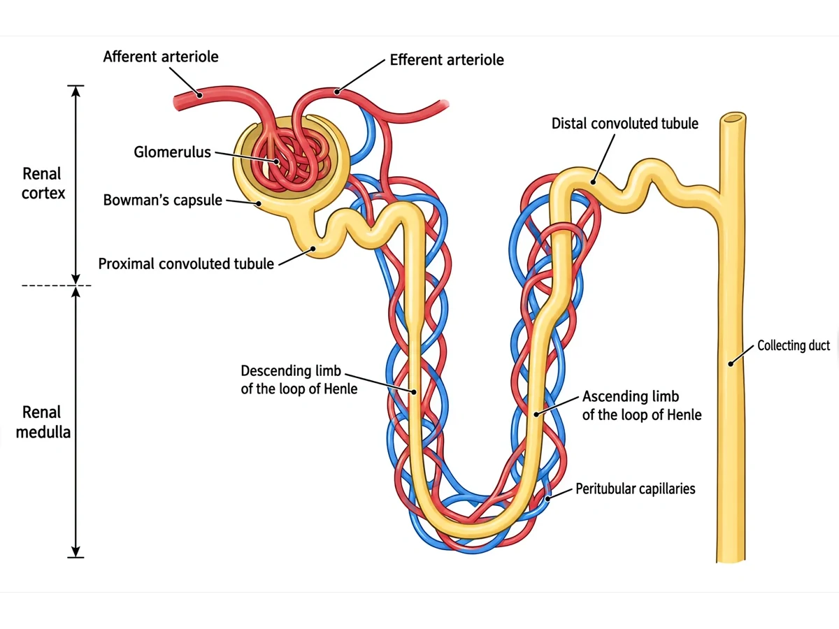
Afferent arteriole, glomerulus, efferent arteriole, and the capillaries around the tubules: the nephron’s own circulation, shown in detail on a nephron diagram.
Veins: cortical radiate, arcuate, and interlobar veins run back beside the arteries and join into the renal vein, which drains into the inferior vena cava.
Where the nephrons sit inside the kidney
Each kidney holds about a million nephrons, far too small to see on a whole-organ section. Their parts are sorted into the two zones. The renal corpuscles and the convoluted tubules lie in the cortex, which is why it looks granular. The loops of Henle dip down into the medulla, and the collecting ducts run through the pyramids in parallel, which gives them their stripes, before opening at the papilla. The nephrons-in-a-pyramid view draws a magnified wedge to show this. For the nephron itself, from the glomerulus and Bowman’s capsule to the collecting duct, use the nephron diagram generator.
Where the kidneys sit in the body
The kidneys lie high against the back wall of the abdomen, one on each side of the spine, at about the level of the twelfth thoracic to the third lumbar vertebra. They are retroperitoneal: they sit behind the lining of the abdominal cavity, not among the intestines. The lowest ribs cover their upper poles from behind.
The right kidney sits slightly lower than the left because the liver takes up the space above it. Each kidney is wrapped in its renal capsule, a cushion of fat, and the renal fascia, which hold it in place. An adrenal gland sits on top of each one. On a front view the person’s right kidney is on the viewer’s left, which is the most common orientation error on student diagrams.
From kidney to bladder: the path of urine
A diagram of the kidney to the bladder follows urine out of the organ. The nephrons make urine and the collecting ducts deliver it to the renal papillae. It drips into the minor calyces, flows into the major calyces and the renal pelvis, and enters the ureter at the hilum. Each ureter is a muscular tube, roughly 25 to 30 cm long, that runs down the back wall of the abdomen into the pelvis and enters the back of the bladder near its base at a slant, so the filling bladder squeezes it shut and stops urine flowing back. The bladder stores the urine until it leaves through the urethra. For the whole system in one front view, use the urinary system diagram generator.
What the kidneys do
Filter the blood: the two kidneys filter about 180 liters of fluid a day and return all but 1 to 2 liters of it, which leaves as urine.
Remove wastes: urea, creatinine, uric acid, and many drugs leave the body in the urine.
Balance water and salts: the kidneys keep the blood’s volume and its sodium, potassium, and calcium levels within narrow limits, under the control of ADH and aldosterone.
Keep the blood’s pH steady: they excrete acid and save bicarbonate.
Help control blood pressure: cells next to each glomerulus release renin when blood pressure falls.
Make hormones: erythropoietin tells the bone marrow to make red blood cells, and the kidneys turn vitamin D into its active form, calcitriol.
Kidney conditions that diagrams often show
Clinical and nursing courses often pair the normal kidney with what can go wrong, drawn on the same section. Kidney stones form from minerals in the urine, usually in the calyces or renal pelvis, and cause severe pain when they move into the ureter and get stuck at one of its narrow points. Hydronephrosis is the swelling of the renal pelvis and calyces when urine cannot drain, for example behind a stone. Polycystic kidney disease, an inherited condition, fills both kidneys with fluid-filled cysts and enlarges them. In chronic kidney disease the kidneys lose nephrons and often shrink. A kidney diagram leaves these out unless you ask for them.
Kidney labels by level
Simple: about six parts, such as cortex, medulla, renal pelvis, ureter, renal artery, and renal vein.
High school: the thirteen standard structures, from the renal capsule, cortex, medulla, pyramids, columns, and papillae through the calyces and pelvis to the ureter and the vessels at the hilum.
College: adds the renal sinus, the renal lobe, and the chain of arteries from segmental to cortical radiate.
Medical: adds the matching veins, the papillary ducts, the medullary rays, the fat and fascia around the kidney, and the poles.
Any level: an unlabeled section with no lines, for coloring or for quizzing aloud.
Using a blank kidney diagram in class
Start blank, end labeled: hand out the blank kidney worksheet, then show the answer key made from the same result so every number matches.
Follow a drop of urine: before naming anything, have students trace the route from a papilla to the ureter with a finger, then label the structures in that order.
Follow a drop of blood: on the blood supply view, trace the arteries from the aorta to the cortex and back out through the veins.
Zoom in, then out: pair the kidney section with the nephron diagram to show what the stripes of a pyramid are, and with the urinary system diagram to show where the ureter goes.
Word bank or not: include one for revision, leave it out for a test. The word bank is alphabetical, so its order gives nothing away.
Most common mistakes on a kidney diagram
These are the errors image models make most often on kidney sections. Check each one before a diagram goes into graded work.
MistakeCorrect
MistakeRenal pyramids with their tips pointing outward
CorrectBases face the cortex; the tips (papillae) point inward to the minor calyces
MistakeCortex and medulla swapped
CorrectThe pale cortex is the outer layer; the striped pyramids of the medulla lie inside it
MistakeRenal columns labeled as medulla
CorrectRenal columns are cortex tissue dipping between the pyramids
MistakePyramids draining straight into the renal pelvis
CorrectPapilla → minor calyx → major calyx → renal pelvis → ureter
MistakeUreter leaving the top or the outer edge of the kidney
CorrectIt leaves the hilum on the inner, concave border and runs down
MistakeRenal vein joined to the aorta, or the artery to the vena cava
CorrectRenal artery from the aorta; renal vein to the inferior vena cava
MistakeAdrenal gland drawn as part of the kidney
CorrectA separate endocrine gland capping the upper pole
MistakeInterlobar arteries running through the middle of a pyramid
CorrectThey run through the renal columns between pyramids, then arch over the bases as arcuate arteries
Renal cortex vs renal medulla
The two regions of a kidney section are the pair students mix up most. This is what each one holds and does.
Renal cortex
Renal medulla
Position
Outer layer, just under the renal capsule
Inner region, deep to the cortex
Appearance in section
Pale and granular
Darker, with striped renal pyramids
Shape
A continuous rim that dips between the pyramids as renal columns
Separate cone-shaped pyramids, bases outward, tips (papillae) inward
Parts of the nephron
Renal corpuscles, proximal and distal convoluted tubules, start of the collecting ducts
Loops of Henle and collecting ducts, ending as papillary ducts
Blood vessels
Cortical radiate arteries, afferent and efferent arterioles, glomeruli, peritubular capillaries
Vasa recta; arcuate vessels run along its border with the cortex
Main job
Filtering the blood and reabsorbing most of the filtrate
Building the salt gradient that concentrates urine, and draining it to the calyces
Kidney Diagram FAQ
A standard kidney diagram is a coronal section, the kidney cut lengthwise from top to bottom. From the outside in it names the renal capsule, the renal cortex, the renal medulla with its renal pyramids, the renal columns between them, the renal papillae, the minor and major calyces, the renal pelvis, and the ureter. At the hilum it adds the renal artery and renal vein. College diagrams add the segmental, interlobar, arcuate, and cortical radiate arteries, the renal sinus, and the fat and fascia around the kidney.
Generate body system diagrams, organ sections and close-ups, blank labeling worksheets with matching answer keys, and revision-ready biology figures with the same SciFigure AI workflow.