Generate a labeled muscular system diagram that names the major muscles on the front and back of the body, or a blank muscular system diagram with numbered lines for students to fill in, plus its answer key. Choose a view and a level, from ten muscles with everyday names to the forearm and calf muscles of an anatomy practical.
Labeled, numbered or blankFront and back viewsMatching answer key
Checked against OpenStax A&P
Worksheet + answer key pairs
Print-ready PNG
AI Muscular System Diagram Generator
Describe the view and the muscles to label, or pick a blank worksheet from the Labels menu.
708 / 2000 characters
Generate a publication-ready PNG from your description.
Preview
Muscular System Diagram Example Previews
Target aspect ratio: 4:3
Sample previews. Describe your diagram to generate your own muscle figure.
Labeled and Blank Muscular System Diagram Examples
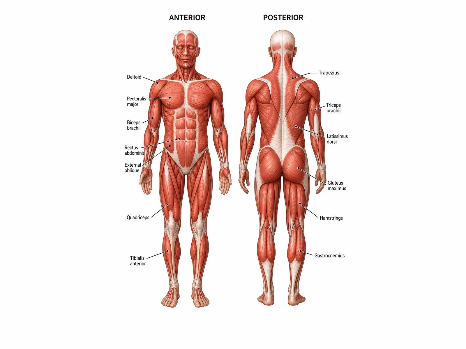
A labeled front-and-back muscle diagram, a blank muscular system worksheet in line art, a numbered color-coded diagram with its matching blank worksheet, and the major muscle groups.
Prompt
Create a labeled human muscular system diagram on a white background with two full-length figures of the same adult side by side, captioned "ANTERIOR" on the left and "POSTERIOR" on the right, both in anatomical position with the palms facing forward. Show the superficial muscles in realistic red with pale tendons and aponeuroses. On the anterior figure label, with thin leader lines on the left: "Deltoid", "Pectoralis major", "Biceps brachii", "Rectus abdominis", "External oblique" (on the side of the abdomen), "Quadriceps" and "Tibialis anterior". On the posterior figure label, with leader lines on the right: "Trapezius", "Triceps brachii", "Latissimus dorsi", "Gluteus maximus", "Hamstrings" and "Gastrocnemius".
Use this example
Prompt
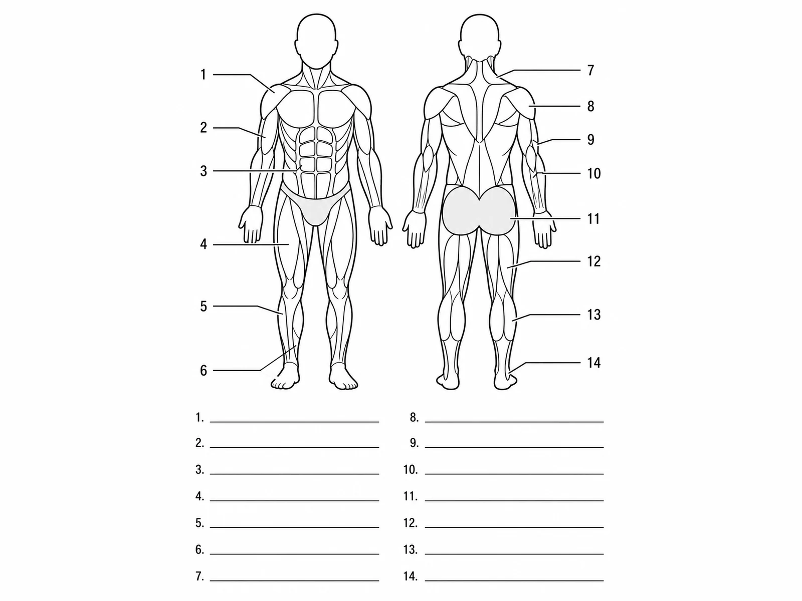
Create a blank muscular system diagram worksheet in clean black line art on a white background: two full-length outline figures side by side, a front view on the left and a back view on the right, with the main surface muscles drawn as outlined shapes and the gluteal muscles lightly shaded. Draw exactly 14 thin leader lines that end on these targets, numbered 1 to 6 on the front figure (numbers on the left) and 7 to 14 on the back figure (numbers on the right): 1 = Deltoid, 2 = Biceps brachii, 3 = Rectus abdominis, 4 = Quadriceps femoris, 5 = Tibialis anterior, 6 = Soleus, 7 = Trapezius, 8 = Deltoid, 9 = Triceps brachii, 10 = Forearm extensors, 11 = Gluteus maximus, 12 = Hamstrings, 13 = Gastrocnemius, 14 = Calcaneal (Achilles) tendon. Below the figures add two columns of numbered answer lines, 1 to 7 and 8 to 14. Put no muscle names anywhere on the image.
Blank worksheet
Use this example
Prompt
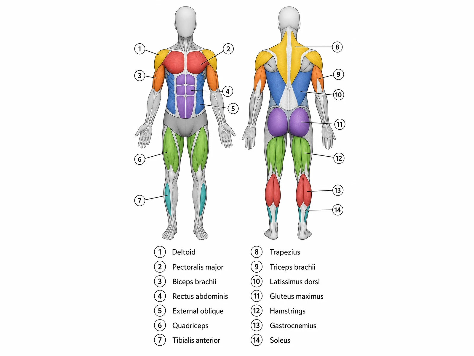
Create a numbered muscular system diagram with a key. Draw two full-length figures of the same adult side by side on a white background, an anterior view on the left and a posterior view on the right, both in anatomical position with the palms facing forward, drawn in pale grey with only the numbered muscles in flat colors. Front: deltoid yellow, pectoralis major red, biceps brachii orange, rectus abdominis purple, external oblique blue, quadriceps green, tibialis anterior teal. Back: trapezius yellow, triceps brachii orange, latissimus dorsi blue, gluteus maximus purple, hamstrings green, gastrocnemius red, soleus teal. Every other muscle, including the forearms and the inner calf seen from the front, stays grey. Each colored muscle gets one thin leader line ending in a small numbered circle, 1 to 7 on the front figure and 8 to 14 on the back: 1 = Deltoid, 2 = Pectoralis major, 3 = Biceps brachii, 4 = Rectus abdominis, 5 = External oblique, 6 = Quadriceps, 7 = Tibialis anterior, 8 = Trapezius, 9 = Triceps brachii, 10 = Latissimus dorsi, 11 = Gluteus maximus, 12 = Hamstrings, 13 = Gastrocnemius, 14 = Soleus. Below the figures add a two-column key listing every number in its circle with the muscle name beside it, 1 to 7 in the left column and 8 to 14 in the right.
Numbered with key
Use this example
Prompt
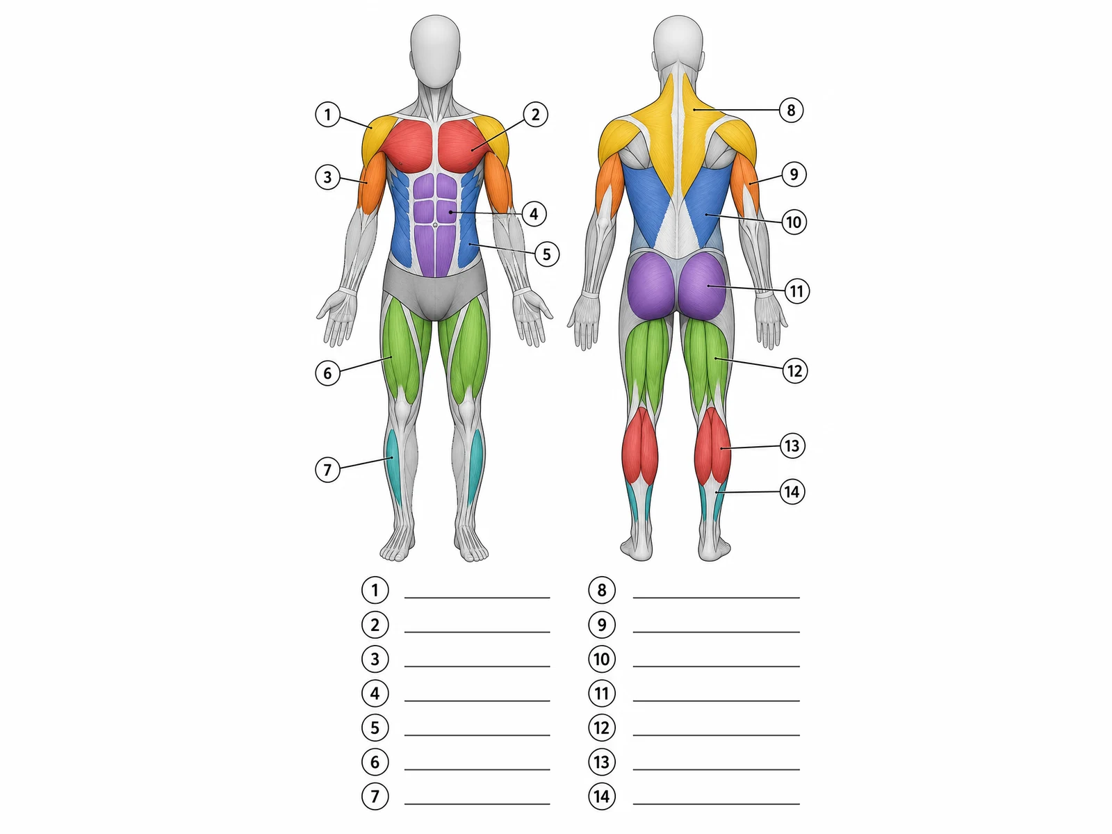
Create the blank worksheet for a numbered muscular system diagram. Draw two full-length figures of the same adult side by side on a white background, an anterior view on the left and a posterior view on the right, both in anatomical position with the palms facing forward, drawn in pale grey with only the numbered muscles in flat colors. Front: deltoid yellow, pectoralis major red, biceps brachii orange, rectus abdominis purple, external oblique blue, quadriceps green, tibialis anterior teal. Back: trapezius yellow, triceps brachii orange, latissimus dorsi blue, gluteus maximus purple, hamstrings green, gastrocnemius red, soleus teal. Every other muscle, including the forearms and the inner calf seen from the front, stays grey. Each colored muscle gets one thin leader line ending in a small numbered circle, 1 to 7 on the front figure and 8 to 14 on the back: 1 = Deltoid, 2 = Pectoralis major, 3 = Biceps brachii, 4 = Rectus abdominis, 5 = External oblique, 6 = Quadriceps, 7 = Tibialis anterior, 8 = Trapezius, 9 = Triceps brachii, 10 = Latissimus dorsi, 11 = Gluteus maximus, 12 = Hamstrings, 13 = Gastrocnemius, 14 = Soleus. Below the figures, where the key would go, add two columns of numbered circles, 1 to 7 and 8 to 14, each followed by an empty ruled answer line. Put no muscle names anywhere on the image.
Blank worksheet
Use this example
Prompt
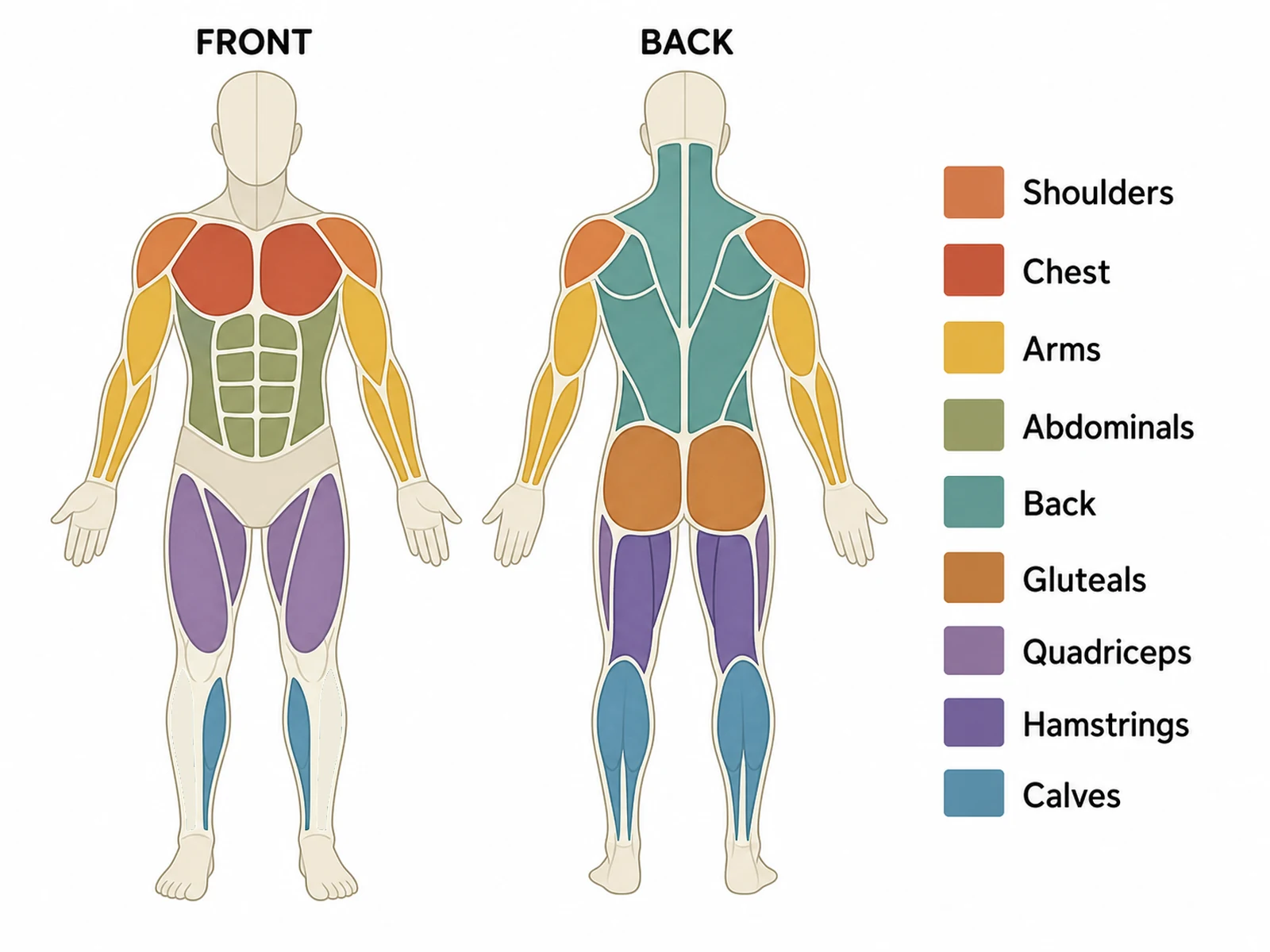
Create a major muscle groups diagram on a white background: two simple full-length figures in a cream fill, captioned "FRONT" and "BACK", with each muscle group shaded in its own flat color and a legend on the right reading "Shoulders", "Chest", "Arms", "Abdominals", "Back", "Gluteals", "Quadriceps", "Hamstrings" and "Calves". Shade the deltoids as shoulders, the pectorals as chest, the upper arms and forearms as arms, the rectus abdominis and obliques as abdominals, and the front of the thighs as quadriceps; on the back shade the trapezius and latissimus dorsi as back, the buttocks as gluteals, the back of the thighs as hamstrings in a darker purple than the quadriceps (the outer strip of each back thigh is vastus lateralis and keeps the quadriceps color) and the calves. Leave the tibialis anterior on the front of the shin unshaded, because it is not a calf muscle.
Major muscle groups
Use this example
How to make a labeled or blank muscular system diagram
1
Pick the view, level, and labels
Front and back, a single view, color-coded muscle groups, or a close-up of the face, arm, or leg. Set the level from middle school to medical, then choose labeled, numbered with a key, a blank worksheet, or a worksheet with a word bank.
2
Generate the muscles
The generator draws the superficial muscles in anatomical position, keeps the biceps and quadriceps on the front and the triceps and hamstrings on the back, and leaves deep muscles unlabeled unless you ask for a deep layer.
3
Make the answer key, check, and export
For a worksheet, make the answer key from the same result so the numbers match. Check the hamstrings, the sartorius, and the oblique fibers against the checklist, then download the PNG.
Muscular system key facts
Skeletal muscles
More than 600
Muscle tissue types
3: skeletal, cardiac, smooth
Skeletal muscle share of body mass
About 38% (men), 31% (women)
Largest muscle
Gluteus maximus
Longest muscle
Sartorius (fibers up to about 30 cm)
Smallest skeletal muscle
Stapedius (middle ear)
Quadriceps femoris
4 muscles
Hamstrings
3 muscles
Rotator cuff
4 muscles (SITS)
Main muscle of breathing
Diaphragm
What is a muscular system diagram?
A muscular system diagram is a labeled drawing of the human body with the skin removed, showing the skeletal muscles that move it. The standard version puts two figures side by side, one from the front and one from the back, because no single view shows all the major muscles: the biceps and quadriceps face forward, while the triceps, hamstrings, and most of the back muscles face away.
In class the figure comes in two forms. A labeled muscular system diagram names each muscle and works as a reference or an answer key. A blank muscular system diagram keeps the drawing and the leader lines but leaves the names off, so students write them in. This page makes both from one description, plus a numbered version with a key, a worksheet with a word bank, and color-coded muscle groups.
Major muscles on the front of the body
An anterior view, read from the head down, shows these surface muscles. They are the core of any labeled front-view muscle diagram.
Head and neck: frontalis over the forehead, orbicularis oculi around each eye, orbicularis oris around the mouth, masseter at the angle of the jaw, and the sternocleidomastoid running diagonally down each side of the neck.
Shoulder and chest: the deltoid capping the shoulder, and the fan-shaped pectoralis major across the chest. The upper edge of the trapezius shows between the neck and shoulder.
Arm: biceps brachii on the front of the upper arm, with the brachialis peeking out at its sides, then the brachioradialis and the forearm flexors below the elbow.
Trunk: serratus anterior in saw-tooth slips below the armpit, rectus abdominis down the middle of the abdomen, and the external oblique on each side.
Hip and thigh: tensor fasciae latae at the outer hip, the long diagonal sartorius, the adductor group and gracilis on the inner thigh, and the quadriceps (rectus femoris, vastus lateralis, vastus medialis) on the front.
Leg: tibialis anterior beside the shin, fibularis (peroneus) longus on the outer leg, and the inner edges of the gastrocnemius and soleus.
Major muscles on the back of the body
A posterior view shows a different set. Most students find the back harder, because several muscles overlap around the shoulder blade.
Neck and upper back: the trapezius, one diamond-shaped sheet from the base of the skull down to the lower thoracic vertebrae and out to both shoulders.
Shoulder blade: the deltoid over the shoulder, and below it, in the gap between the trapezius, deltoid, and latissimus dorsi, the infraspinatus, teres minor, and teres major.
Lower and side back: the latissimus dorsi, the widest muscle of the back, sweeping from the lower spine and pelvis up into the armpit.
Arm: triceps brachii on the back of the upper arm, with its long and lateral heads visible, and the forearm extensors below the elbow.
Hip: gluteus maximus forming the buttock, and gluteus medius above and to the side of it.
Thigh and leg: the hamstrings (biceps femoris on the outer side, semitendinosus and semimembranosus on the inner side), then the two heads of the gastrocnemius, the soleus below them, and the calcaneal (Achilles) tendon into the heel.
The major muscle groups and what they do
Coaches, physical education classes, and introductory biology often teach muscles as groups rather than one by one. A color-coded muscle group diagram shades each group and matches it to a legend.
Shoulders (deltoids): lift the arm forward, out to the side, and back.
Chest (pectorals): bring the arm forward and across the body, as in a push-up.
Upper back (trapezius, rhomboids, latissimus dorsi): move the shoulder blades and pull the arms down and back, as in rowing or a pull-up.
Arms (biceps and triceps): the biceps bend the elbow; the triceps straighten it. They are the textbook antagonistic pair.
Core (rectus abdominis, obliques, erector spinae): bend, twist, and steady the trunk; the erector spinae, deep along the spine, keeps it upright.
Gluteals: extend the hip when climbing stairs or standing up, and steady the pelvis when walking.
Quadriceps and hamstrings: the quadriceps straighten the knee; the hamstrings bend it and extend the hip.
Calves (gastrocnemius and soleus): point the foot down, lifting the heel when walking, running, and jumping.
Superficial and deep muscles: what a surface view can show
Muscles lie in layers. A surface diagram shows only the outer layer, so some well-known muscles never appear on it. The vastus intermedius hides under the rectus femoris, the pectoralis minor under the pectoralis major, the rhomboids and supraspinatus under the trapezius, and the internal oblique and transversus abdominis under the external oblique. The subscapularis lies on the front of the shoulder blade, against the ribs, and cannot be seen from either side.
Textbooks solve this by drawing the superficial layer on one side of the body and the deep layer on the other, or with a cut-away window. Ask for that layout when a worksheet needs deep muscles. Labeling a hidden muscle on the surface is one of the most common errors on muscle diagrams, and the generator is told to avoid it.
Muscles of the head and neck
Muscles of facial expression attach to the skin rather than moving bones: frontalis raises the eyebrows, orbicularis oculi closes the eye, orbicularis oris closes and purses the lips, zygomaticus major pulls the corner of the mouth up in a smile, and buccinator holds the cheek against the teeth.
Chewing muscles close the jaw: masseter, over the angle of the jaw, and temporalis, a fan over the side of the skull. Both raise the mandible.
Sternocleidomastoid: runs from the sternum and clavicle up to the mastoid process behind the ear. One side turns the head to the opposite side; both together flex the neck.
Trapezius: its upper fibers lift the shoulders in a shrug and form the slope between the neck and shoulder.
Muscles of the trunk: chest, abdomen, and back
Pectoralis major: the fan-shaped chest muscle, converging on the upper humerus. It flexes, adducts, and medially rotates the arm.
Rectus abdominis: two vertical straps beside the white linea alba, crossed by tendinous intersections that give the "six-pack" outline. It flexes the trunk.
Abdominal wall layers: external oblique on the outside with fibers running down and forward, internal oblique under it running roughly at right angles, and transversus abdominis deepest, running horizontally. Each layer’s fibers cross the one above, which makes the wall strong.
Diaphragm: a dome of muscle under the lungs. When it contracts it flattens and draws air in, which makes it the main muscle of breathing. It does not show on a surface view.
Latissimus dorsi: the broad lower-back sheet that pulls the arm down and back, attached at its lower end by a flat aponeurosis.
Erector spinae: three columns of muscle (iliocostalis, longissimus, spinalis) deep along each side of the spine that keep the back upright.
Muscles of the shoulder, arm, and forearm
Deltoid: caps the shoulder in three parts. All three together raise the arm out to the side; the front part also lifts it forward and the back part draws it back.
Rotator cuff: supraspinatus, infraspinatus, teres minor, and subscapularis wrap the shoulder joint and hold the head of the humerus in its shallow socket.
Biceps brachii: two heads on the front of the upper arm. It bends the elbow and turns the palm up. The brachialis under it bends the elbow whichever way the palm faces.
Triceps brachii: three heads on the back of the upper arm, ending in one tendon at the olecranon. It straightens the elbow.
Forearm: the flexors on the palm side bend the wrist and fingers, the extensors on the back straighten them, and the brachioradialis on the thumb side helps bend the elbow.
Muscles of the hip, thigh, and leg
Gluteal muscles: gluteus maximus, the largest muscle in the body, extends the hip; gluteus medius and minimus abduct it and keep the pelvis level when standing on one leg.
Iliopsoas: the main hip flexor, running from the lumbar spine and inside of the pelvis to the femur. Most of it is hidden inside the abdomen and pelvis.
Quadriceps femoris: rectus femoris, vastus lateralis, vastus medialis, and the hidden vastus intermedius share one tendon around the patella to the tibia, and straighten the knee.
Sartorius: the longest muscle, crossing the thigh diagonally. It helps flex, abduct, and laterally rotate the hip, as when sitting cross-legged.
Adductors: adductor longus, brevis, magnus, and the gracilis on the inner thigh pull the leg toward the midline.
Hamstrings: biceps femoris, semitendinosus, and semimembranosus bend the knee and extend the hip.
Leg: tibialis anterior lifts the foot (dorsiflexion); gastrocnemius and soleus point it down (plantar flexion) through the calcaneal tendon; the fibularis muscles on the outer leg turn the sole outward.
How skeletal muscles are named
Muscle names look hard, but most are descriptions in Latin, and a student who reads them can often find the muscle on the diagram.
Location: temporalis over the temporal bone, tibialis anterior on the front of the tibia, intercostals between the ribs.
A skeletal muscle usually crosses a joint and is attached by tendons to bones on either side. The origin is the attachment on the bone that stays still; the insertion is the attachment on the bone that moves. When a muscle contracts it shortens and pulls the insertion toward the origin. Muscles can only pull, never push.
That is why muscles come in opposing pairs. The prime mover, or agonist, does most of the work of a movement; the antagonist has the opposite action and relaxes to let it happen; synergists help the prime mover; and fixators hold the origin bone steady. Bending the elbow uses the biceps brachii as the prime mover, the brachialis as a synergist, and the triceps brachii as the antagonist. Straightening it reverses the roles.
Inside a skeletal muscle: from fascicle to sarcomere
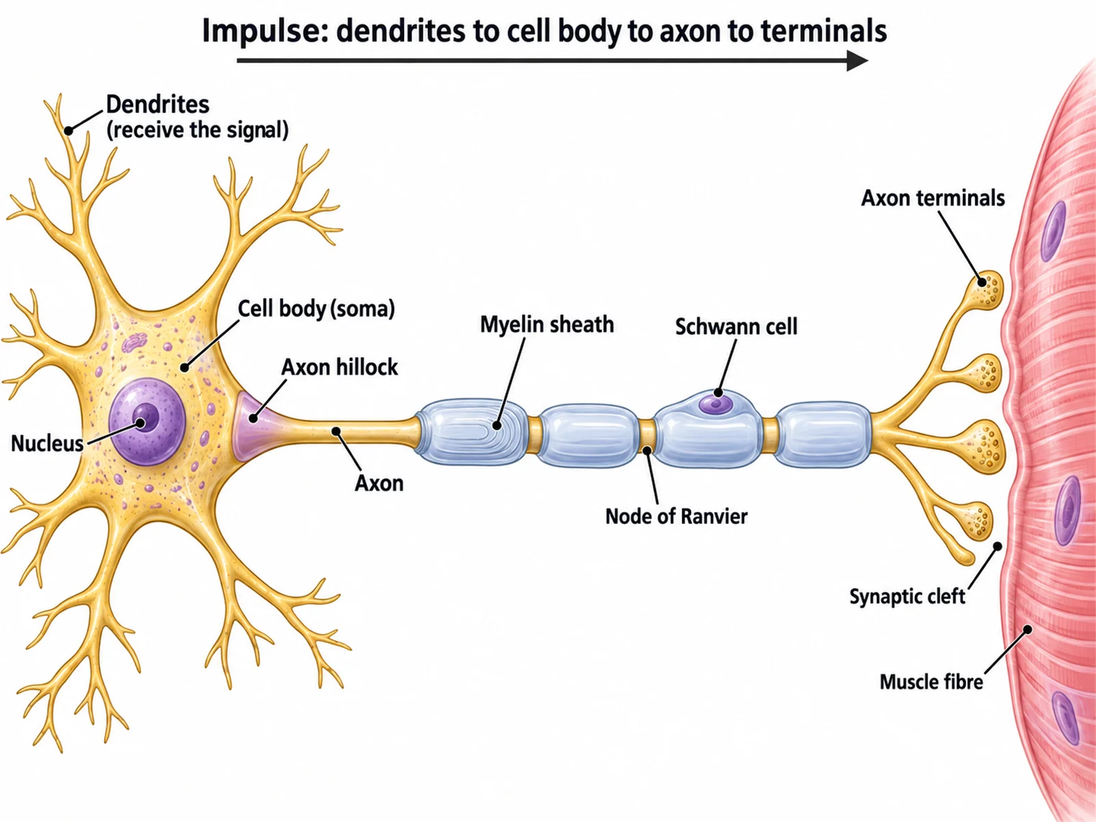
Anatomy and physiology courses pair the whole-body diagram with a zoom into one muscle. The muscle structure view draws this sequence.
Whole muscle: wrapped in a connective tissue layer called the epimysium, which runs on into the tendon that attaches to bone.
Fascicle: a bundle of muscle fibers inside the muscle, wrapped in perimysium. How fascicles are arranged gives muscles their shapes: parallel, fusiform, circular (around the mouth and eyes), convergent (pectoralis major), or pennate like a feather (the deltoid is multipennate).
Muscle fiber: a single muscle cell, wrapped in endomysium. It forms when many cells fuse, so it has many nuclei. Fibers in the sartorius can reach about 30 cm long.
Myofibril: one of hundreds to thousands of rod-like strands packed inside each fiber, surrounded by the sarcoplasmic reticulum, which stores the calcium that triggers contraction.
Sarcomere: the repeating unit of a myofibril, about 2 micrometers long, from one Z disc to the next. Thin actin filaments attach to the Z discs and thick myosin filaments sit in the middle. When the muscle contracts, myosin pulls the actin inward and the sarcomere shortens: the sliding filament model.
Muscles to label on a blank muscular system diagram, by level
The right number of labels depends on the class. These are the sets the level menu uses when your description does not list its own muscles.
Middle school: about ten big muscles with everyday names, such as biceps, triceps, deltoid, chest, abdominals, quadriceps, hamstrings, gluteus maximus, and calf.
High school: about twenty standard surface muscles across the front and back, from the sternocleidomastoid and trapezius down to tibialis anterior, gastrocnemius, and the calcaneal tendon.
College: adds the face and chewing muscles, serratus anterior, the forearm groups, the individual hamstrings and adductors, the small shoulder-blade muscles, and the soleus.
Medical: adds the parts and heads of muscles and the named forearm muscles that an anatomy practical tests.
Any level: an unlabeled figure with no lines, for coloring or for quizzing aloud.
Using a blank muscular system diagram in class
Start blank, end labeled: hand out the blank muscular system worksheet, then show the answer key made from the same result so every number matches.
Front first, then back: the front muscles are easier, so a two-lesson sequence can label the anterior view first and the posterior view next.
Name the action as well as the muscle: once a student labels the biceps and triceps, ask which bends the elbow and which straightens it.
Word bank or not: include one for a scaffolded lesson, leave it out for a test. The word bank is alphabetical, so its order gives nothing away.
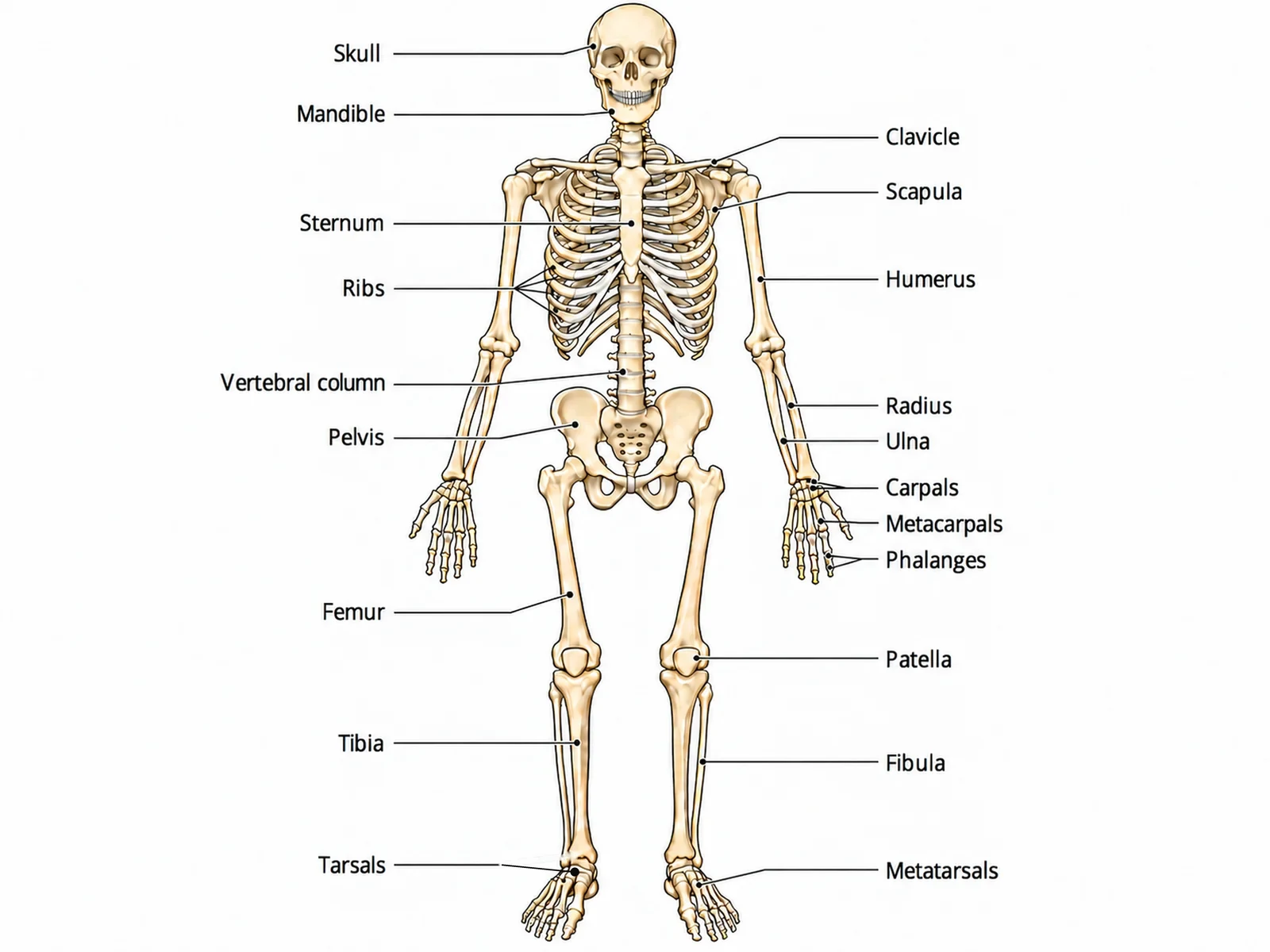
Pair it with the skeleton: the muscles on this page attach to the bones on the skeletal system diagram, so the two worksheets together show origins and insertions.
Most common mistakes on a muscular system diagram
These are the errors image models make most often on muscle figures. Check each one before a diagram goes into graded work.
MistakeCorrect
MistakeVastus intermedius labeled on the surface of the thigh
CorrectIt lies under the rectus femoris; a surface view shows only rectus femoris, vastus lateralis and vastus medialis
MistakeBiceps femoris on the inner side of the thigh
CorrectBiceps femoris is the outer (lateral) hamstring; semitendinosus and semimembranosus are on the inner side
MistakeTriceps brachii drawn on the front of the arm
CorrectBiceps brachii on the front of the upper arm, triceps brachii on the back
MistakeSartorius running straight down the thigh
CorrectIt crosses the thigh diagonally, from the front of the hip bone to the inner side of the knee
MistakeExternal oblique fibers sloping up toward the midline
CorrectThey run down and forward toward the midline, like hands in front pockets
MistakeTeres major listed as a rotator cuff muscle
CorrectThe rotator cuff is supraspinatus, infraspinatus, teres minor and subscapularis
MistakeThe trapezius drawn as two separate shoulder blobs
CorrectOne diamond-shaped sheet from the skull to the lower thoracic spine and out to both shoulders
MistakeThe Achilles tendon or iliotibial tract labeled as a muscle
CorrectBoth are connective tissue: the calcaneal tendon joins the calf muscles to the heel; the iliotibial tract runs down the outer thigh
Skeletal vs cardiac vs smooth muscle
A muscular system diagram shows skeletal muscle, but most worksheets also ask students to tell the three muscle tissues apart.
Skeletal muscle
Cardiac muscle
Smooth muscle
Where it is
Attached to bones by tendons
Wall of the heart
Walls of blood vessels, gut, bladder, uterus, airways
Under the microscope
Striated (striped)
Striated
Not striated
Cell shape
Long cylindrical fibers
Short branched cells
Spindle-shaped cells
Nuclei per cell
Many, at the edge of the fiber
One or two, central
One, central
Control
Voluntary
Involuntary, sets its own rhythm
Involuntary
Special feature
Organized into named muscles with origins and insertions
Intercalated discs link cells so the heart contracts as one unit
Contracts slowly and can stay contracted for long periods
Muscular System Diagram FAQ
A standard front-and-back diagram names the big surface muscles a biology course expects. On the front: sternocleidomastoid, deltoid, pectoralis major, biceps brachii, rectus abdominis, external oblique, sartorius, the quadriceps (rectus femoris, vastus lateralis, vastus medialis), the adductors, tibialis anterior, and the calf. On the back: trapezius, deltoid, latissimus dorsi, triceps brachii, gluteus maximus, the hamstrings, gastrocnemius, and the calcaneal (Achilles) tendon. Anatomy courses add the face, forearm, and deeper muscles.
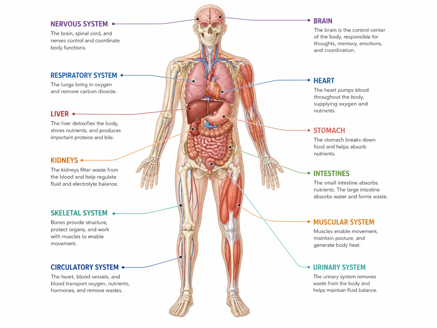
Generate body system diagrams, muscle and organ close-ups, blank labeling worksheets with matching answer keys, and revision-ready biology figures with the same SciFigure AI workflow.