Generate a labeled leaf cross section diagram with every layer in the right order — waxy cuticle, upper epidermis, palisade mesophyll, spongy mesophyll with air spaces, and the lower epidermis with stomata and guard cells — and a vein with xylem above phloem. Choose a dicot, monocot, C4, or dry-habitat leaf, add gas exchange arrows, or make a blank worksheet with its matching answer key.
Cuticle to lower epidermisXylem above phloemLabeled, simple, or blank
Checked against OpenStax Biology 2e
Xylem above phloem
Labeled, numbered, or blank
Print-ready PNG
AI Leaf Cross Section Diagram Generator
Describe the leaf type, the layers to label, and the level, or ask for a blank worksheet.
616 / 2000 characters
Generate a publication-ready PNG from your description.
Preview
Leaf Cross Section Example Previews
Target aspect ratio: 4:3
Sample previews. Describe your diagram to generate your own leaf cross section.
Leaf Cross Section Diagram Examples
Use these as visual directions for a labeled leaf cross section, gas exchange and transport, a simple version for younger classes, dicot vs monocot leaves, a stoma and guard cell close-up, and a blank worksheet.
Prompt
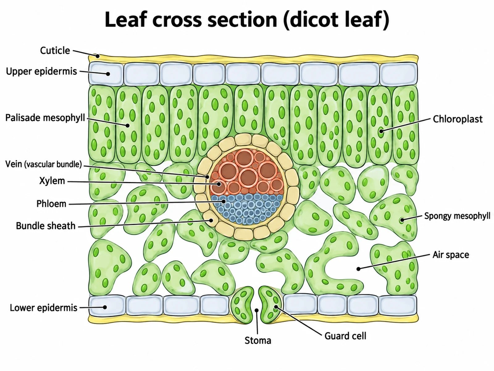
Create a fully labeled leaf cross section diagram of a typical dicot leaf on a white background in a clean biology textbook style. Draw a wide rectangular block of leaf seen in section. From top to bottom: a thin smooth waxy film, then one row of flat clear cells with no chloroplasts, then one row of tall column-shaped cells packed with green chloroplasts, then irregular loosely packed rounded cells with large white air spaces between them and fewer chloroplasts, then one row of flat clear cells containing one stoma: a gap between two bean-shaped guard cells that contain chloroplasts, opening into an air space above it; then a thinner waxy film. In the middle, where the column-shaped and loose layers meet, draw one round vein: larger thick-walled xylem cells on its upper half, smaller phloem cells on its lower half, and a ring of bundle sheath cells around it. Label exactly these thirteen targets with thin leader lines, listed here from top to bottom: "Cuticle", "Upper epidermis", "Palisade mesophyll", "Chloroplast", "Vein (vascular bundle)", "Xylem", "Phloem", "Bundle sheath", "Spongy mesophyll", "Air space", "Lower epidermis", "Guard cell", "Stoma".
Use this example
Prompt
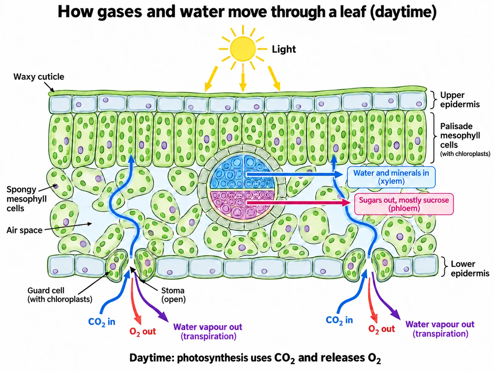
Create a leaf cross section diagram showing gas exchange and transport in daylight on a white background. Draw a typical dicot leaf in section: cuticle, upper epidermis, tall palisade cells with chloroplasts, loose spongy cells with large air spaces, and a lower epidermis with two open stomata between guard cells. Add a small sun above the leaf with an arrow labeled "Light". At each stoma draw a blue arrow pointing IN labeled "CO2 in" that continues through the air spaces to a palisade cell, and two arrows pointing OUT labeled "O2 out" and "Water vapour out (transpiration)". In the vein, draw xylem on the upper side with an arrow labeled "Water and minerals in (xylem)" and phloem on the lower side with an arrow labeled "Sugars out, mostly sucrose (phloem)". Caption: "Daytime: photosynthesis uses CO2 and releases O2".
Use this example
Prompt
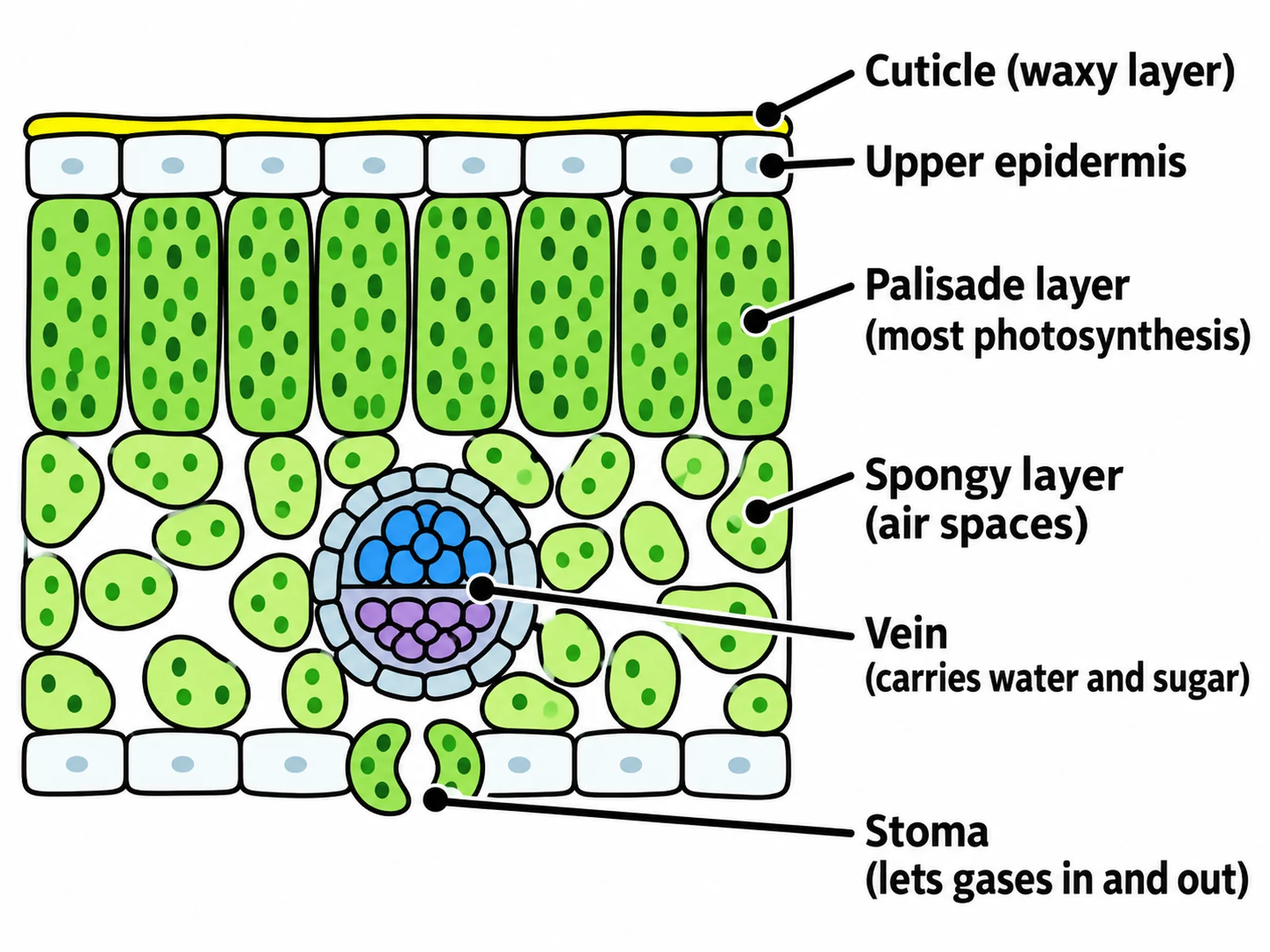
Create a simple leaf cross section diagram for younger students, easy to copy by hand, with bold clear colours, thick dark outlines, large text, and plenty of white space on a white background. Draw a rectangular slice of leaf with, from top to bottom: a thin shiny top line, a row of flat clear cells, a row of tall green column-shaped cells with green dots, a layer of loose round cells with big gaps, a round vein in that loose layer, and a bottom row of flat cells with one stoma between two bean-shaped guard cells. Use exactly six labels: "Cuticle (waxy layer)", "Upper epidermis", "Palisade layer (most photosynthesis)", "Spongy layer (air spaces)", "Vein (carries water and sugar)", and "Stoma (lets gases in and out)". No other labels.
Use this example
Prompt
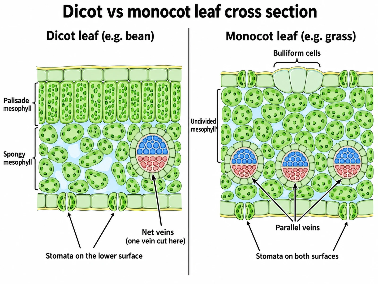
Create a side-by-side comparison of two leaf cross sections on a white background titled "Dicot vs monocot leaf cross section". Left panel, headed "Dicot leaf (e.g. bean)": cuticle, upper epidermis, tall palisade cells, loose spongy cells with air spaces, one vein with xylem on its upper side and phloem on its lower side, and a lower epidermis with stomata only on the underside; labels "Palisade mesophyll", "Spongy mesophyll", "Stomata on the lower surface", "Net veins". Right panel, headed "Monocot leaf (e.g. grass)": cuticle, upper epidermis with a group of large fan-shaped "Bulliform cells", mesophyll of similar rounded cells not divided into layers labeled "Undivided mesophyll", a row of three similar veins each with xylem on its upper side and phloem below labeled "Parallel veins", and stomata in both the upper and lower epidermis labeled "Stomata on both surfaces".
Use this example
Prompt
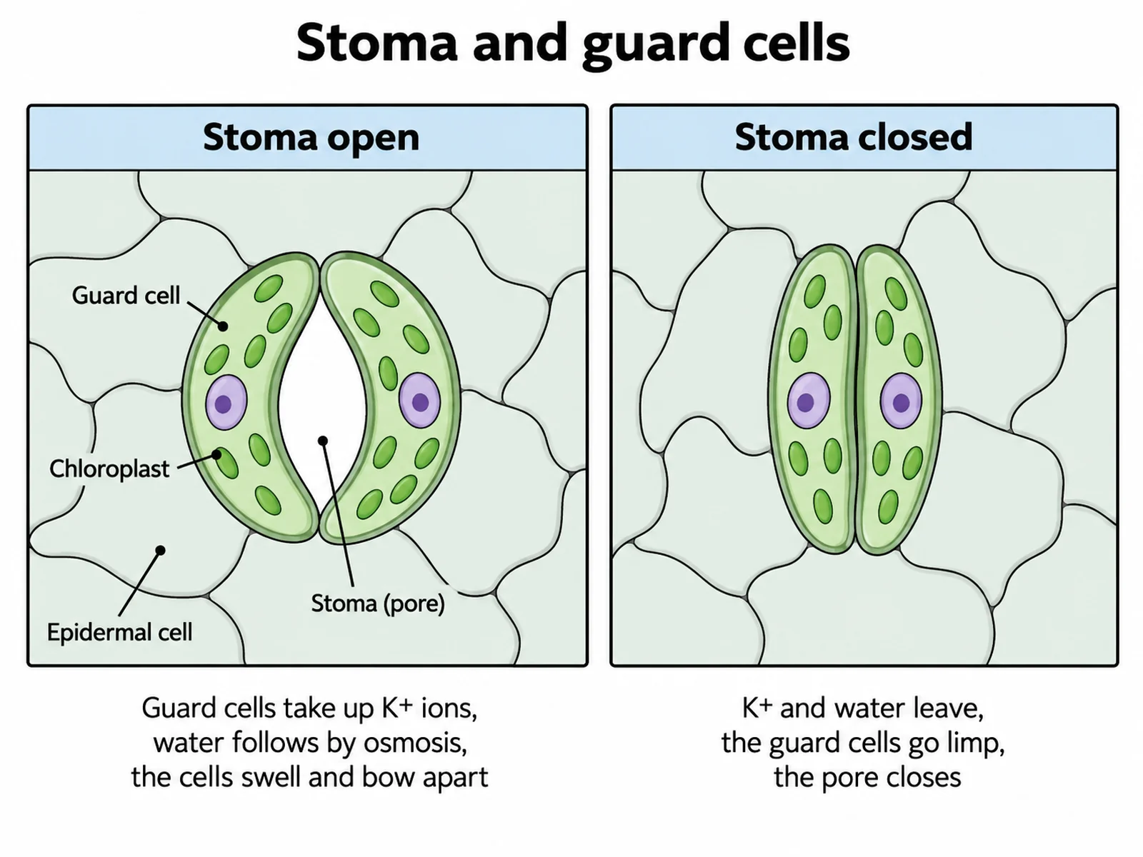
Create a two-panel close-up of a stoma on a white background, seen from below as a surface view of the lower epidermis. Each panel shows two bean-shaped guard cells with green chloroplasts and a visible nucleus, surrounded by irregular clear epidermal cells with no chloroplasts. Left panel, headed "Stoma open": the guard cells are swollen and curved apart, leaving an open pore labeled "Stoma (pore)", with the note "Guard cells take up K+ ions, water follows by osmosis, the cells swell and bow apart". Right panel, headed "Stoma closed": the guard cells are limp and straight and the pore is shut, with the note "K+ and water leave, the guard cells go limp, the pore closes". Label "Guard cell", "Chloroplast", and "Epidermal cell" in the left panel.
Use this example
Prompt
Create a blank leaf cross section worksheet that is the same drawing as a labeled dicot leaf cross section with the label words removed. Draw a wide rectangular block of leaf in section: a thin waxy film on top, one row of flat clear cells, one row of tall column-shaped cells with chloroplasts, irregular loosely packed cells with large air spaces, one round vein where the two middle layers meet with larger xylem cells on its upper half, smaller phloem cells on its lower half and a ring of bundle sheath cells, a bottom row of flat clear cells with one stoma between two bean-shaped guard cells, and a thinner film below. Draw thirteen thin leader lines ending in numbered empty answer lines, numbered top to bottom: 1 top film, 2 top row of cells, 3 column-shaped cells, 4 one chloroplast, 5 the whole vein, 6 the upper half of the vein, 7 the lower half of the vein, 8 the ring around the vein, 9 the loose cells, 10 one air space, 11 the bottom row of cells, 12 one guard cell, 13 the pore. Add a word bank: "cuticle, upper epidermis, palisade mesophyll, chloroplast, vein (vascular bundle), xylem, phloem, bundle sheath, spongy mesophyll, air space, lower epidermis, guard cell, stoma".
Blank worksheet
Use this example
How to make a labeled leaf cross section diagram
1
Pick the leaf and the labels
Choose a dicot, monocot, C4, or dry-habitat leaf and a level, then choose labeled, numbered with a key, or a blank worksheet. Ask for gas exchange arrows or a stoma close-up only if you need them.
2
Generate the section
The generator keeps the layers in order from the cuticle down, packs the chloroplasts into the palisade cells, puts the stomata and guard cells in the lower epidermis, and draws the vein with xylem above phloem.
3
Check it, then export
Check the layer order, the stomata, the chloroplasts in the guard cells, and the vein, then download the PNG — or make the matching blank worksheet or answer key from the same image.
A leaf cross section diagram shows what a leaf looks like inside when it is cut across and viewed edge-on, the way a thin slice of leaf appears under a microscope. Instead of the outline and veins you see from above, it shows the leaf as a sandwich of layers, each made of a different kind of cell doing a different job: a waterproof skin on both faces, a layer of tall cells built to catch light, a looser layer full of air spaces built for gas exchange, and veins that bring water in and carry sugar out.
That is why a diagram of a leaf cross section is one of the standard figures in plant biology. It connects structure to function in a single picture: you can see why photosynthesis happens mostly near the top surface, how carbon dioxide gets from the air to the cells that need it, where water is lost, and how the leaf is plumbed into the rest of the plant. A good leaf cross section labeled diagram makes those connections visible by getting the order of the layers, the position of the stomata, and the layout of the vein exactly right.
Leaf cross section labeled diagram: the parts to label
Cuticle: the waxy, non-cellular film on the outer surface, thicker on top.
Upper epidermis: one row of flat, tightly fitting, clear cells.
Palisade mesophyll: one or two rows of tall, column-shaped cells packed with chloroplasts.
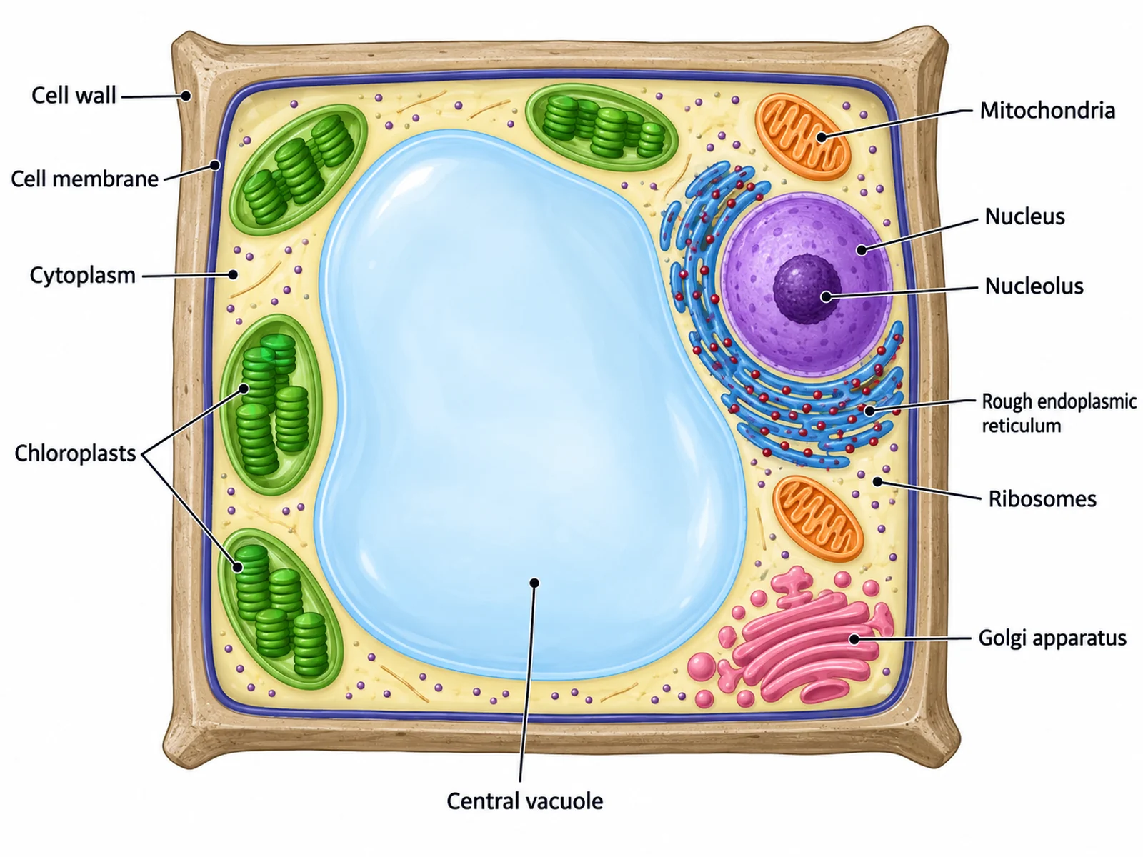
Chloroplasts: most visible in the palisade cells, fewer in the spongy cells, and present in the guard cells.
Spongy mesophyll: irregular, loosely arranged cells with fewer chloroplasts.
Air spaces: the connected gaps between the spongy cells, largest just behind each stoma.
Vein (vascular bundle): in the mesophyll, usually where the palisade and spongy layers meet.
Xylem: on the upper side of the vein, carrying water and minerals into the leaf.
Phloem: on the lower side of the vein, carrying sugars out of the leaf.
Bundle sheath: the ring of cells wrapped around the vein.
Lower epidermis: the bottom row of clear cells, where most stomata are.
Stoma and guard cells: a pore between two bean-shaped guard cells that contain chloroplasts.
The layers of a leaf from top to bottom
The top of the leaf is covered by the cuticle, a waxy layer secreted by the epidermal cells. It is not alive and not made of cells; it is a film that cuts water loss through the leaf surface, and it is usually thicker on the upper side, which faces the sun. Under it is the upper epidermis, normally a single row of flat, tightly joined cells. They contain no chloroplasts and are clear, so light passes straight through them into the leaf, and they protect the tissue below.
Next comes the palisade mesophyll, the photosynthetic powerhouse of the leaf. Its cells are long and column-shaped, standing on end in one or two tightly packed rows, and they are crowded with chloroplasts. Below them is the spongy mesophyll, made of rounded, irregular cells arranged loosely so that large air spaces open between them. Spongy cells also photosynthesise, but they have fewer chloroplasts, and their main job is to provide a moist surface and open pathways for gases.
The bottom of the leaf is closed by the lower epidermis, another row of clear cells broken by stomata, each flanked by two guard cells, and covered by a thinner cuticle. Running through the mesophyll are the veins. In a section through the middle of a dicot leaf the largest vein is the midrib, which often makes the leaf thicker there and may carry supporting tissue above and below it.
Stomata and guard cells
Stomata are the leaf’s adjustable pores. Each one is a gap between two guard cells, and it opens into an air space in the spongy mesophyll just behind it, sometimes called the substomatal cavity. When the stomata are open, carbon dioxide can diffuse in for photosynthesis — but water vapour diffuses out at the same time, so every open stoma is a trade-off between gaining carbon and losing water.
Guard cells control that trade-off by changing shape. When they take up potassium ions, water follows by osmosis, the cells swell, and because their inner walls are thicker and less stretchy than their outer walls, they bow apart and open the pore. When potassium and water leave, the guard cells go limp and the pore closes. They usually open in light and close in the dark or when the plant is short of water. Guard cells are the only epidermal cells with chloroplasts, and in most dicot leaves the stomata are concentrated on the underside, where the leaf is shaded and cooler and loses less water.
The vein: xylem, phloem, and the bundle sheath
Every vein in a leaf is a vascular bundle containing both xylem and phloem, wrapped in a bundle sheath. In a leaf section the xylem is on the upper side of the bundle and the phloem on the lower side; OpenStax describes the larger xylem cells at the top of the bundle and the smaller phloem cells at the bottom. The arrangement is inherited from the stem, where xylem lies on the inner side of each bundle: when a bundle bends out into a leaf, its inner side ends up facing the upper surface. Xylem cells are dead at maturity, with thick, strengthened walls, and they bring water and dissolved minerals into the leaf, pulled up by transpiration. Phloem is living tissue that carries the sugars made in the leaf, mainly sucrose, to the parts of the plant that need them, such as roots, fruits, and growing tips. The bundle sheath cells around the vein regulate what passes between the vein and the mesophyll; in C4 plants they become the site of the Calvin cycle.
How a leaf works: gas exchange and transport in a cross section of leaf diagram
Follow the raw materials of photosynthesis through a cross section of leaf diagram and every structure makes sense. Light enters through the clear cuticle and upper epidermis and is absorbed mostly by the chloroplasts of the palisade cells. Water arrives from the roots in the xylem of the veins and spreads out into the mesophyll cells. Carbon dioxide diffuses from the air into the leaf through the open stomata, spreads through the air spaces of the spongy layer, and dissolves in the film of water on the mesophyll cells before entering them.
The products leave by the same routes in reverse. Oxygen from photosynthesis diffuses out of the cells, through the air spaces, and out of the stomata, along with water vapour evaporating from the moist mesophyll surfaces — the transpiration that pulls more water up the xylem. Sugars are loaded into the phloem and carried out of the leaf. In a diagram with arrows, daytime gas arrows point CO₂ in and O₂ and water vapour out; at night, when only respiration continues and most stomata close, the small net exchange runs the other way.
Dicot vs monocot leaf cross section
Most textbook leaf cross sections show a dicot leaf, such as a bean, sunflower, or privet leaf, with its palisade layer above its spongy layer. The leaf is dorsiventral: its top and bottom surfaces are different, because it is held flat and lit from above. A monocot leaf, such as the blade of a grass, is usually held more upright and lit from both sides, so it is isobilateral. Its mesophyll is not divided into palisade and spongy layers, its stomata are on both surfaces, and its parallel veins appear in a section as a row of similar bundles running side by side. Grasses also have groups of large, thin-walled bulliform cells in the upper epidermis, which lose water in dry weather and let the leaf roll or fold to cut water loss. When a worksheet asks you to compare a dicot and a monocot leaf, those four differences — mesophyll, stomata, veins, and bulliform cells — are what the marker is looking for.
C4 Kranz anatomy and leaves adapted to dry or wet places
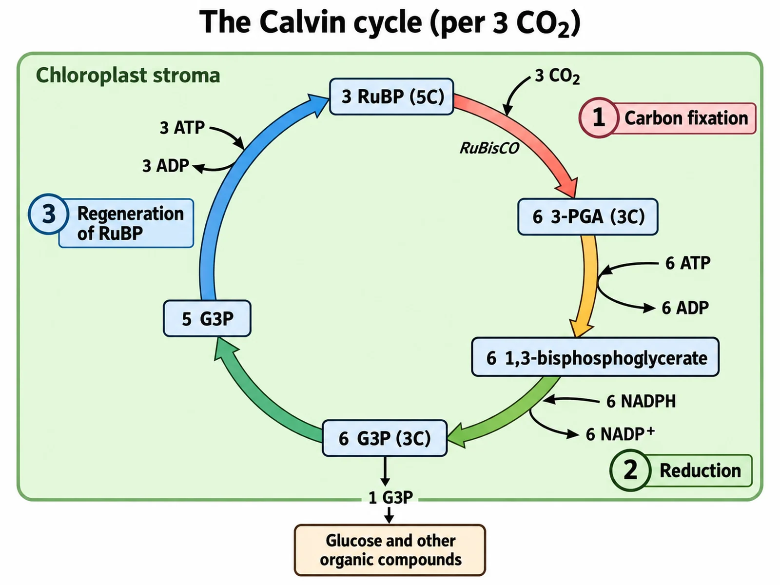
Many grasses from hot climates, including maize, sugarcane, and sorghum, are C4 plants, and their leaf cross section looks different again. Each vein is surrounded by a ring of large bundle sheath cells packed with chloroplasts, and the mesophyll cells are arranged around that ring like the spokes of a wheel. This is Kranz anatomy, from the German word for wreath. The mesophyll cells capture carbon dioxide into a four-carbon acid, which moves into the bundle sheath cells and releases CO₂ there at high concentration for RuBisCO and the Calvin cycle, which cuts photorespiration in hot, bright conditions. The veins of a C4 leaf are also packed closer together.
Habitat changes leaf anatomy too. Leaves of xerophytes, plants of dry places, cut water loss with a thick cuticle, stomata sunken in pits or grooves and often fringed with hairs that trap still, humid air, and in some species a leaf that rolls up with the stomata on the inside; OpenStax gives the needle-like leaves of conifers, with sunken stomata and a small surface area, as an example. Floating leaves of aquatic plants do the opposite: their stomata are on the upper surface, since the lower surface is under water, and their mesophyll has large air spaces that keep the leaf afloat.
How to draw a leaf cross section
To draw a cross section leaf diagram by hand, start with a long horizontal rectangle and divide it into bands before drawing any cells. Draw a thin line along the top for the cuticle and a row of flat brick-like boxes below it for the upper epidermis. Under that, draw one or two rows of tall upright rectangles touching each other, and fill them with small green dots for chloroplasts — the palisade layer. Below the palisade, draw rounded, irregular cells with plenty of white space between them for the spongy layer, and place a circle in it, touching the bottom of the palisade, for the vein: draw larger cells in its upper half for xylem and smaller ones in its lower half for phloem, with a ring of cells around the whole. Finish with a row of flat boxes for the lower epidermis, leave one gap in it, and draw two bean-shaped guard cells with green dots on either side of the gap, with a large air space just above it. Add a thin cuticle line along the bottom.
A diagram of leaf cross section under the microscope
A prepared slide of a leaf cross section, often a privet or similar dicot leaf, shows the same layers as a diagram but is harder to read at first. Under low power the leaf appears as a long, thin band, thicker at the midrib. The palisade layer stands out as a dense, dark green row near one surface — that surface is the top of the leaf — while the spongy layer looks pale and patchy because of its air spaces. The epidermis is a single clear line of cells on each face, and the stomata appear as small breaks in the lower epidermis flanked by two darker guard cells. The veins are cut at various angles, so some appear round and others elongated. Labeling a micrograph and then generating a clean labeled diagram of the same leaf is a good way to connect what students see with what they learn.
Cross section of a leaf with labels: worksheet tips
A labeling worksheet is only fair if every leader line has exactly one answer. Point each line at the middle of the thing it names — the centre of the palisade row, not the boundary between two layers — and give layers and individual cells separate lines: one to the palisade mesophyll, another to a single chloroplast inside it. Number the lines from the top surface down so the answer key reads in the same order as the leaf. For the vein, use three lines — the whole vein, its upper half for xylem, and its lower half for phloem — so students have to know the arrangement, not just the word. If you add a word bank, list exactly as many terms as there are lines. Generating the labeled diagram first and then using the blank edit on that image keeps the worksheet and the answer key identical, line for line.
Leaf cross section images: what to check before you use one
Chloroplasts: packed in the palisade, fewer in the spongy layer, present in guard cells, absent from other epidermal cells.
Stomata: in the lower epidermis of a dicot leaf, each between two guard cells.
Vein: in the mesophyll, xylem on top and phloem below, with a bundle sheath around it.
Cuticle: a smooth film, never a row of cells.
Arrows: in daylight CO₂ in, O₂ and water vapour out.
Most common leaf cross section mistakes
These are the errors that turn up most often in student drawings, in images found online, and in AI output. Check for each one before a diagram goes on a worksheet.
MistakeCorrect
MistakeThe spongy layer is drawn above the palisade layer.
CorrectPalisade mesophyll sits directly under the upper epidermis; spongy mesophyll is below it.
MistakeA dicot leaf has its stomata on the upper surface only.
CorrectMost dicot stomata are in the lower epidermis. Upper-surface stomata belong to floating leaves.
MistakeGuard cells are drawn without chloroplasts.
CorrectGuard cells contain chloroplasts — they are the only epidermal cells that do.
MistakeThe whole epidermis is filled with chloroplasts.
CorrectEpidermal cells are clear so light reaches the palisade layer.
MistakePhloem is drawn above xylem in the vein.
CorrectXylem is on the upper side of a leaf vein and phloem on the lower side.
MistakeThe cuticle is drawn as a row of cells.
CorrectThe cuticle is a waxy, non-cellular film secreted onto the epidermis.
MistakeThe palisade layer has bigger air spaces than the spongy layer.
CorrectPalisade cells are tightly packed; the large connected air spaces are in the spongy layer.
MistakeDaytime arrows show O₂ going in and CO₂ coming out.
CorrectIn daylight CO₂ diffuses in through the stomata, and O₂ and water vapour diffuse out.
Dicot vs monocot vs C4 leaf cross section
The same tissues appear in all three. What changes is how the mesophyll is arranged, where the stomata are, and how the veins are spaced.
Dicot leaf (e.g. bean)
Monocot leaf (e.g. wheat)
C4 leaf, Kranz anatomy (e.g. maize)
Mesophyll
Palisade layer above a spongy layer
Not divided into palisade and spongy
Mesophyll cells arranged radially around each bundle sheath
Stomata
Mostly on the lower surface
On both surfaces
On both surfaces
Veins in section
A midrib and smaller veins of a branching net
A row of similar parallel veins
Many parallel veins, close together
Bundle sheath
Thin layer around each vein
Present around each vein
Large cells packed with chloroplasts, the "wreath"
Where the Calvin cycle runs
Mesophyll cells
Mesophyll cells
Bundle sheath cells
Special features
Often hairs (trichomes)
Bulliform cells that let the leaf roll
Bulliform cells in grasses such as maize
Top and bottom
Different (dorsiventral)
Similar (isobilateral)
Similar (isobilateral)
Leaf Cross Section Diagram FAQ
From the top surface down: the waxy cuticle, the upper epidermis, the palisade mesophyll, the spongy mesophyll with its air spaces, the lower epidermis with stomata and guard cells, and a thinner lower cuticle. Running through the mesophyll is a vein, or vascular bundle, with xylem on its upper side, phloem on its lower side, and a bundle sheath around it. A labeled leaf cross section diagram normally also points out the chloroplasts in the palisade cells and the air space behind each stoma.
Generate plant cell, chloroplast, photosynthesis, and flower diagrams, plus worksheet-ready biology illustrations, with the same SciFigure AI workflow.