Female Reproductive System Diagram: Labeled & Blank
Generate a labeled female reproductive system diagram in front or side view, naming the ovaries, uterine tubes, uterus, cervix, and vagina, or an unlabeled, blank version for students to label with a matching answer key. Add the ovarian follicles, the menstrual cycle, or the path from ovulation to implantation, in a clear textbook style.
Labeled, numbered or unlabeledFront and side viewsMatching answer key
Checked against OpenStax A&P
Worksheet + answer key pairs
Print-ready PNG
AI Female Reproductive System Diagram Generator
Describe the view and the structures to label, or pick an unlabeled or blank version from the Labels menu.
584 / 2000 characters
Generate a publication-ready PNG from your description.
Preview
Female Reproductive System Diagram Example Previews
Target aspect ratio: 4:3
Sample previews. Describe your diagram to generate your own reproductive anatomy figure.
Labeled and Unlabeled Female Reproductive System Diagram Examples
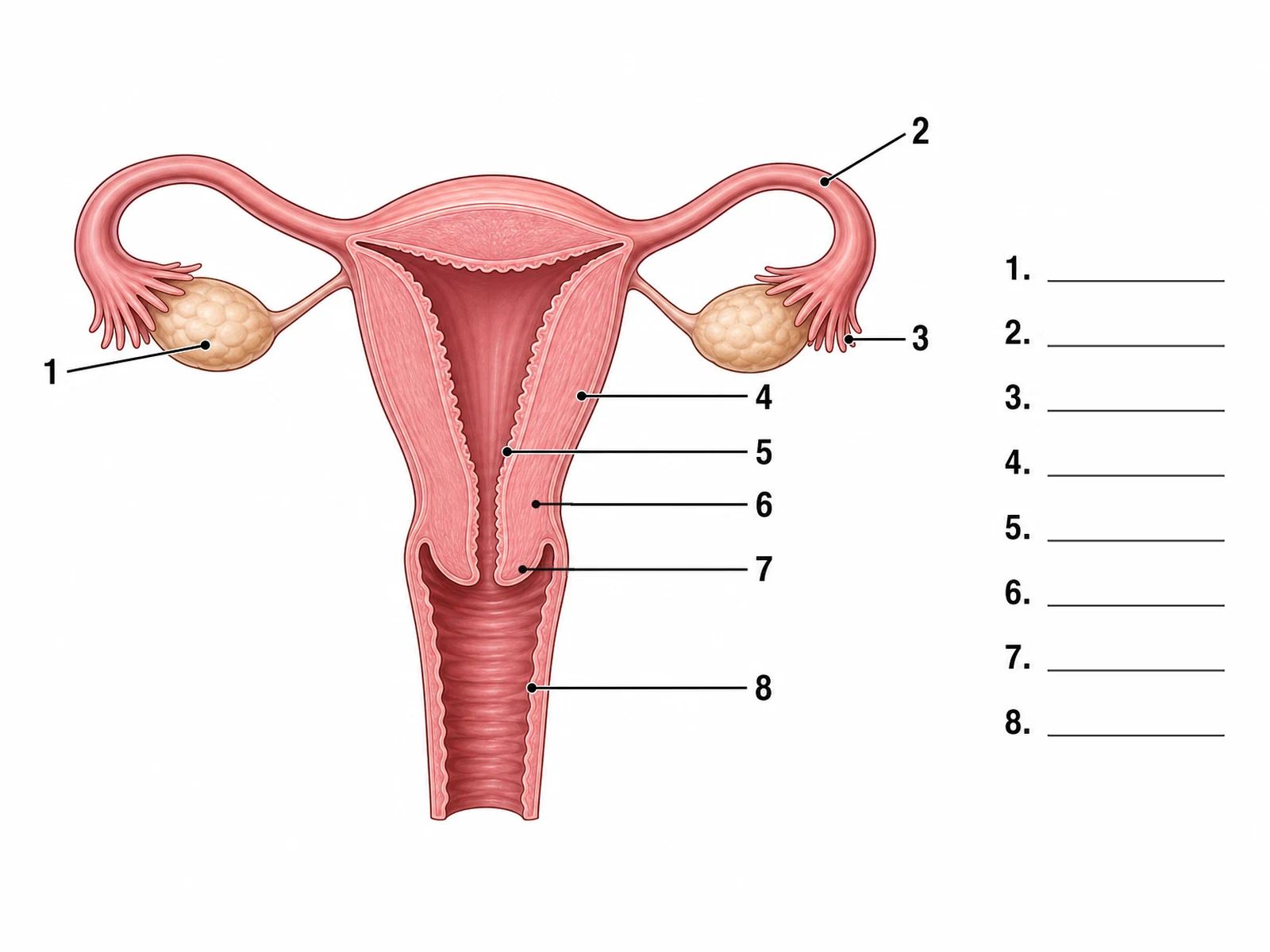
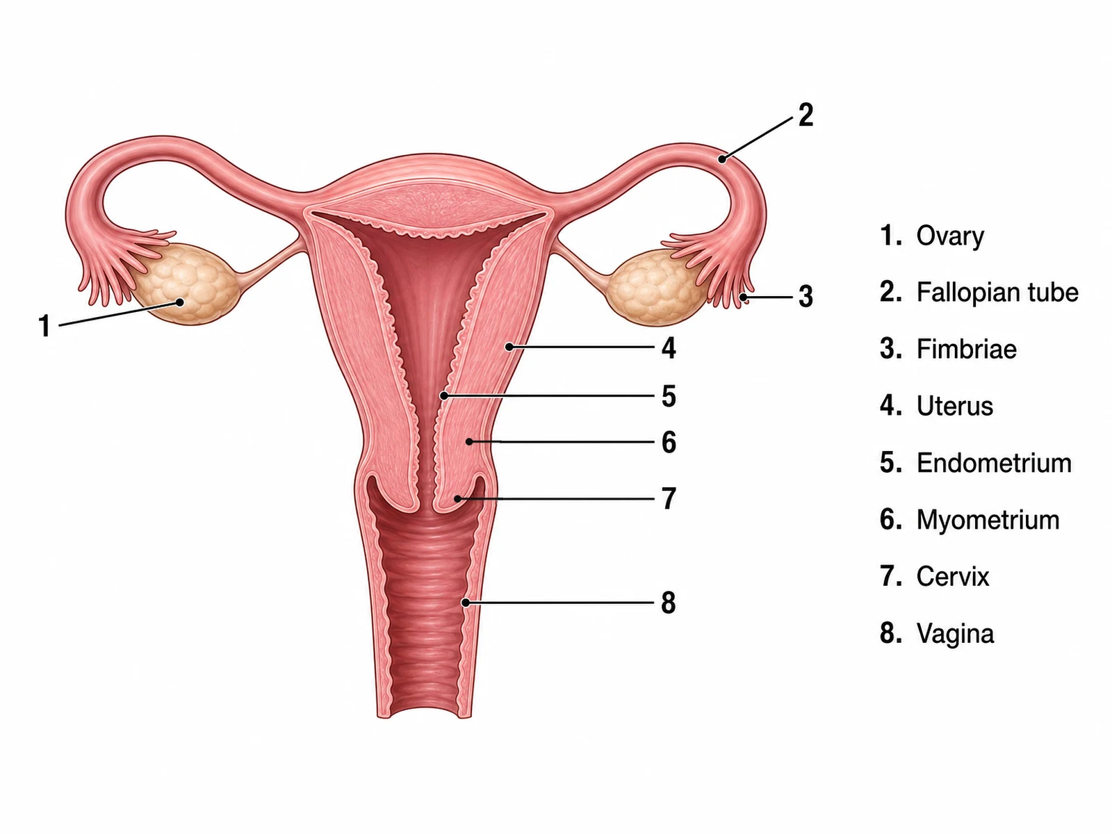
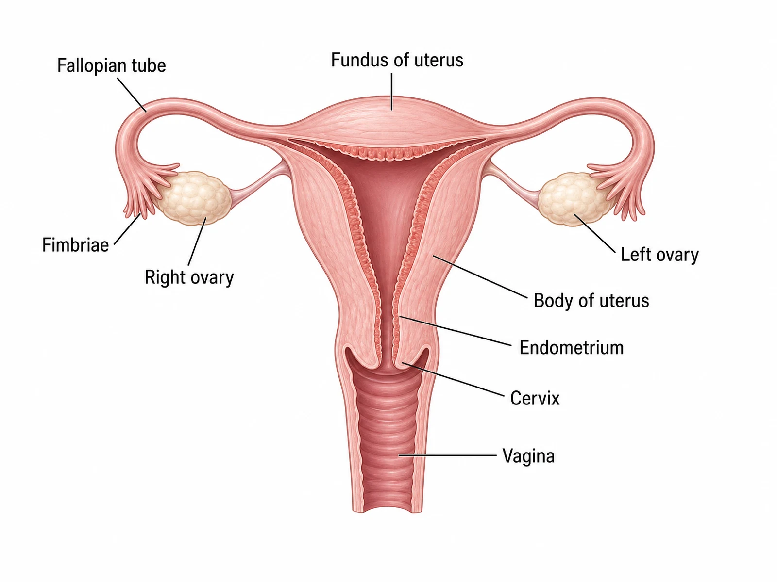
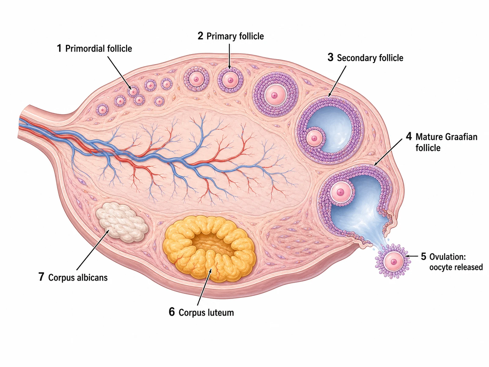
A labeled front view, an unlabeled worksheet with its matching answer key, ovarian follicle development, a menstrual cycle chart, and the path from ovulation to implantation.
Prompt
Create a labeled female reproductive system diagram in a soft pink medical textbook style on a white background: a front view with the uterus, cervix and vagina cut open down the middle to show the uterine cavity, the frilled endometrium lining it and the thick muscular wall, and a uterine tube curving out from each upper corner of the uterus to fimbriae that drape over an almond-shaped ovary without joining it. Seen from the front, the person’s right side is on the viewer’s left. Label "Fallopian tube", "Fimbriae" and "Right ovary" on the viewer’s left, "Fundus of uterus" at the top, and "Left ovary", "Body of uterus", "Endometrium" (on the inner lining), "Cervix" and "Vagina" on the viewer’s right, each with a thin leader line.
Use this example
Prompt
Create an unlabeled female reproductive system diagram worksheet for labeling practice. Draw a front view of the female reproductive organs in a soft pink medical textbook style on a white background: the uterus, cervix and upper vagina cut open down the middle to show the triangular uterine cavity, the thin frilled endometrium lining it and the thick myometrium around it; a uterine tube curving out from each upper corner of the uterus and ending in fimbriae that drape over an almond-shaped ovary without joining it; each ovary tied to the uterus by its ligament. Draw eight thin leader lines, each ending in a number: 1 on the ovary on the viewer’s left, 2 on the top of the tube and 3 on the fimbriae on the viewer’s right, then 4 to 8 down the right side of the uterus and vagina, in this order: 1 = Ovary, 2 = Fallopian tube, 3 = Fimbriae, 4 = Uterus, 5 = Endometrium, 6 = Myometrium, 7 = Cervix, 8 = Vagina. On the right add a column of numbered answer lines, 1 to 8, each an empty ruled line. Put no structure names anywhere on the image.
Blank worksheet
Use this example
Prompt
Create the answer key for an unlabeled female reproductive system worksheet. Draw a front view of the female reproductive organs in a soft pink medical textbook style on a white background: the uterus, cervix and upper vagina cut open down the middle to show the triangular uterine cavity, the thin frilled endometrium lining it and the thick myometrium around it; a uterine tube curving out from each upper corner of the uterus and ending in fimbriae that drape over an almond-shaped ovary without joining it; each ovary tied to the uterus by its ligament. Draw eight thin leader lines, each ending in a number: 1 on the ovary on the viewer’s left, 2 on the top of the tube and 3 on the fimbriae on the viewer’s right, then 4 to 8 down the right side of the uterus and vagina, in this order: 1 = Ovary, 2 = Fallopian tube, 3 = Fimbriae, 4 = Uterus, 5 = Endometrium, 6 = Myometrium, 7 = Cervix, 8 = Vagina. On the right add a key listing each number with its structure name, so it matches the worksheet line for line.
Numbered with key
Use this example
Prompt
Create a labeled diagram of one ovary cut in section, in a soft medical illustration style on a white background, with blood vessels branching through the central medulla and the follicles arranged clockwise around the cortex in order of development, numbered: "1 Primordial follicle" (a cluster of small follicles with one flat cell layer), "2 Primary follicle", "3 Secondary follicle" (with a fluid-filled antrum, as many histology books name it), "4 Mature Graafian follicle" bulging at the surface, "5 Ovulation: oocyte released" with the oocyte and its corona radiata leaving the ovary, "6 Corpus luteum" as a large yellow folded body, and "7 Corpus albicans" as a small pale scar.
Ovary and follicles
Use this example
Prompt
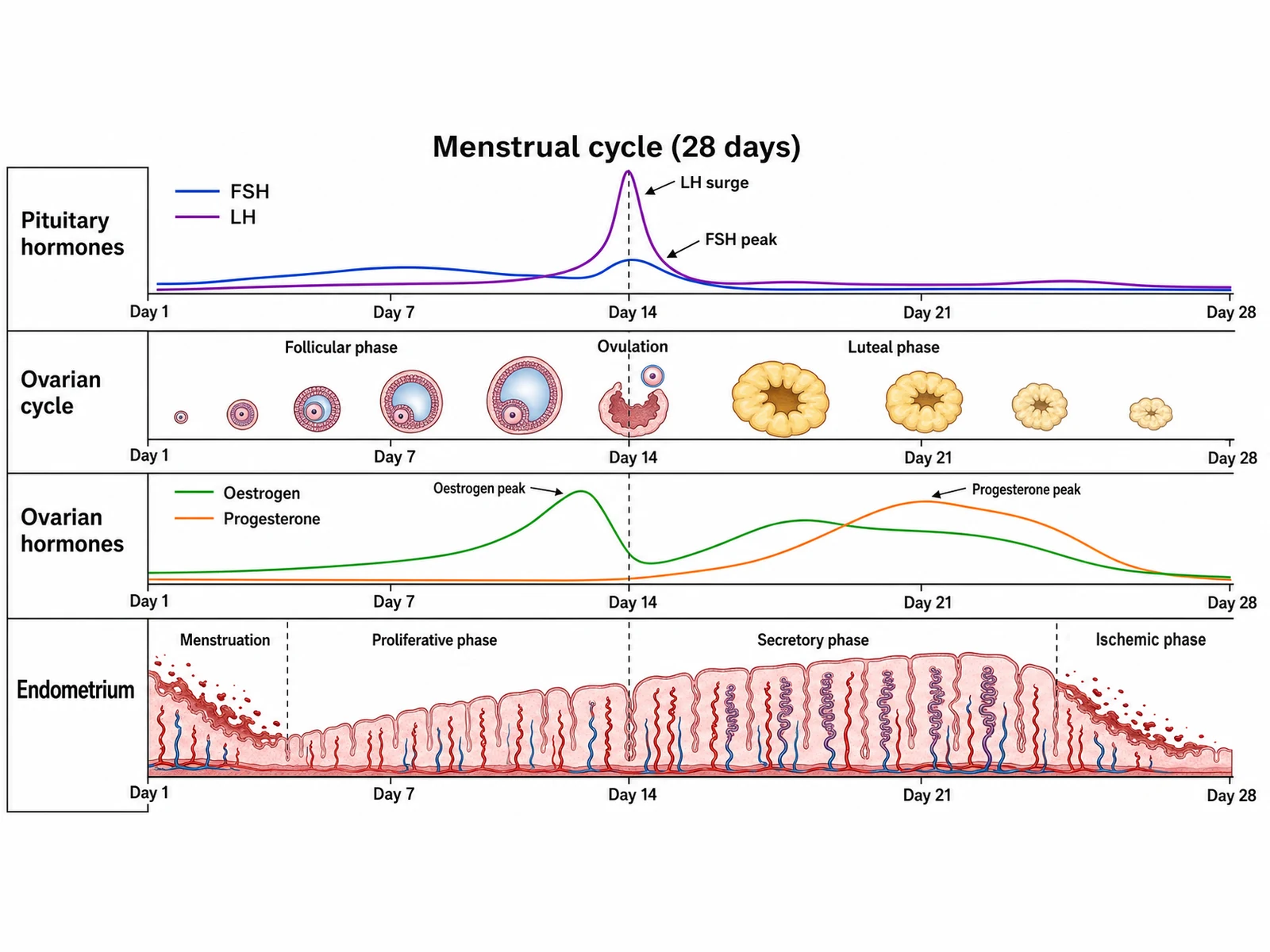
Create a menstrual cycle chart titled "Menstrual cycle (28 days)" with four stacked rows sharing one axis marked Day 1, Day 7, Day 14, Day 21 and Day 28, and a dashed vertical line at day 14. Row 1 "Pituitary hormones": FSH and LH curves with the LH surge and a smaller FSH peak at day 14. Row 2 "Ovarian cycle": follicles growing through the follicular phase, the follicle rupturing at ovulation, then the corpus luteum growing and shrinking through the luteal phase. Row 3 "Ovarian hormones": estrogen peaking just before day 14, dipping, then a second smaller rise, and progesterone low until ovulation and peaking near day 21. Row 4 "Endometrium": the lining shed during menstruation, rebuilt in the proliferative phase, thickest with coiled arteries in the secretory phase, and breaking down in a final ischemic phase.
Menstrual cycle chart
Use this example
Prompt
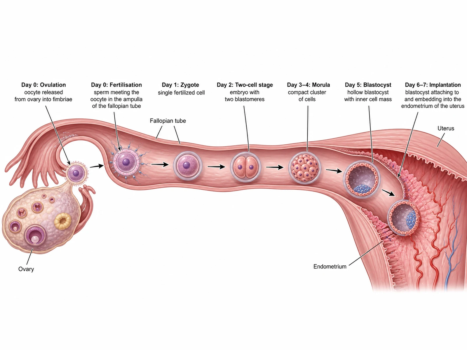
Create a labeled fertilization and implantation diagram on a white background: an ovary on the left with follicles, a fallopian tube running to the right with its fimbriae over the ovary, and the uterus on the right with a thick glandular endometrium and spiral arteries. Along the tube, left to right with arrows between them and captions above: "Day 0: Ovulation" (an oocyte leaving the ovary into the fimbriae), "Day 0: Fertilisation" (sperm meeting the oocyte in the ampulla), "Day 1: Zygote", "Day 2: Two-cell stage", "Day 3–4: Morula", "Day 5: Blastocyst" with its inner cell mass, and "Day 6–7: Implantation" with the blastocyst embedding into the endometrium. Also label "Ovary", "Fallopian tube", "Uterus" and "Endometrium".
Fertilization and implantation
Use this example
How to make a labeled or unlabeled female reproductive system diagram
1
Pick the view, level, and labels
A front view, a side (sagittal) section, both, the ovary and follicles, the menstrual cycle, fertilization and implantation, or the uterine wall. Then choose labeled, numbered, blank, a word bank, or fully unlabeled.
2
Generate the diagram
The generator draws the organs in a clean textbook style, keeps the tube open beside the ovary, puts the parts of the tube and the layers of the uterine wall in order, and places the bladder in front and the rectum behind in a side view.
3
Make the answer key, check, and export
For a worksheet, make the answer key from the same result so the numbers match. Check the tube segments, the wall layers, and the side-view neighbors, then download the PNG.
Female reproductive system key facts
Ovaries
2, each about 2–3 cm long
Uterine tube
About 10–12 cm long
Uterus (not pregnant)
About 7 cm long, 5 cm wide
Vagina
About 10 cm long
Oocytes at birth / puberty
About 1–2 million / 400,000
Average cycle
28 days, ovulation about day 14
Usual site of fertilization
Ampulla of the uterine tube
Oocyte lifespan after ovulation
About 24 hours
Corpus luteum without pregnancy
Lasts about 10–12 days
Implantation begins
End of the first week after fertilization
What is a female reproductive system diagram?
A female reproductive system diagram is a labeled drawing of the organs that make oocytes and hormones, carry the oocyte to be fertilized, and hold and nourish a pregnancy. The standard version is a front view of the internal organs: the uterus in the middle, a uterine tube and an ovary on each side, and the vagina below. A side (sagittal) section adds the bladder in front and the rectum behind, which shows how the organs sit in the pelvis.
In class the figure comes in two forms. A labeled diagram names each structure and works as a reference or an answer key. An unlabeled diagram of the female reproductive system, or a blank one with numbered lines, is for labeling practice. This page makes both, along with figures of the ovarian follicles, the menstrual cycle, and the path from ovulation to implantation.
Parts of the female reproductive system
Ovaries: two almond-sized glands that hold the follicles, release an oocyte at ovulation, and make estrogen and progesterone.
Uterine (fallopian) tubes: two tubes that catch the oocyte, provide the place where fertilization usually happens, and carry the early embryo to the uterus.
Uterus: the hollow, muscular organ where an embryo implants and grows, made of the fundus, body, and cervix.
Cervix: the narrow lower part of the uterus, with a canal that opens into the vagina.
Vagina: the muscular canal, about 10 cm long, from the cervix to the outside of the body.
External genitalia (vulva): the structures around the vaginal and urethral openings, described in a section below.
Mammary glands: often grouped with the reproductive system because they make milk after birth, though they are not part of the pelvic organs.
The ovaries
Each ovary is about 2 to 3 cm long, roughly the size of an almond, and lies on the side wall of the pelvis, near the open end of its uterine tube. It is held in place by the ovarian ligament, which joins it to the side of the uterus; the suspensory ligament, which carries its blood vessels and lymphatics from the pelvic wall; and the mesovarium, a fold of the broad ligament. In section an ovary has an outer cortex, where the follicles develop, and an inner medulla of connective tissue and blood vessels. The ovaries have two jobs: releasing an oocyte in each cycle and making the hormones estrogen and progesterone.
The uterine tubes: infundibulum, ampulla, isthmus
Each uterine tube, also called a fallopian tube or oviduct, runs from the upper corner of the uterus out toward the ovary. Its parts are always listed in the same order, and a labeled diagram should keep them that way.
Infundibulum: the funnel-shaped outer end, open to the pelvic cavity and fringed with finger-like fimbriae that sweep over the ovary at ovulation.
Ampulla: the widest and longest part, curving over the ovary. Fertilization usually happens here.
Isthmus: the narrow, thick-walled part next to the uterus.
Intramural (uterine) part: the short stretch that passes through the uterine wall into the cavity.
Lining: ciliated cells and smooth muscle work together to move the oocyte, or the early embryo, toward the uterus over several days.
The uterus: fundus, body, and cervix
The uterus is shaped like an upside-down pear, about 7 cm long and 5 cm wide when not pregnant. The fundus is the rounded top, above the points where the tubes enter. The body (corpus) is the main part, around a narrow cavity. The cervix is the narrow lower part. It projects into the top of the vagina, and its cervical canal runs from the internal os, where it meets the uterine cavity, to the external os, which opens into the vagina. The uterus is held by the broad ligament along its sides, the round ligament, which runs from the upper corner down through the groin, and the uterosacral ligaments behind. In most people it tilts forward over the bladder.
The uterine wall: endometrium, myometrium, perimetrium
Perimetrium: the thin outer covering, a layer of the peritoneum.
Myometrium: the thick middle layer of smooth muscle, by far the thickest layer of the wall. It stretches enormously in pregnancy and contracts strongly in labor.
Endometrium: the inner lining of the cavity, rich in glands and blood vessels. Its deep layer, the stratum basalis, stays in place. Its upper layer, the stratum functionalis, thickens in each cycle and is shed during menstruation if no embryo implants.
Spiral arteries: coiled vessels in the functional layer that constrict at the end of a cycle, cutting off its blood supply so it breaks down and is shed.
The vagina and the external structures
The vagina is a muscular canal, about 10 cm long, running up and back from its opening to the cervix. It lies behind the bladder and urethra and in front of the rectum. The recesses around the cervix at its top are the vaginal fornices, the posterior one deepest. The external genitalia, together called the vulva, include the mons pubis, the labia majora and labia minora, the clitoris, and the vestibule, the area between the labia minora into which both the urethral opening and the vaginal opening lead, the urethral opening in front. Most school diagrams show the internal organs only, and this generator does the same unless a clinical diagram of the vulva is requested.
Reading a side (sagittal) view
A midsagittal section cuts the pelvis down the middle, so it shows the female reproductive organs among their neighbors. From front to back the order is the pubic symphysis, the urinary bladder with the urethra running down from it, the uterus and vagina, and the rectum in front of the sacrum and coccyx. The uterus usually leans forward over the top of the bladder (anteverted) and is bent forward on itself at the cervix (anteflexed).
The peritoneum drapes over the top of the organs and dips down between them, forming two pouches: a shallow one between the uterus and the bladder, and a deeper one, the recto-uterine pouch, between the uterus and the rectum. Only one ovary and one tube appear in a sagittal view, and their position is approximate. A diagram with the bladder behind the uterus, or the urethra opening into the vagina, has the order wrong.
Ovarian follicles: from primordial follicle to corpus albicans
An ovary diagram shows the stages one follicle passes through, usually arranged around the ovary so that they can be read in order.
Primordial follicle: an oocyte with a single flat layer of cells around it. A girl is born with about one to two million oocytes in follicles like this, and about 400,000 remain at puberty.
Primary follicle: the surrounding cells become cube-shaped granulosa cells, and a clear coat, the zona pellucida, forms around the oocyte.
Secondary follicle: several layers of granulosa cells and small pockets of fluid.
Tertiary (antral) follicle: the pockets merge into one large fluid-filled antrum, with the oocyte on a mound of cells. In each cycle usually only one of these becomes dominant.
Ovulation: the dominant follicle bursts at the surface of the ovary and releases a secondary oocyte, still surrounded by the zona pellucida and a ring of cells, the corona radiata.
Corpus luteum: the empty follicle becomes a yellowish body that makes progesterone and estrogen.
Corpus albicans: if there is no pregnancy the corpus luteum breaks down after 10 to 12 days into a pale scar, which slowly disappears over months.
Naming differs between books. OpenStax calls a follicle with several cell layers a secondary follicle and the one with a large antrum a tertiary (antral) follicle. Many histology texts call the antral follicle a secondary follicle and keep "mature" or "Graafian" for the largest one just before ovulation. Use the scheme your course uses, and say which in your description.
The menstrual cycle: the ovarian and uterine cycles
The menstrual cycle is two cycles running together. In the ovary, the follicular phase grows a dominant follicle, ovulation releases its oocyte, and the luteal phase is the life of the corpus luteum. In the uterus, menstruation sheds the old functional layer, the proliferative phase rebuilds it under estrogen, and the secretory phase readies it for an embryo under progesterone.
Textbooks describe an average cycle of 28 days with ovulation around day 14, counting day 1 as the first day of menstruation. Real cycles vary, between people and from month to month, and most of the difference falls in the follicular phase before ovulation. A cycle chart lines the hormones, the ovary, and the endometrium up on one time axis, which is how this generator draws it. Some charts also mark the last days of the cycle as an ischemic phase: as progesterone falls, the spiral arteries constrict and the functional layer starts to break down, just before menstruation begins on the next day 1.
The hormones of the cycle
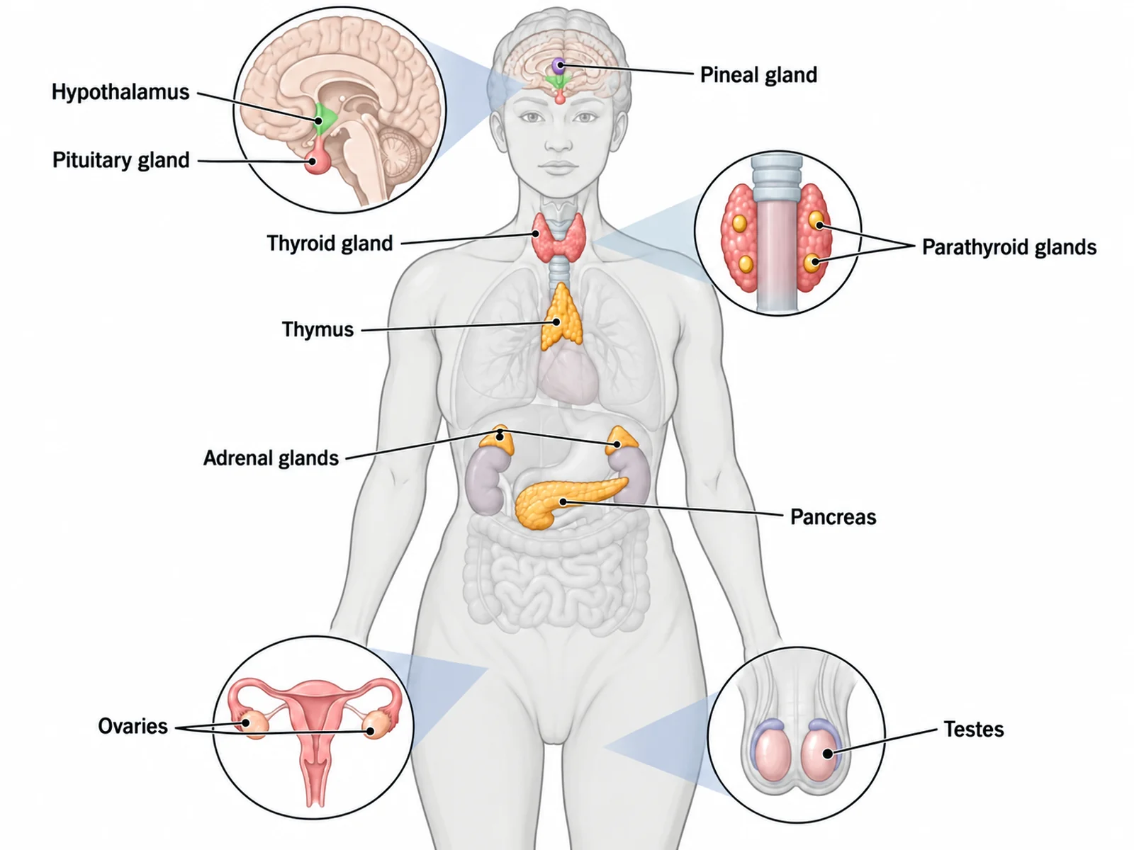
The female reproductive system is run by the endocrine system, through a chain from the brain to the ovary.
GnRH: released by the hypothalamus in pulses, it tells the anterior pituitary to release FSH and LH.
FSH (follicle-stimulating hormone): makes a group of follicles grow at the start of each cycle.
Estrogen: made by the growing follicles. At first it holds FSH and LH down. When the dominant follicle makes enough of it for long enough, it switches to triggering the LH surge.
LH (luteinizing hormone): its surge triggers ovulation and turns the empty follicle into the corpus luteum.
Progesterone: made by the corpus luteum, it turns the endometrium secretory and ready for implantation. When the corpus luteum breaks down, progesterone falls and menstruation starts.
hCG (human chorionic gonadotropin): made by the early placenta after implantation, it keeps the corpus luteum making progesterone. Pregnancy tests detect it.
Fertilization and implantation: the first week
Ovulation: the secondary oocyte is released into the pelvic cavity and swept into the tube by the fimbriae. It can be fertilized for only about a day.
Fertilization: usually in the ampulla. A sperm crosses the corona radiata and the zona pellucida, the oocyte completes its second meiotic division, and the two nuclei combine into a diploid zygote.
Cleavage: the zygote divides as it moves down the tube, into 2, 4, and 8 cells and then a solid ball, the morula, which reaches the uterus about three days after fertilization.
Blastocyst: in the uterus the cells form a hollow ball, with an inner cell mass that will become the embryo and an outer layer, the trophoblast, that will help form the placenta.
Implantation: the blastocyst sheds its zona pellucida and begins to burrow into the endometrium at the end of the first week, usually in the fundus or the back wall of the uterus. It is completely embedded by the middle of the second week.
Ectopic pregnancy: if the embryo implants outside the uterus, most often in a uterine tube, the pregnancy cannot continue normally and needs urgent medical care.
Female reproductive system labels by level
Simple: about six parts, such as ovary, fallopian tube, uterus, endometrium, cervix, and vagina.
High school: the ovary, uterine tube, fimbriae, fundus, body of the uterus, endometrium, myometrium, cervix, and vagina, plus the bladder, urethra, and rectum in a side view.
College: adds the three named parts of the tube, the uterine cavity, the perimetrium, the cervical canal and external os, and the ovarian and broad ligaments.
Medical: adds the suspensory and round ligaments, the mesovarium, the internal os, the vaginal fornices, the two layers of the endometrium, and the pouches of the pelvic cavity.
Using an unlabeled female reproductive system diagram in class
Start blank, end labeled: hand out the unlabeled worksheet, then show the answer key made from the same result so every number matches.
Follow the oocyte: before naming anything, have students trace the route from the ovary across the gap into the infundibulum, along the ampulla and isthmus, and into the uterus.
Pair the views: label the front view first, then the side view, so students see the same uterus from two directions and learn its neighbors.
Line up the cycle: give the cycle chart as a second sheet and ask which hormone is highest on day 13 and on day 21.
Link the systems: the hormones on this page come from the hypothalamus, the pituitary, and the ovaries, so pair it with the endocrine system diagram.
Most common mistakes on a female reproductive system diagram
These are the errors image models make most often on these diagrams. Check each one before a diagram goes into graded work.
MistakeCorrect
MistakeFimbriae fused to the ovary, as if the tube were a pipe from the ovary
CorrectThe tube opens beside the ovary; the oocyte is swept into it across a small gap
MistakeAmpulla and isthmus swapped
CorrectThe ampulla is the wide outer part near the ovary; the isthmus is the narrow part next to the uterus
MistakeFertilization marked in the uterus
CorrectFertilization usually happens in the ampulla of the uterine tube
MistakeEndometrium labeled as the outer layer of the uterus
CorrectFrom the cavity outward: endometrium, myometrium, perimetrium
MistakeFundus labeled at the bottom, or the cervix at the top
CorrectThe fundus is the dome above the tubes; the cervix is the narrow lower part in the vagina
MistakeBladder drawn behind the uterus in a side view
CorrectFront to back: pubic symphysis, bladder, uterus and vagina, rectum
MistakeUrethra opening into the vagina
CorrectSeparate tubes and openings; the urethra lies in front of the vagina
MistakeOvulation on day 1 or a progesterone peak before ovulation
CorrectDay 1 is the first day of menstruation; ovulation is about day 14; progesterone peaks in the luteal phase
The phases of a 28-day cycle
Worksheets often ask students to line up what happens in the ovary with what happens in the uterus. In an average cycle the two run side by side like this.
Menses (days 1–5)
Proliferative (days 6–14)
Ovulation (about day 14)
Secretory (days 15–28)
Ovarian phase
Follicular
Follicular
Ovulation
Luteal
In the ovary
A group of follicles starts to grow
One dominant follicle matures
The mature follicle releases a secondary oocyte
The corpus luteum forms, then breaks down if there is no pregnancy
Leading hormone
FSH rising; estrogen and progesterone low
Estrogen
LH surge (with FSH)
Progesterone, with estrogen
Endometrium
Functional layer shed
Rebuilt from the basal layer and thickening
Thick and well supplied with blood
Glands secrete; thickest and ready for implantation
Female Reproductive System Diagram FAQ
The standard front view names the two ovaries, the two uterine (fallopian) tubes with their fimbriae, the uterus with its fundus, body, and cervix, the layers of the uterine wall (endometrium, myometrium, perimetrium), and the vagina. College diagrams split each tube into the infundibulum, ampulla, and isthmus, and add the ovarian, suspensory, broad, and round ligaments. A side (sagittal) view adds the neighbors: the bladder and urethra in front, the rectum behind, and the pubic symphysis.
Generate body system diagrams, organ sections and close-ups, blank labeling worksheets with matching answer keys, and revision-ready biology figures with the same SciFigure AI workflow.