Generate a labeled nephron diagram that follows the filtrate from the glomerulus and Bowman’s capsule through the tubules and loop of Henle to the collecting duct, or an unlabeled, blank nephron for labeling practice with a matching answer key. Add the blood supply, the processes, or the renal corpuscle up close.
Labeled or unlabeledCortex and medulla zonesMatching answer key
Checked against OpenStax A&P
Worksheet + answer key pairs
Print-ready PNG
AI Nephron Diagram Generator
Describe the parts and processes to show, or pick an unlabeled or blank version from the Labels menu.
567 / 2000 characters
Generate a publication-ready PNG from your description.
Preview
Nephron Diagram Example Previews
Target aspect ratio: 3:4
Sample previews. Describe your diagram to generate your own nephron figure.
Labeled and Unlabeled Nephron Diagram Examples
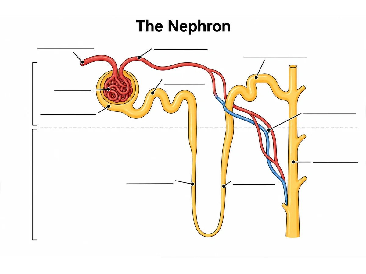
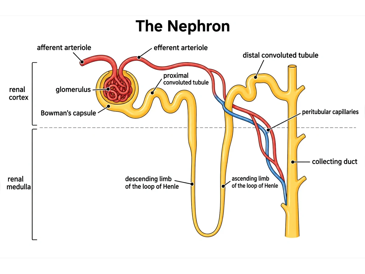
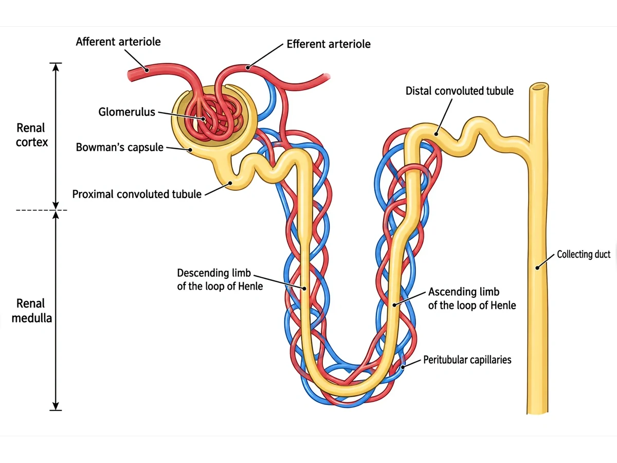
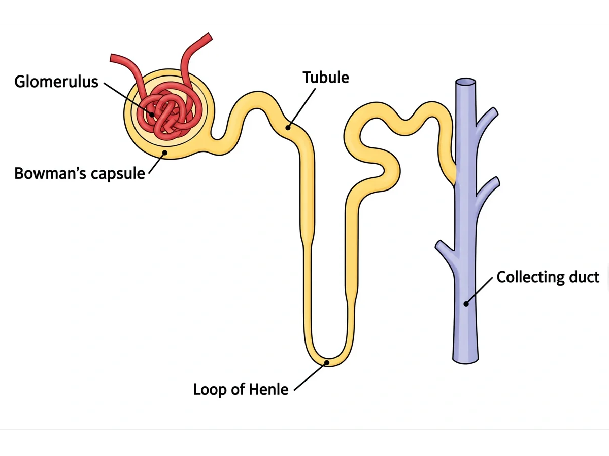
A labeled nephron, an unlabeled nephron worksheet with its answer key, a simple nephron diagram, a renal corpuscle close-up, and cortical and juxtamedullary nephrons in the kidney.
Prompt
Create a labeled nephron diagram on a white background, with a pale band across the top labeled "Renal cortex" and a darker band below labeled "Renal medulla". In the cortex draw the glomerulus cupped inside Bowman’s capsule, with a wider afferent arteriole entering and a narrower efferent arteriole leaving at the same side, the efferent arteriole branching into peritubular capillaries. From the opposite side of the capsule run a long coiled proximal convoluted tubule, then a loop of Henle whose descending limb dips into the medulla and whose ascending limb climbs back, then a shorter distal convoluted tubule that touches its own afferent arteriole, then a collecting duct running straight down through the medulla. Label: "Afferent arteriole", "Efferent arteriole", "Glomerulus", "Bowman’s capsule", "Proximal convoluted tubule", "Descending limb", "Ascending limb", "Loop of Henle", "Distal convoluted tubule", "Collecting duct", "Peritubular capillaries".
Use this example
Prompt
Create a blank, unlabeled nephron diagram worksheet for labeling practice. Draw a single nephron on a white background, with a pale band across the upper part of the image for the renal cortex and a slightly darker band below it for the renal medulla, separated by a faint line. In the cortex: a round renal corpuscle made of a ball of capillaries, the glomerulus, cupped inside a double-walled Bowman’s capsule; a wider afferent arteriole entering the glomerulus and a narrower efferent arteriole leaving it at the same side; the efferent arteriole branching into a net of peritubular capillaries around the tubules. From the opposite side of the capsule, a long, tightly coiled proximal convoluted tubule, then a loop of Henle whose descending limb runs straight down into the medulla, turns in a hairpin, and whose ascending limb runs back up into the cortex; then a shorter distal convoluted tubule that passes close against the afferent arteriole of its own corpuscle; then a short connecting piece into a straight collecting duct, shared with other nephrons, running down through the medulla toward the bottom of the image. Put two brackets on the viewer’s left for the two regions, and ten thin leader lines to the parts, each bracket and line ending at a short horizontal ruled answer line with a number beside it and no text, in this order: 1 = Renal cortex (bracket on the left), 2 = Renal medulla (bracket on the left), 3 = Afferent arteriole, 4 = Efferent arteriole, 5 = Glomerulus, 6 = Bowman’s capsule, 7 = Proximal convoluted tubule, 8 = Distal convoluted tubule, 9 = Peritubular capillaries, 10 = Descending limb of the loop of Henle, 11 = Ascending limb of the loop of Henle, 12 = Collecting duct. Put no names, no key and no word bank anywhere on the image.
Blank worksheet
Use this example
Prompt
Create the answer key for a blank nephron worksheet. Draw a single nephron on a white background, with a pale band across the upper part of the image for the renal cortex and a slightly darker band below it for the renal medulla, separated by a faint line. In the cortex: a round renal corpuscle made of a ball of capillaries, the glomerulus, cupped inside a double-walled Bowman’s capsule; a wider afferent arteriole entering the glomerulus and a narrower efferent arteriole leaving it at the same side; the efferent arteriole branching into a net of peritubular capillaries around the tubules. From the opposite side of the capsule, a long, tightly coiled proximal convoluted tubule, then a loop of Henle whose descending limb runs straight down into the medulla, turns in a hairpin, and whose ascending limb runs back up into the cortex; then a shorter distal convoluted tubule that passes close against the afferent arteriole of its own corpuscle; then a short connecting piece into a straight collecting duct, shared with other nephrons, running down through the medulla toward the bottom of the image. Put two brackets on the viewer’s left for the two regions, and ten thin leader lines to the parts, each ending at a short ruled line with its number, and write the answer on each line in dark sans-serif text: 1 = Renal cortex (bracket on the left), 2 = Renal medulla (bracket on the left), 3 = Afferent arteriole, 4 = Efferent arteriole, 5 = Glomerulus, 6 = Bowman’s capsule, 7 = Proximal convoluted tubule, 8 = Distal convoluted tubule, 9 = Peritubular capillaries, 10 = Descending limb of the loop of Henle, 11 = Ascending limb of the loop of Henle, 12 = Collecting duct. Keep the numbering in this order so it matches the worksheet line for line.
Use this example
Prompt
Create a simple nephron diagram for beginners on a white background, with large text and plenty of white space. Draw a glomerulus cupped inside Bowman’s capsule, one tubule that coils away from the capsule, runs down into a long U-shaped loop of Henle and back up, coils again and joins a collecting duct. Label only "Glomerulus", "Bowman’s capsule", "Tubule", "Loop of Henle" and "Collecting duct".
Use this example
Prompt
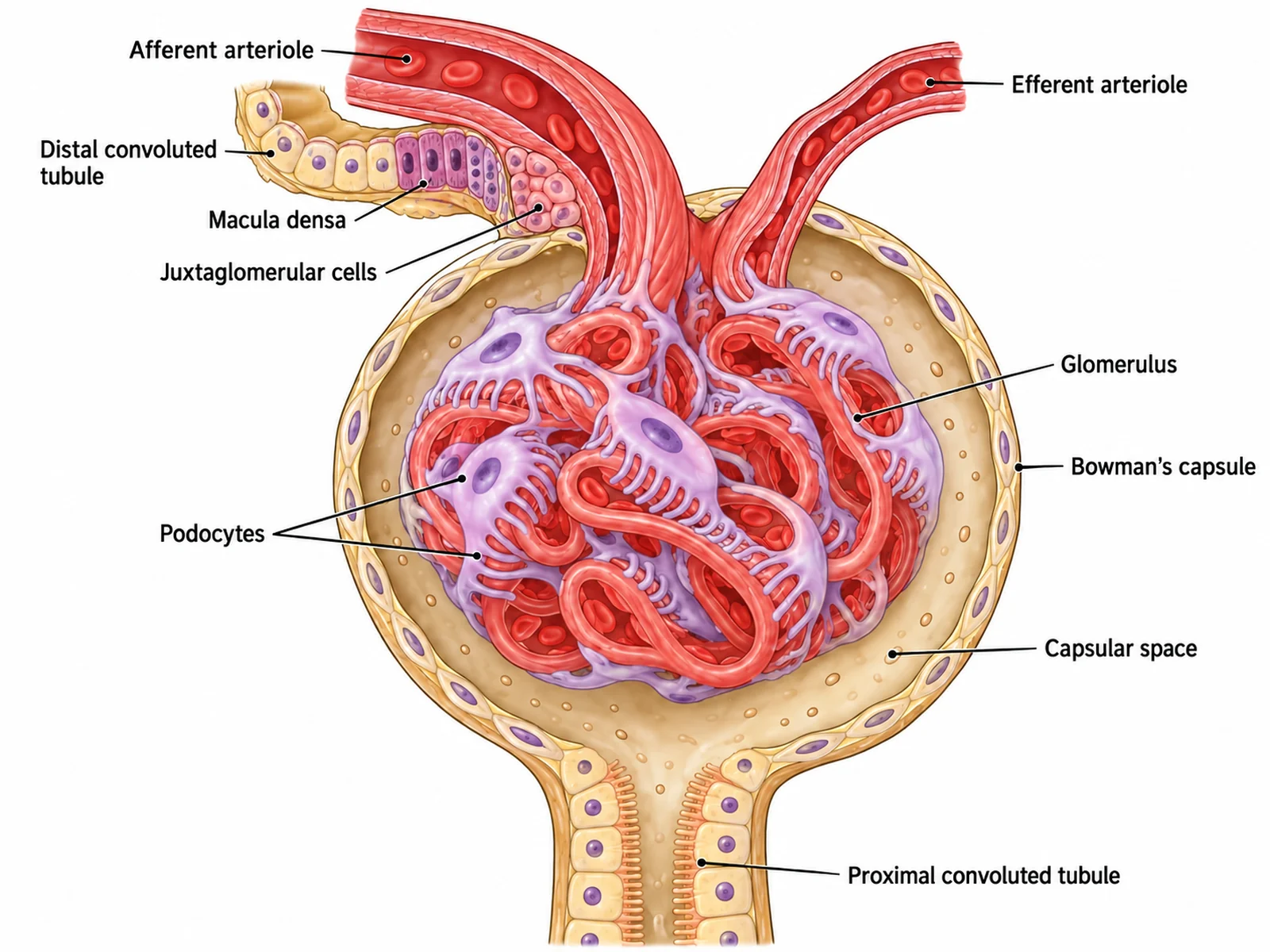
Create a labeled close-up of a renal corpuscle on a white background. Draw a tuft of looping glomerular capillaries cupped inside a double-walled Bowman’s capsule: the outer parietal layer, the capsular space, and the inner layer of podocytes wrapping the capillaries with foot processes. At the vascular pole draw a WIDER afferent arteriole entering and a NARROWER efferent arteriole leaving, side by side. At the opposite pole draw the start of the proximal convoluted tubule. Show the distal convoluted tubule passing against the afferent arteriole, with its darker macula densa cells and the juxtaglomerular cells in the arteriole wall. Label "Glomerulus", "Bowman’s capsule", "Capsular space", "Podocytes", "Afferent arteriole", "Efferent arteriole", "Proximal convoluted tubule", "Macula densa", "Juxtaglomerular cells".
Renal corpuscle close-up
Use this example
Prompt
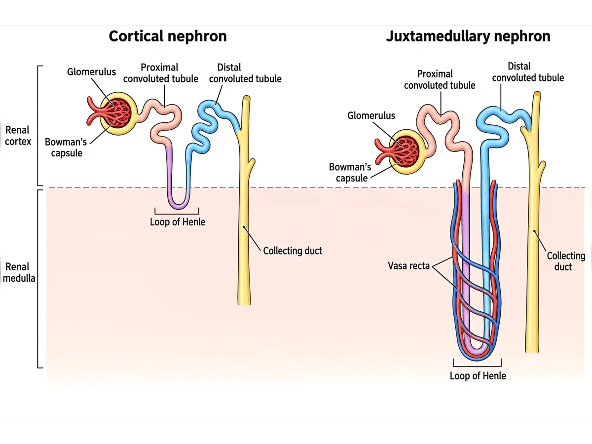
Create a labeled diagram of nephrons in position in the kidney on a white background. Draw a vertical wedge of kidney from the renal capsule at the top, through the cortex, to the tip of a renal pyramid (the papilla) at the bottom, with the cortex and the medulla shaded differently. Draw a cortical nephron with its corpuscle high in the cortex and a short loop of Henle that only just enters the medulla, and a juxtamedullary nephron with its corpuscle near the cortex–medulla border and a long loop reaching deep into the medulla, with straight vasa recta beside it. Both drain into one collecting duct that runs down to open at the papilla. Add a small inset of a whole kidney cut lengthwise marking where the wedge comes from. Label "Renal capsule", "Cortex", "Medulla", "Cortical nephron", "Juxtamedullary nephron", "Vasa recta", "Collecting duct", "Renal papilla".
Nephrons in the kidney
Use this example
How to make a labeled or unlabeled nephron diagram
1
Pick the view, level, and labels
A whole nephron, the processes, the renal corpuscle, the loop and collecting duct, or nephrons in the kidney. Then choose labeled, numbered, blank, a word bank, or a fully unlabeled drawing.
2
Generate the nephron
The generator keeps the parts in filtrate order, the corpuscle and convoluted tubules in the cortex, the loop in the medulla, and the afferent arteriole wider than the efferent.
3
Make the answer key, check, and export
For a worksheet, make the answer key from the same result so the numbers match. Check the order of the tubules and the zones, then download the PNG.
Nephron key facts
Nephrons per kidney
About 1 million
Filtrate made per day
About 150 to 180 liters
Urine per day
About 1 to 2 liters
Renal corpuscle
Glomerulus + Bowman’s capsule
Water and Na+ reabsorbed in the PCT
About two thirds
Cortical / juxtamedullary
About 85% / 15%
Hormone for water in the collecting duct
ADH
Hormone for Na+ in the DCT and duct
Aldosterone
What is a nephron diagram?
A nephron diagram is a magnified drawing of one of the microscopic filtering units of the kidney, laid out so the path of the filtrate can be followed from start to finish. It runs from the glomerulus and Bowman’s capsule, through the proximal convoluted tubule, the loop of Henle, and the distal convoluted tubule, to the collecting duct. Most versions add the blood vessels that supply it and shade the background to show which parts lie in the renal cortex and which in the medulla.
It is one of the most labeled figures in biology and physiology, so it comes in several forms. A labeled nephron is the reference and answer key. An unlabeled nephron, or a blank nephron diagram with numbered lines, is for labeling practice. Process diagrams add arrows for filtration, reabsorption, and secretion. This page makes all of them from one description.
Parts of the nephron, in order
Afferent arteriole: brings blood from the cortical radiate artery into the glomerulus. It is wider than the vessel leaving.
Glomerulus: a tuft of leaky capillaries where blood is filtered.
Bowman’s (glomerular) capsule: the double-walled cup around the glomerulus that collects the filtrate. Glomerulus and capsule together are the renal corpuscle.
Efferent arteriole: carries blood out of the glomerulus. It is narrower than the afferent arteriole.
Proximal convoluted tubule: the long, coiled first stretch of tubule. Its cells have a brush border of microvilli for reabsorption.
Loop of Henle: a hairpin with a descending limb running down into the medulla and an ascending limb running back up. The ascending limb has a thin lower part and a thick upper part.
Distal convoluted tubule: a shorter coiled stretch back in the cortex, which passes against its own afferent arteriole.
Collecting duct: receives the fluid from many nephrons through connecting tubules and carries it down through the medulla to the papilla.
Peritubular capillaries and vasa recta: the second capillary bed, fed by the efferent arteriole, that takes back what the tubules reabsorb.
How a nephron makes urine
Urine formation has three steps, and a process diagram puts each in its place.
Glomerular filtration: blood pressure in the glomerulus pushes water and small solutes (glucose, amino acids, salts, urea) through the capillary wall into Bowman’s capsule. Blood cells and most proteins stay behind. The two kidneys together filter around 150 to 180 liters a day.
Tubular reabsorption: as the filtrate flows on, useful substances move back into the peritubular capillaries. The proximal tubule takes back about two thirds of the water and sodium and, normally, all the glucose and amino acids. The loop, the distal tubule, and the collecting duct recover most of the rest.
Tubular secretion: substances the body needs to remove are moved from the capillaries into the tubule, including hydrogen ions, potassium, and many drugs. This happens mainly in the proximal and distal tubules.
What is left, about 1 to 2 liters a day, drains from the collecting ducts into the renal pelvis as urine.
The renal corpuscle and the filtration barrier
The renal corpuscle has two poles. At the vascular pole the afferent arteriole enters and the efferent arteriole leaves, side by side. At the opposite urinary pole the capsular space opens into the proximal tubule. Bowman’s capsule has an outer parietal layer of flat cells and an inner visceral layer of podocytes. These are octopus-like cells whose interlocking foot processes wrap each glomerular capillary and leave narrow filtration slits between them.
Anything filtered has to cross three layers: the capillary wall, which is full of small pores; the basement membrane; and the slits between the podocyte feet. Together they let water, ions, glucose, and urea through but hold back blood cells and most plasma proteins. That is why protein or blood in the urine points to damaged glomeruli. Filtration is driven by blood pressure, which is kept high because the efferent arteriole is narrower than the afferent one.
The loop of Henle and concentrated urine
The loop of Henle exists to make the medulla salty, so that the kidney can save water when it needs to. The thick ascending limb actively pumps sodium and chloride out into the surrounding tissue but does not let water follow. That raises the salt concentration of the medulla, highest at the tip of the pyramids. The descending limb is permeable to water, so water leaves it into the salty tissue and the fluid inside becomes more concentrated on the way down.
Because fluid flows down one limb and up the other, the effect builds along the length of the loop. This is called the countercurrent multiplier. The vasa recta, running in hairpins alongside, carry away the reabsorbed water without washing out the salt. When the body is short of water, ADH puts water channels into the collecting duct, and as the duct passes down through the salty medulla, water is drawn out and the urine becomes concentrated. Without ADH the duct stays watertight and the urine stays dilute.
The blood supply: two capillary beds in a row
Blood reaches each nephron through a chain of narrowing arteries. The renal artery branches into segmental, interlobar, arcuate, and cortical radiate arteries, and each cortical radiate artery gives off afferent arterioles. The nephron’s blood supply is unusual in having two capillary beds one after the other. The first is the glomerulus, between the afferent and efferent arterioles, where pressure is high and fluid is filtered out. The second is the peritubular capillaries, or the vasa recta for juxtamedullary nephrons, where pressure is low and fluid is taken back in. Diagrams that connect the glomerulus straight to a vein, or leave out the second capillary bed, miss half of how the nephron works.
Cortical and juxtamedullary nephrons
Not all nephrons are the same shape. About 85 percent are cortical nephrons. Their corpuscles sit in the outer cortex and their short loops of Henle barely enter the outer medulla, and they are wrapped in peritubular capillaries. The other 15 percent or so are juxtamedullary nephrons. Their corpuscles lie deep in the cortex, next to the medulla, and their long loops reach far down into the inner medulla, accompanied by the vasa recta. The juxtamedullary nephrons build most of the salt gradient that concentrates urine. A standard nephron diagram shows one generic nephron; the nephrons-in-the-kidney view shows the two types side by side.
Hormones that act on the nephron
ADH (antidiuretic hormone): released by the posterior pituitary when the blood is too concentrated. It inserts water channels into the collecting duct so more water is reabsorbed.
Aldosterone: released by the adrenal cortex. It increases sodium reabsorption and potassium secretion in the distal tubule and collecting duct; water follows the sodium.
Renin and angiotensin: when blood pressure or the salt reaching the macula densa falls, the juxtaglomerular cells release renin. That starts a cascade producing angiotensin II, which constricts blood vessels and triggers aldosterone release.
Atrial natriuretic peptide: released by the heart when the blood volume is high. It reduces sodium reabsorption, so more salt and water are lost.
Parathyroid hormone: increases calcium reabsorption in the distal tubule.
What the fluid contains at each stage
Following the composition of the fluid down the tubule explains why each part of a nephron diagram is labeled the way it is.
In Bowman’s capsule the filtrate is essentially blood plasma without its large proteins. It holds water, glucose, amino acids, salts, and urea at about the same concentrations as the plasma.
By the end of the proximal tubule about two thirds of the water and salt have gone back to the blood, along with normally all of the glucose and amino acids. The volume is much smaller, but the fluid is still about as concentrated as plasma.
At the bottom of the loop of Henle the fluid is at its most concentrated, because water has left the descending limb into the salty medulla.
At the top of the thick ascending limb it is more dilute than plasma, because salt has been pumped out but water could not follow.
In the collecting duct the final concentration depends on ADH: with ADH water is drawn out and the urine is concentrated, without it the urine stays dilute.
Urine leaving the duct carries water, urea, creatinine, excess salts, and hydrogen ions. Glucose in the urine is a warning sign. In untreated diabetes the filtered glucose exceeds what the proximal tubule can carry back, and the excess spills over.
Where the nephron sits in the urinary system
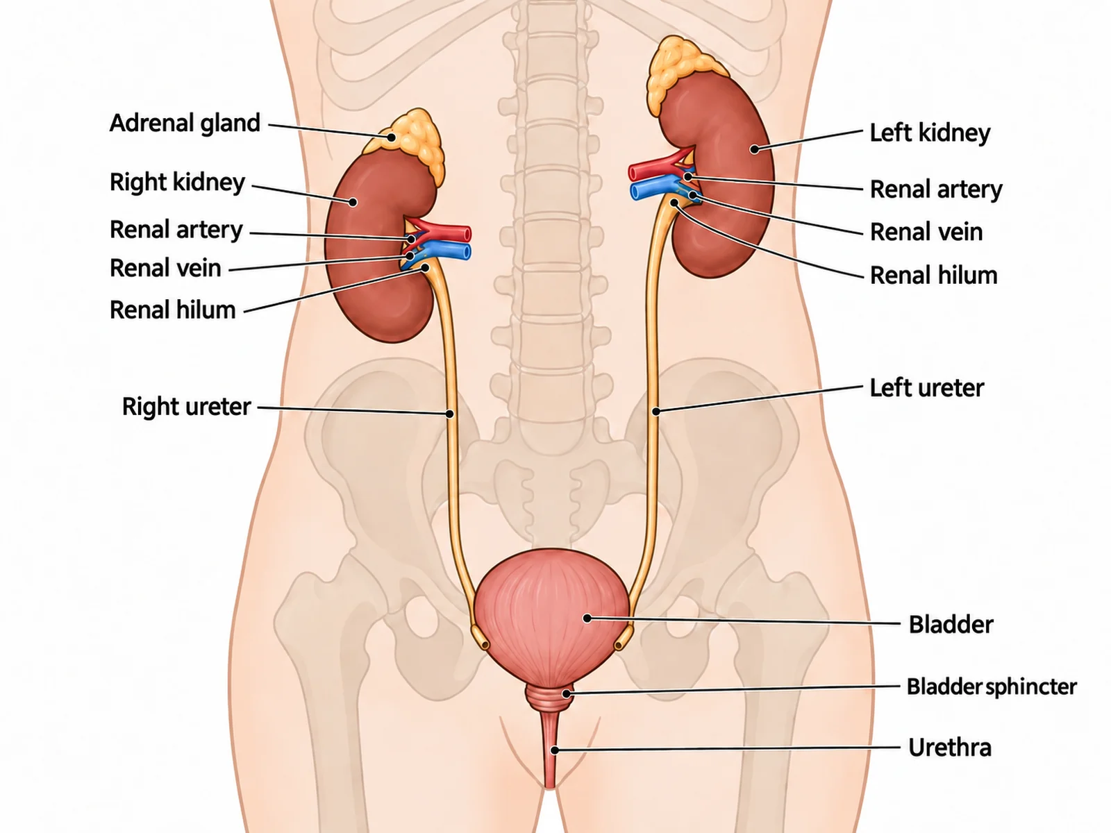
A nephron diagram is a zoom into one tiny part of the kidney, and it helps to know the route back out. The renal corpuscles and convoluted tubules make up the granular-looking cortex. The loops of Henle and the collecting ducts, lying side by side, give the medulla its striped renal pyramids. Collecting ducts merge into larger papillary ducts that open at the tip of each pyramid, the renal papilla, into a minor calyx. Minor calyces join into major calyces, the major calyces into the funnel-shaped renal pelvis, and the renal pelvis into the ureter, which carries urine down to the bladder. For the organ-level picture of the kidneys, ureters, bladder, and urethra, use the urinary system diagram generator. This page covers the microscopic unit inside it.
Reading a nephron diagram: orientation and scale
Nephron diagrams follow a shared convention. The kidney’s outer surface is at the top, the cortex below it, and the medulla below that, with the loop of Henle hanging down toward the papilla at the bottom. A diagram drawn upside down or sideways is not wrong as anatomy, but it confuses students who learned the usual layout, so this generator keeps it unless you ask otherwise.
The figure is a schematic, not a scale drawing. A renal corpuscle is about a fifth of a millimetre across, barely visible to the naked eye. The real tubule is far longer and more tangled than any diagram shows, and its two convoluted parts wind around each other close to their own corpuscle. Diagrams stretch the nephron out so that each part can be seen and labeled. What should stay true is the order of the parts, which zone each part lies in, and where the vessels connect.
When nephrons are damaged
Clinical courses often pair the nephron diagram with what happens when it fails. Nephrons do not regrow, so kidney function is measured as the glomerular filtration rate, the volume of filtrate made per minute, and chronic kidney disease is staged by how far it has fallen. Long-term high blood pressure and diabetes are the most common causes; both damage the glomeruli. Damaged glomeruli start to let protein through, so protein in the urine is an early sign. When too few working nephrons remain, waste such as urea and creatinine builds up in the blood, and dialysis or a transplant has to take over the filtering. A disease panel like this is easy to add to a diagram, but only when you ask for it.
Nephron labels by level
Simple: about six parts with plain names, such as glomerulus, Bowman’s capsule, tubule, loop of Henle, collecting duct, and blood vessels.
High school: the ten standard parts, from both arterioles through the two convoluted tubules to the collecting duct and the peritubular capillaries.
College: adds the separate limbs of the loop with the thick ascending limb, the renal corpuscle as a unit, the juxtaglomerular apparatus, and the cortex and medulla.
Medical: adds the macula densa, the juxtaglomerular cells, the podocytes, the connecting tubule, the vasa recta, and the thin and thick segments of each limb.
Nephron diagram, unlabeled: labeling practice in class
Blank first, key second: hand out the unlabeled nephron worksheet, then show the answer key made from the same result so the numbers match.
Twelve labels is a good size: the cortex and medulla brackets, both arterioles, the glomerulus, Bowman’s capsule, both convoluted tubules, both limbs of the loop, the capillaries, and the collecting duct.
Follow the drop: have students trace one drop of filtrate in order before they write any names.
Use the process view as a second sheet, asking students to add filtration, reabsorption, and secretion arrows in the right places.
Word bank or not: include one for revision, leave it out for a test. It is printed in alphabetical order.
Most common mistakes on a nephron diagram
These are the errors image models make most often on nephrons. Check each one before a diagram goes into graded work.
MistakeCorrect
MistakeProximal and distal tubules swapped
CorrectThe proximal tubule leaves the capsule first and is longer and more coiled
MistakeEfferent arteriole wider than the afferent
CorrectAfferent (in) is wider; efferent (out) is narrower
MistakeGlomerulus beside the capsule instead of inside it
MistakeLoop of Henle in the cortex, or the corpuscle in the medulla
CorrectCorpuscle and convoluted tubules in the cortex; the loop dips into the medulla
MistakeCollecting duct looping back up or joining the loop
CorrectIt runs straight down through the medulla to the papilla
MistakeWater leaving the thick ascending limb
CorrectWater leaves the descending limb; the thick ascending limb pumps out salt only
MistakeDistal tubule ending far from its corpuscle
CorrectIt returns to touch its own afferent arteriole (the juxtaglomerular apparatus)
MistakeFiltration arrows along the tubules
CorrectFiltration happens only at the glomerulus
Proximal vs distal convoluted tubule
The two convoluted tubules look alike on a diagram and are the pair most often swapped.
Proximal convoluted tubule
Distal convoluted tubule
Position in the sequence
Right after Bowman’s capsule
After the loop of Henle, before the collecting duct
Length and coiling
Longer, more tightly coiled
Shorter, less coiled
Lining
Cuboidal cells with a brush border of microvilli
Cuboidal cells without a brush border
Main reabsorption
Most water and Na+, all glucose and amino acids
Fine-tuning of Na+, Cl− and Ca2+
Hormone control
Mostly unregulated
Aldosterone and parathyroid hormone
Landmark
Starts at the urinary pole, opposite the arterioles
Touches its own afferent arteriole at the macula densa
Nephron Diagram FAQ
In order along the path of the filtrate: the glomerulus inside Bowman’s (glomerular) capsule, which together form the renal corpuscle; the proximal convoluted tubule; the loop of Henle with its descending and ascending limbs; the distal convoluted tubule; and the collecting duct. Most diagrams also label the afferent and efferent arterioles and the peritubular capillaries, and many mark where the cortex ends and the medulla begins.
Generate body system diagrams, organ and tissue close-ups, blank labeling worksheets with matching answer keys, and revision-ready biology figures with the same SciFigure AI workflow.