Generate a labeled endocrine system diagram showing the glands and the hormones they release, an endocrine system chart, or a blank, unlabeled diagram for students to label with a matching answer key. Choose the body, a gland and hormone chart, a close-up, or a feedback flow chart.
Labeled, chart or blankGlands and hormonesMatching answer key
Checked against OpenStax A&P
Worksheet + answer key pairs
Print-ready PNG
AI Endocrine System Diagram Generator
Describe the glands, hormones, and view, or pick a blank worksheet from the Labels menu.
563 / 2000 characters
Generate a publication-ready PNG from your description.
Preview
Endocrine System Diagram Example Previews
Target aspect ratio: 3:4
Sample previews. Describe your diagram to generate your own endocrine system figure.
Labeled and Blank Endocrine System Diagram Examples
A labeled endocrine system diagram, a blank, unlabeled worksheet with its answer key, an endocrine system chart of glands and hormones, a hypothalamus and pituitary close-up, and a feedback flow chart.
Prompt
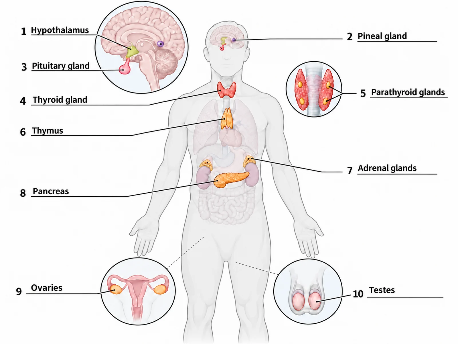
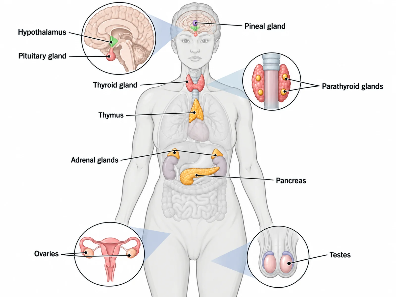
Create a labeled human endocrine system diagram on a white background: a front-facing body outline in pale grey, the person’s right side on the viewer’s left, with the glands in soft colors and other organs faint. Beside the head, a round magnified inset shows a midline section of the brain with the hypothalamus, the pituitary hanging below it on its stalk, and the pineal gland further back. The thyroid wraps the front of the windpipe just below the larynx, and a small inset shows it from behind with four parathyroid glands on its back. The thymus sits behind the breastbone above the heart, an adrenal gland caps each faint kidney, and the pancreas lies behind the stomach with its head on the viewer’s left. Two small insets show the ovaries and the testes. Label with thin leader lines: "Hypothalamus", "Pineal gland", "Pituitary gland", "Thyroid gland", "Parathyroid glands", "Thymus", "Adrenal glands", "Pancreas", "Ovaries", "Testes".
Use this example
Prompt
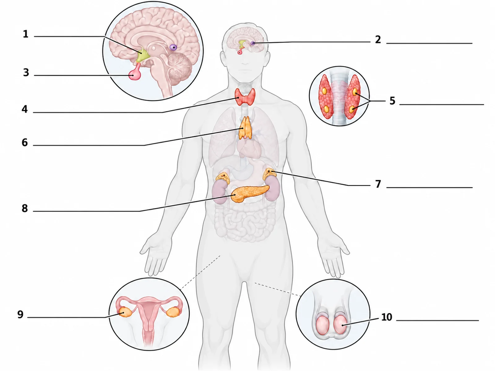
Create a blank, unlabeled endocrine system worksheet for students to label, on a white background. Draw a front-facing human body outline in pale grey, the person’s right side on the viewer’s left, with the endocrine glands drawn in soft colors and every other organ left out or drawn faint. Beside the head, a round magnified inset shows a midline section of the brain facing the viewer’s left, with the hypothalamus at the base of the brain, the pea-sized pituitary gland hanging below it on a thin stalk, and the small pineal gland further back near the middle of the brain. The butterfly-shaped thyroid gland wraps the front of the windpipe just below the larynx; a small inset beside the neck shows the thyroid from behind with four small parathyroid glands on its back surface. The thymus sits in the upper chest behind the breastbone, above the heart. An adrenal gland caps the top of each faintly drawn kidney. The pancreas lies across the upper abdomen behind a faint stomach, its head on the person’s right (viewer’s left) and its tail toward the person’s left. Two small insets at the bottom show the ovaries beside a faint uterus and the testes in a faint scrotum. Draw exactly 10 thin leader lines, each ending at a short horizontal ruled answer line with a number beside it and no text, pointing at these targets in this order: 1 = Hypothalamus, 2 = Pineal gland, 3 = Pituitary gland, 4 = Thyroid gland, 5 = Parathyroid glands, 6 = Thymus, 7 = Adrenal glands, 8 = Pancreas, 9 = Ovaries, 10 = Testes. Put no gland names, no hormone names, no key and no word bank anywhere on the image. Leave generous white space.
Blank worksheet
Use this example
Prompt
Create the answer key for a blank endocrine system worksheet, on a white background. Draw a front-facing human body outline in pale grey, the person’s right side on the viewer’s left, with the endocrine glands drawn in soft colors and every other organ left out or drawn faint. Beside the head, a round magnified inset shows a midline section of the brain facing the viewer’s left, with the hypothalamus at the base of the brain, the pea-sized pituitary gland hanging below it on a thin stalk, and the small pineal gland further back near the middle of the brain. The butterfly-shaped thyroid gland wraps the front of the windpipe just below the larynx; a small inset beside the neck shows the thyroid from behind with four small parathyroid glands on its back surface. The thymus sits in the upper chest behind the breastbone, above the heart. An adrenal gland caps the top of each faintly drawn kidney. The pancreas lies across the upper abdomen behind a faint stomach, its head on the person’s right (viewer’s left) and its tail toward the person’s left. Two small insets at the bottom show the ovaries beside a faint uterus and the testes in a faint scrotum. Draw exactly 10 thin leader lines, each ending at a short horizontal ruled line with its number, and write the answer on each line in dark sans-serif text: 1 = Hypothalamus, 2 = Pineal gland, 3 = Pituitary gland, 4 = Thyroid gland, 5 = Parathyroid glands, 6 = Thymus, 7 = Adrenal glands, 8 = Pancreas, 9 = Ovaries, 10 = Testes. Keep the numbering in this order so it matches the worksheet line for line, and add no hormone names.
Use this example
Prompt
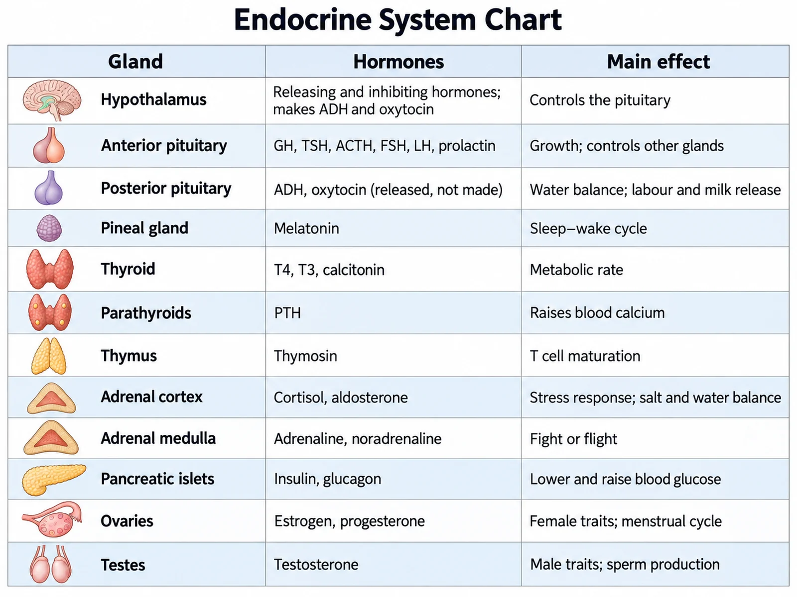
Create an endocrine system chart on a white background with three columns headed "Gland", "Hormones" and "Main effect", and a small icon of each gland in the first column. Rows, in this order: Hypothalamus | releasing and inhibiting hormones; makes ADH and oxytocin | controls the pituitary. Anterior pituitary | GH, TSH, ACTH, FSH, LH, prolactin | growth; controls other glands. Posterior pituitary | ADH, oxytocin (released, not made) | water balance; labour and milk release. Pineal gland | melatonin | sleep–wake cycle. Thyroid | T4, T3, calcitonin | metabolic rate. Parathyroids | PTH | raises blood calcium. Thymus | thymosin | T cell maturation. Adrenal cortex | cortisol, aldosterone | stress response; salt and water balance. Adrenal medulla | adrenaline, noradrenaline | fight or flight. Pancreatic islets | insulin, glucagon | lower and raise blood glucose. Ovaries | estrogen, progesterone | female traits; menstrual cycle. Testes | testosterone | male traits; sperm production.
Gland and hormone chart
Use this example
Prompt
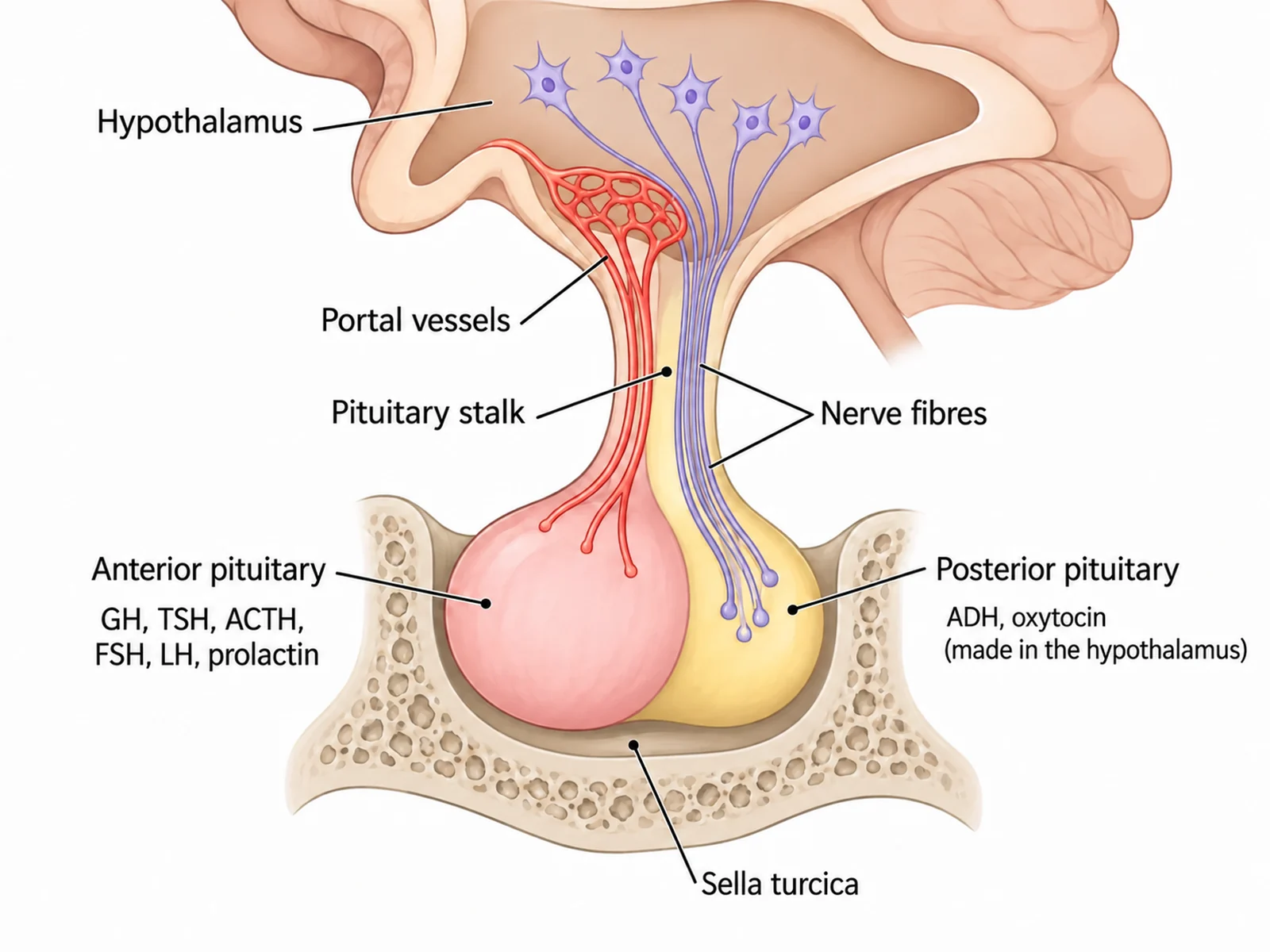
Create a labeled close-up of the hypothalamus and pituitary gland on a white background, drawn as a midline section facing the viewer’s left. Show the hypothalamus at the base of the brain, the pituitary stalk (infundibulum) below it, the anterior pituitary at the front, and the posterior pituitary behind it, the pituitary sitting in its bony pocket (sella turcica). Draw small blood vessels, the hypophyseal portal system, running from the hypothalamus down the stalk to the anterior pituitary, and nerve fibres running from hypothalamic neurons down the stalk into the posterior pituitary. List "GH, TSH, ACTH, FSH, LH, prolactin" beside the anterior pituitary and "ADH, oxytocin (made in the hypothalamus)" beside the posterior pituitary. Label "Hypothalamus", "Pituitary stalk", "Portal vessels", "Anterior pituitary", "Posterior pituitary", "Sella turcica".
Hypothalamus and pituitary
Use this example
Prompt
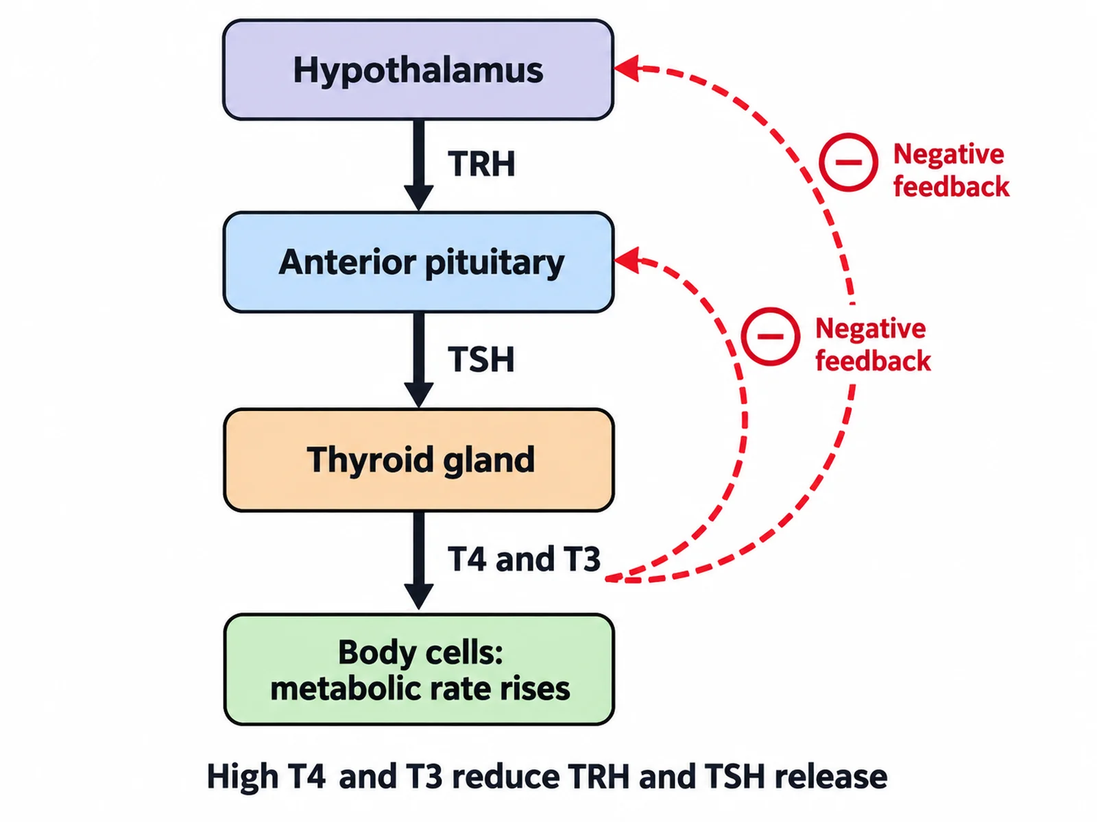
Create an endocrine system flow chart of the hypothalamus–pituitary–thyroid axis on a white background. Draw rounded boxes connected by downward arrows: "Hypothalamus" → arrow labeled "TRH" → "Anterior pituitary" → arrow labeled "TSH" → "Thyroid gland" → arrow labeled "T4 and T3" → "Body cells: metabolic rate rises". Draw two curved dashed arrows from the "T4 and T3" arrow back up to the anterior pituitary and the hypothalamus, each marked with a circled minus sign and labeled "Negative feedback". Add a caption reading "High T4 and T3 reduce TRH and TSH release".
Feedback flow chart
Use this example
How to make a labeled or blank endocrine system diagram
1
Pick the view, figure, and labels
Full body, a gland and hormone chart, a pituitary or thyroid close-up, or a feedback flow chart. Choose a neutral, female, or male figure, then labeled, numbered, blank, or a word bank.
2
Generate the diagram
The generator places each gland at its real level, keeps the parathyroids behind the thyroid and the adrenals on top of the kidneys, and writes hormones only with the gland that makes them.
3
Make the answer key, check, and export
For a worksheet, make the answer key from the same result so the numbers match. Check the thyroid level, the pancreas orientation, and any hormone names, then download the PNG.
An endocrine system diagram is a labeled drawing of the glands that release hormones into the blood. The standard version shows a front-facing body with the glands placed where they really sit: the hypothalamus, pituitary, and pineal in the head, the thyroid and parathyroids in the neck, the thymus in the chest, the adrenal glands and pancreas in the abdomen, and the ovaries or testes in the pelvis.
The same figure is used three ways. A labeled diagram of the endocrine system is a reference. An unlabeled or blank version with numbered lines is a worksheet. An endocrine system chart, sometimes called an endocrine system map, sets the glands beside their hormones and effects. This page makes all three, with an answer key that matches the worksheet line for line.
The major endocrine glands and where they are
Hypothalamus: a small region at the base of the brain, above the pituitary. It is part of the brain and also the endocrine system’s control centre.
Pituitary gland: pea-sized, hanging from the hypothalamus on a stalk and sitting in a bony pocket of the skull base (the sella turcica), roughly behind the eyes. It has an anterior and a posterior lobe.
Pineal gland: a tiny pine-cone-shaped gland deep in the brain near its centre, behind the hypothalamus.
Thyroid gland: butterfly-shaped, in the front of the neck below the larynx, wrapped around the windpipe.
Parathyroid glands: usually four small glands on the back of the thyroid.
Thymus: in the upper chest behind the breastbone, above the heart. It is large in children and shrinks after puberty.
Adrenal glands: one on top of each kidney, each with an outer cortex and an inner medulla.
Pancreas: behind the stomach, across the back of the upper abdomen. Its hormone-making cells are the pancreatic islets.
Gonads: the ovaries in the female pelvis and the testes in the male scrotum.
Endocrine system chart: glands, hormones, and effects
When a diagram adds hormones, these are the standard pairings an introductory course expects. Each hormone belongs to one gland; a chart that moves one is wrong.
Hypothalamus: releasing and inhibiting hormones that control the anterior pituitary (TRH, CRH, GnRH, GHRH, and inhibiting hormones such as somatostatin). It also makes ADH and oxytocin.
Posterior pituitary: releases ADH (antidiuretic hormone, which tells the kidneys to keep water) and oxytocin (uterine contractions in labour, milk release).
Pineal gland: melatonin, which helps set the sleep–wake cycle.
Thyroid gland: thyroxine (T4) and triiodothyronine (T3), which set the metabolic rate, and calcitonin, which can lower blood calcium.
Parathyroid glands: parathyroid hormone (PTH), which raises blood calcium.
Thymus: thymosins, which help T cells mature.
Adrenal cortex: cortisol (stress, blood glucose) and aldosterone (sodium and water balance). Adrenal medulla: adrenaline and noradrenaline for the fight-or-flight response.
Pancreatic islets: insulin lowers blood glucose and glucagon raises it.
Ovaries: estrogen and progesterone. Testes: testosterone.
The hypothalamus and pituitary: the control centre
The hypothalamus and pituitary sit together at the base of the brain and control much of the rest of the system. The hypothalamus releases small amounts of releasing and inhibiting hormones into a short network of portal blood vessels that runs down the pituitary stalk to the anterior pituitary. In response, the anterior pituitary releases its own hormones into the general circulation. Three of them, TSH, ACTH, and FSH with LH, act on other endocrine glands: the thyroid, the adrenal cortex, and the gonads.
The posterior pituitary works differently. It is an extension of the brain rather than a gland in its own right. Neurons whose cell bodies lie in the hypothalamus make ADH and oxytocin and send them down their axons through the stalk to be stored and released in the posterior lobe. A close-up diagram should show both routes: blood vessels to the anterior lobe and nerve fibres to the posterior lobe.
Reading a front-view endocrine diagram
Like every anatomical figure, a front-view endocrine diagram shows the person facing you, so their right side is on your left. That matters most for the pancreas. Its head sits in the C-shaped curve of the duodenum on the person’s right, which is your left, and its tail stretches across toward the spleen on the person’s left.
Level matters as much as side. The pituitary is roughly level with the eyes, not in the neck. The thyroid is below the larynx, not at the jaw. The thymus is behind the upper breastbone, not in the neck. The adrenal glands sit on the upper poles of the kidneys, which lie high against the back wall of the abdomen. Because the hypothalamus, pituitary, and pineal lie one behind another on the midline, a good diagram shows them in a magnified section of the brain rather than stacking them on a front view.
Inside the adrenal gland: cortex and medulla
An adrenal close-up is a common second figure, because each adrenal gland is really two glands in one. The outer cortex is built in three layers, each making a different class of steroid hormone. Under the capsule, the zona glomerulosa makes aldosterone, which tells the kidneys to keep sodium and water. The middle zona fasciculata, the thickest layer, makes cortisol, which raises blood glucose and shapes the stress response. The inner zona reticularis makes small amounts of androgens. The medulla at the centre is different tissue altogether. Its cells are modified nerve cells of the sympathetic nervous system, and they release adrenaline and noradrenaline within seconds of a nerve signal. A diagram of the gland should show the cortex as a rim around the medulla, with the three zones in that order from the outside in.
Three kinds of hormone
More detailed charts group hormones by their chemistry, because that decides how they reach their target.
Steroid hormones are made from cholesterol: cortisol, aldosterone, estrogen, progesterone, and testosterone. They are fat-soluble, pass through cell membranes, and act on receptors inside the cell, often switching genes on or off.
Peptide and protein hormones are chains of amino acids: insulin, glucagon, growth hormone, ADH, oxytocin, and the pituitary hormones. They cannot cross the membrane, so they bind receptors on the cell surface and act through second messengers inside it.
Amine hormones are made from a single amino acid: adrenaline and noradrenaline, melatonin, and the thyroid hormones T4 and T3. The thyroid hormones are the exception in their group, because they enter cells and act on receptors inside, like steroids.
Labels by level
Middle school: about seven glands with no hormones, such as pituitary, thyroid, thymus, adrenal glands, pancreas, and ovaries or testes.
High school: the ten standard glands, adding the hypothalamus, pineal gland, and parathyroids.
College: the ten glands with their main hormones written under each name, plus the feedback flow chart.
Medical: subdivisions as well as glands, such as the anterior and posterior pituitary, the thyroid follicles and parafollicular cells, the three zones of the adrenal cortex and the medulla, and the cell types of the pancreatic islets.
Organs with endocrine tissue that diagrams often leave out
Hormones are not made only by the classic glands. Many organs have endocrine cells as a second job. Introductory diagrams usually leave them out; fuller ones list them in a side panel.
Heart: atrial natriuretic peptide, released when the atria are stretched, which helps lower blood volume.
Kidneys: erythropoietin, which drives red blood cell production, and the active form of vitamin D. They also release renin.
Stomach and small intestine: gastrin, secretin, and cholecystokinin, which coordinate digestion.
Adipose tissue: leptin, which signals the body’s energy stores.
Skin: makes a precursor of vitamin D when exposed to sunlight.
Placenta: during pregnancy, hormones including human chorionic gonadotropin (hCG).
Feedback loops and the endocrine system flow chart
Most hormone levels are held steady by negative feedback, which is why a flow chart of the endocrine system is a common figure. The hypothalamus–pituitary–thyroid axis is the standard example. The hypothalamus releases TRH, the anterior pituitary responds with TSH, and the thyroid responds with T4 and T3. Rising T4 and T3 then inhibit both the hypothalamus and the pituitary, so less TRH and TSH are released and thyroid output falls back. The cortisol axis (CRH, ACTH, cortisol) follows the same pattern.
Blood glucose is regulated without the pituitary. After a meal, rising glucose triggers insulin from the pancreatic islets, and glucose moves into cells. Between meals, falling glucose triggers glucagon, and the liver releases glucose. Positive feedback is rarer. Oxytocin in childbirth is the textbook case: contractions push the baby against the cervix, which triggers more oxytocin and stronger contractions until birth.
Endocrine vs exocrine glands
An endocrine gland has no duct. It releases hormones into the fluid around its cells, and the blood carries them to any cell with a matching receptor. An exocrine gland releases its product through a duct onto a surface or into a cavity, like sweat onto the skin or saliva into the mouth. The pancreas does both: most of it is exocrine tissue making digestive enzymes that drain into the duodenum, with endocrine islets scattered through it. The ovaries and testes release hormones into the blood and also release eggs or sperm. That is why both appear on an endocrine diagram even though they belong to the reproductive system too.
Male and female endocrine diagrams
Apart from the gonads, the endocrine system is the same in both sexes, so a diagram has three honest options. A female figure shows the ovaries in the pelvis on either side of the uterus. A male figure shows the testes in the scrotum. A neutral figure shows the two gonads as small separate insets. The neutral version is the most reusable worksheet, because one sheet serves the whole class. What a diagram must never do is draw both ovaries and testes inside one body, a mistake image models make often enough that this page’s rules forbid it.
Using a blank endocrine system diagram in class
Glands first, hormones second: label the glands on the blank body, then fill in a blank chart of hormones as a second step.
Ten labels is a good size: hypothalamus, pineal, pituitary, thyroid, parathyroids, thymus, adrenals, pancreas, ovaries, and testes.
Make the key from the same result, so the numbering on the worksheet and the answer key match exactly.
Use the feedback flow chart for the "how" question: give students the boxes and have them draw the stimulating and inhibiting arrows.
Word bank or not: include one for revision, leave it out for a test. It is printed in alphabetical order.
How the endocrine system changes across a lifetime
A diagram is a snapshot of one age, and a few glands change enough to matter. The thymus is largest relative to body size in childhood, when it is busiest training T cells; after puberty it shrinks and is gradually replaced by fat, so an older adult’s thymus is hard to find. Puberty begins when the hypothalamus increases its pulses of GnRH, which raises FSH and LH from the pituitary and switches on the ovaries or testes. Growth hormone output is highest in childhood and adolescence and falls with age. At menopause the ovaries stop responding and estrogen and progesterone fall, while FSH and LH rise because negative feedback is lost. If a worksheet is about a child or an older adult, say so in the description so the thymus is drawn at the right size.
Common disorders linked to each gland
Pancreatic islets: type 1 diabetes, where the insulin-making beta cells are destroyed, and type 2 diabetes, where the body responds poorly to insulin.
Thyroid: hypothyroidism (too little T4 and T3, with tiredness and weight gain) and hyperthyroidism (too much, with weight loss and a fast heart rate). A goitre is an enlarged thyroid, classically from iodine deficiency.
Adrenal cortex: Addison’s disease (too little cortisol and aldosterone) and Cushing’s syndrome (too much cortisol).
Pituitary: too much growth hormone causes gigantism in children and acromegaly in adults; too little in childhood causes short stature.
Posterior pituitary: too little ADH causes diabetes insipidus, with large volumes of dilute urine.
Most common mistakes on an endocrine system diagram
These are the errors image models make most often on endocrine diagrams. Check each one before a diagram goes into graded work.
MistakeCorrect
MistakeThyroid at the jaw or on the larynx
CorrectBelow the larynx, wrapped around the front of the windpipe
MistakeParathyroids on the front of the thyroid
CorrectUsually four, on the back surface of the thyroid
MistakePituitary in the neck or loose in the skull
CorrectHanging from the hypothalamus on a stalk, in a bony pocket at the skull base
MistakeAdrenal glands drawn as part of the kidney
CorrectSeparate glands sitting on top of each kidney
MistakeThymus in the neck
CorrectUpper chest, behind the breastbone, above the heart
MistakePancreas mirrored or in front of the stomach
CorrectBehind the stomach, head on the person’s right in the duodenal curve
MistakeOne body with both ovaries and testes
CorrectA female figure, a male figure, or a neutral figure with two insets
MistakeADH or oxytocin credited to the anterior pituitary
CorrectMade in the hypothalamus, released by the posterior pituitary
Endocrine system vs nervous system
Both systems coordinate the body, and exam questions often ask how they differ.
Endocrine system
Nervous system
Messenger
Hormones carried in the blood
Electrical impulses and neurotransmitters
Speed
Seconds to hours
Milliseconds
Duration of effect
Often long-lasting
Usually brief
Reach
Any cell with the right receptor, anywhere in the body
The specific cells a neuron connects to
Typical jobs
Growth, metabolism, reproduction, water and salt balance
Movement, sensation, rapid reflexes
Where they meet
The hypothalamus sends hormones to the pituitary
The hypothalamus is part of the brain
Endocrine System Diagram FAQ
The major endocrine glands: hypothalamus, pituitary, pineal, thyroid, parathyroids, thymus, adrenal glands, pancreas, and the gonads (ovaries or testes). Many diagrams also name the main hormone of each gland, which turns the figure into a combined diagram and chart. Fuller versions add the organs with endocrine tissue, such as the heart, kidneys, stomach, small intestine, and adipose tissue.
Generate body system diagrams, organ close-ups, blank labeling worksheets with matching answer keys, and revision-ready biology figures with the same SciFigure AI workflow.