Generate a labeled synapse diagram that follows the signal one way across the gap: an action potential opens voltage-gated Ca²⁺ channels, synaptic vesicles release neurotransmitter, it crosses a 20–40 nm synaptic cleft, and it binds receptors on the postsynaptic membrane. Switch to an electrical synapse, a neuromuscular junction, or excitatory vs inhibitory synapses, or make a blank worksheet with its answer key.
Chemical, electrical, or NMJCa²⁺ in, transmitter acrossLabeled or blank worksheet
Checked against OpenStax Biology 2e
Ca²⁺ in, transmitter one way
Labeled, numbered, or blank
Print-ready PNG
AI Synapse Diagram Generator
Describe the synapse type, the parts to label, and the level, or ask for a blank worksheet.
612 / 2000 characters
Generate a publication-ready PNG from your description.
Preview
Synapse Diagram Example Previews
Target aspect ratio: 4:3
Sample previews. Describe your diagram to generate your own synapse figure.
Synapse Diagram Examples
Use these as visual directions for a labeled synapse, the steps of synaptic transmission, a chemical vs electrical synapse, the neuromuscular junction, excitatory vs inhibitory synapses, and a blank worksheet.
Prompt
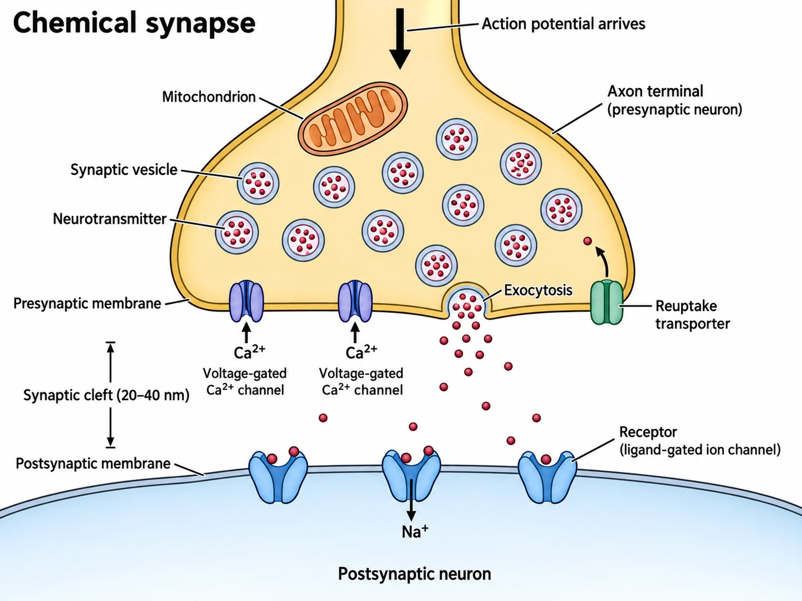
Create a fully labeled chemical synapse diagram on a white background in clean biology textbook style. At the top draw the rounded end of an axon labeled "Axon terminal (presynaptic neuron)", with a downward arrow entering it labeled "Action potential arrives". Inside it draw a "Mitochondrion" and many small round "Synaptic vesicle"s filled with dots labeled "Neurotransmitter". In its lower membrane, labeled "Presynaptic membrane", draw two "Voltage-gated Ca2+ channel"s with small arrows labeled "Ca2+" pointing INTO the terminal, and one vesicle fusing with the membrane and spilling dots, labeled "Exocytosis". Below, leave a clear open gap labeled "Synaptic cleft (20–40 nm)". At the bottom draw a curved "Postsynaptic membrane" of a "Postsynaptic neuron" with cup-shaped "Receptor (ligand-gated ion channel)" proteins holding transmitter dots and an arrow labeled "Na+" entering. On the presynaptic membrane add one "Reuptake transporter" with an arrow carrying a dot back in. The two cells never touch.
Use this example
Prompt
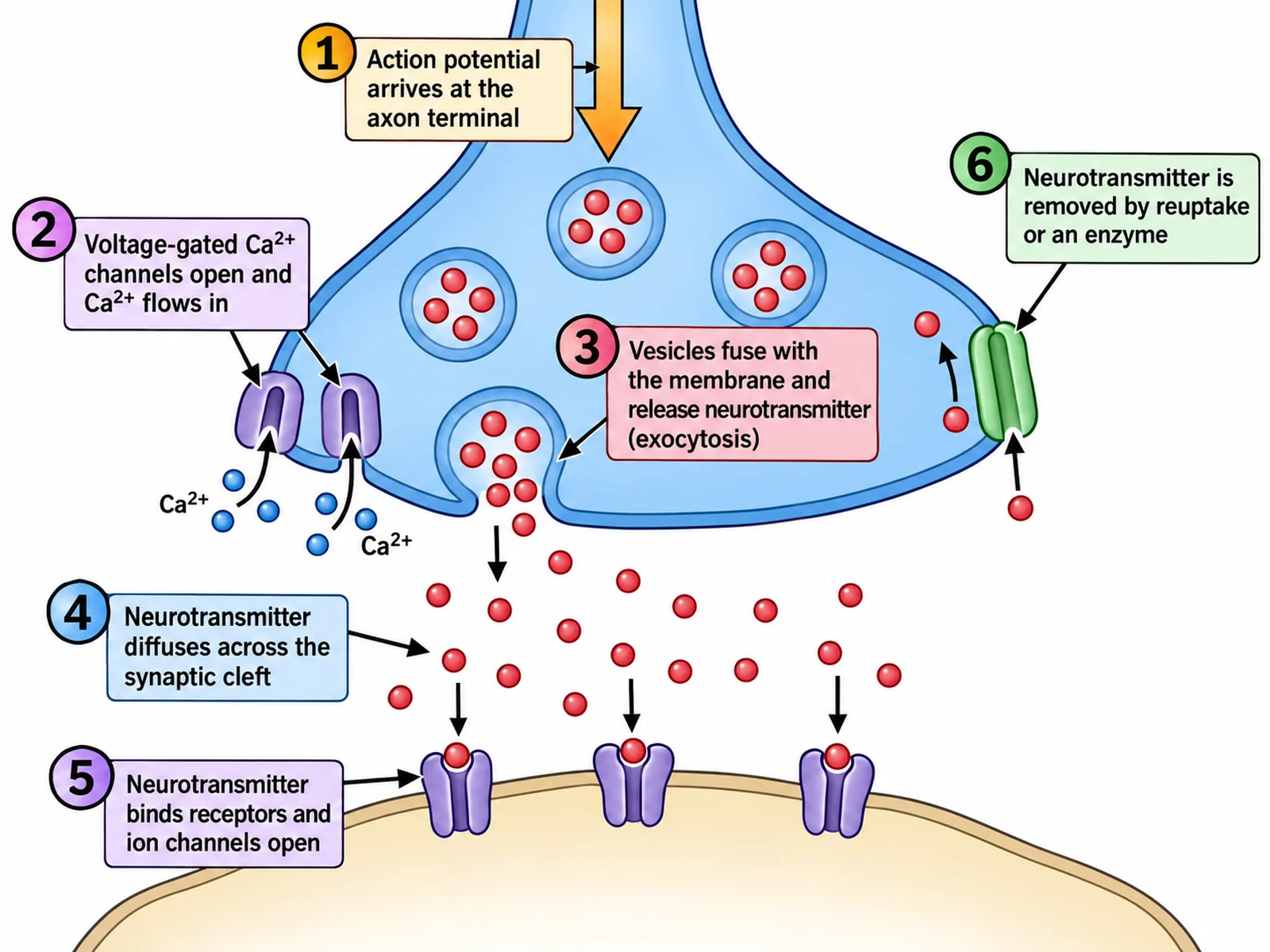
Create a diagram of the synapse that numbers the steps of synaptic transmission 1 to 6 on a white background. Draw an axon terminal at the top, an open synaptic cleft, and a postsynaptic membrane at the bottom. Place large numbered circles with short captions: "1 Action potential arrives at the axon terminal"; "2 Voltage-gated Ca2+ channels open and Ca2+ flows in" with arrows pointing into the terminal; "3 Vesicles fuse with the membrane and release neurotransmitter (exocytosis)"; "4 Neurotransmitter diffuses across the synaptic cleft"; "5 Neurotransmitter binds receptors and ion channels open"; "6 Neurotransmitter is removed by reuptake or an enzyme" with a transporter on the presynaptic membrane carrying it back in. Every arrow across the cleft points down, from the presynaptic to the postsynaptic side.
Use this example
Prompt
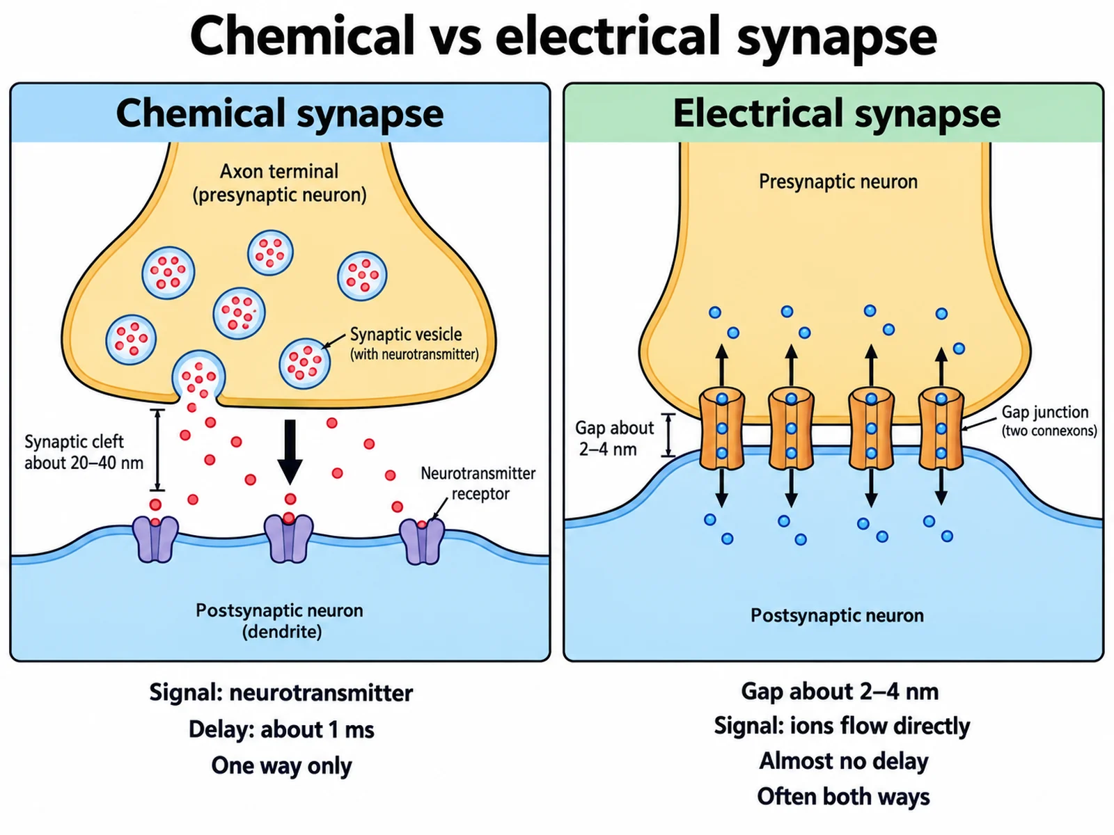
Create a two-panel comparison on a white background titled "Chemical vs electrical synapse". Left panel "Chemical synapse": an axon terminal with synaptic vesicles releasing neurotransmitter into an open gap labeled "Synaptic cleft about 20–40 nm", receptors on the postsynaptic membrane, one downward arrow, and captions "Signal: neurotransmitter", "Delay: about 1 ms", "One way only". Right panel "Electrical synapse": two cell membranes much closer together, joined by several barrel-shaped channels labeled "Gap junction (two connexons)", with small ion dots passing straight through and arrows pointing both ways, and captions "Gap about 2–4 nm", "Signal: ions flow directly", "Almost no delay", "Often both ways". No vesicles in the right panel.
Use this example
Prompt
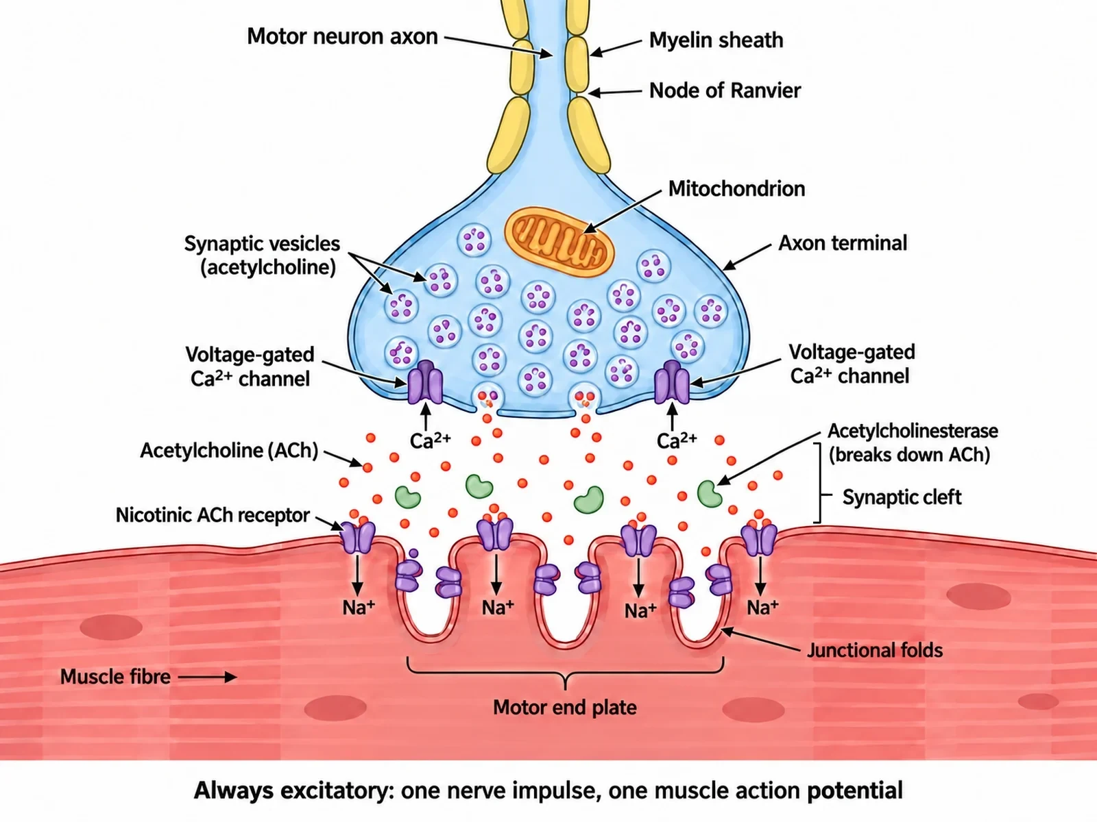
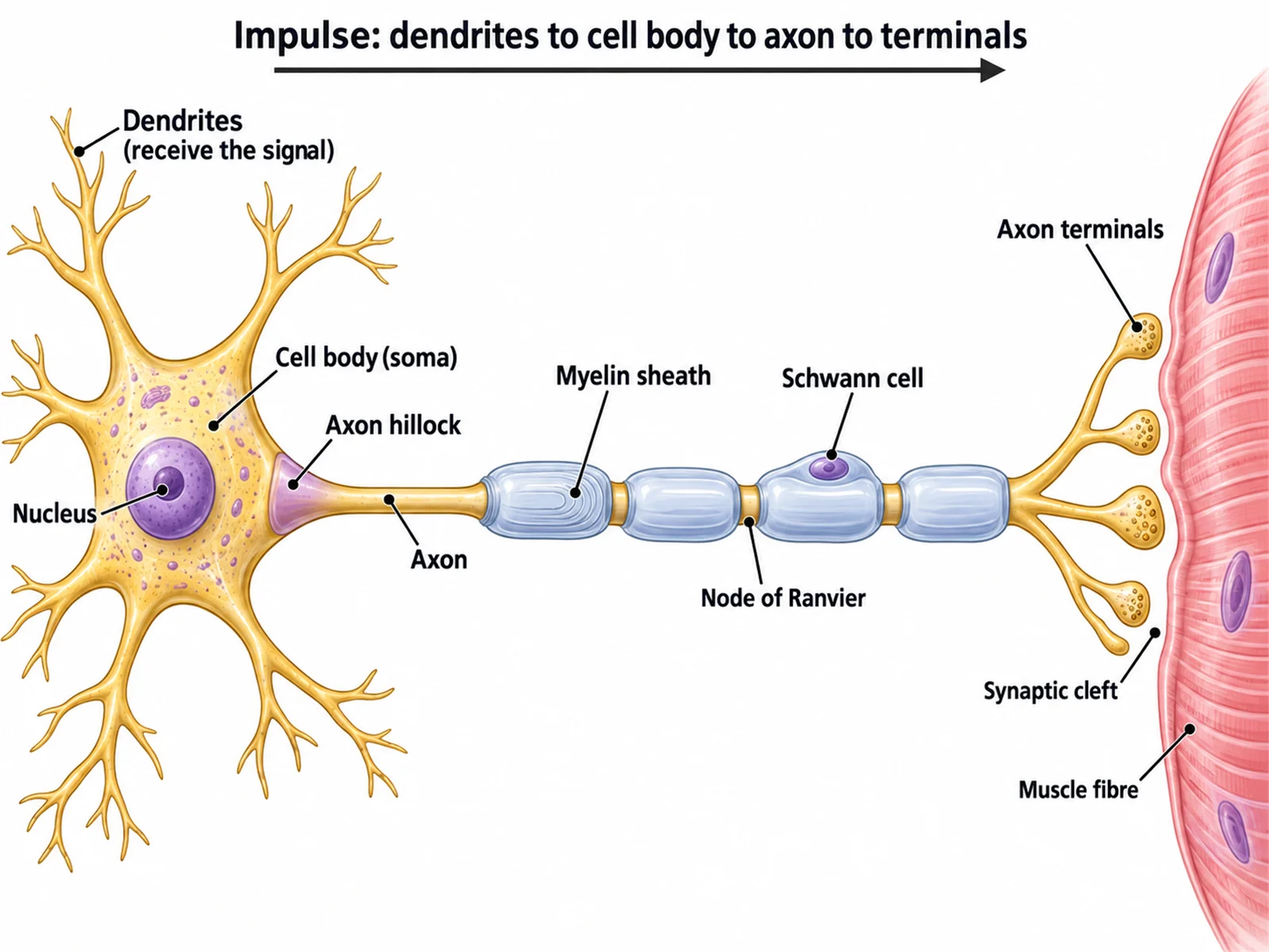
Create a labeled neuromuscular junction diagram on a white background. At the top draw a myelinated "Motor neuron axon" ending in an "Axon terminal" that contains "Synaptic vesicles (acetylcholine)" and a mitochondrion, with "Voltage-gated Ca2+ channel"s and Ca2+ arrows pointing into the terminal. Below it draw a skeletal "Muscle fibre" whose membrane under the terminal is folded into deep "Junctional folds", labeled "Motor end plate". Show "Acetylcholine (ACh)" dots crossing the open "Synaptic cleft" and binding "Nicotinic ACh receptor"s on the folds, with an arrow labeled "Na+" entering the muscle fibre. Draw small scissor-shaped enzymes in the cleft labeled "Acetylcholinesterase (breaks down ACh)". Caption: "Always excitatory: one nerve impulse, one muscle action potential".
Use this example
Prompt
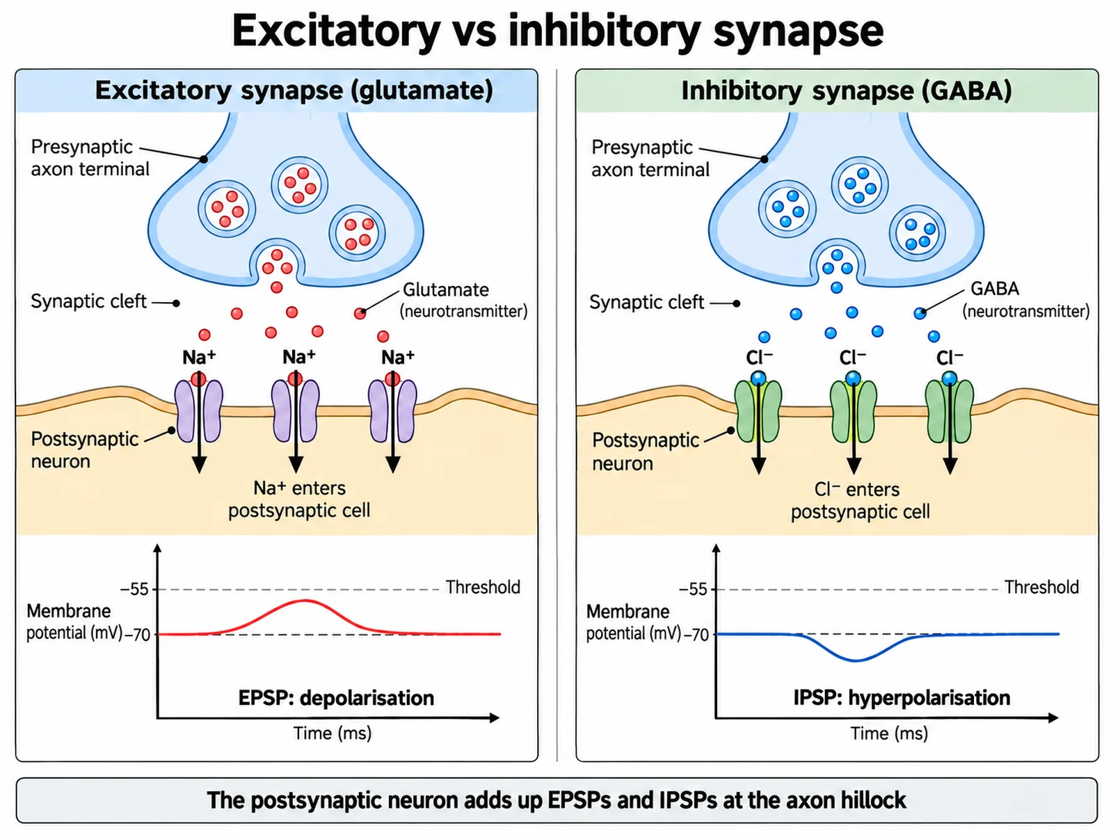
Create a two-panel comparison on a white background titled "Excitatory vs inhibitory synapse". Left panel "Excitatory synapse (glutamate)": an axon terminal releasing transmitter across an open cleft onto receptors whose channels let "Na+" flow INTO the postsynaptic cell, with a small trace below rising from −70 mV toward threshold labeled "EPSP: depolarisation". Right panel "Inhibitory synapse (GABA)": the same layout, but the receptor channels let "Cl-" flow INTO the postsynaptic cell, with a small trace below dipping below −70 mV labeled "IPSP: hyperpolarisation". Under both panels add one caption: "The postsynaptic neuron adds up EPSPs and IPSPs at the axon hillock".
Use this example
Prompt
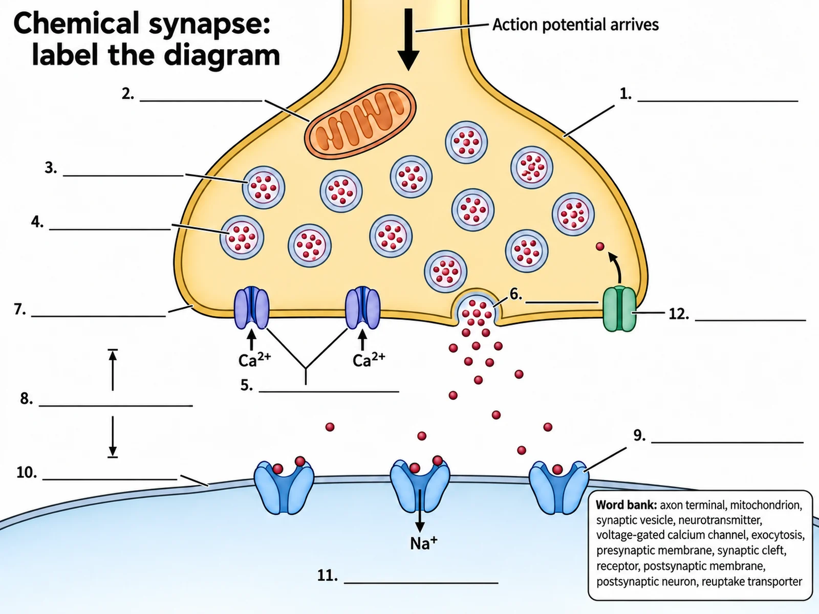
Create a blank synapse worksheet that is the same drawing as a labeled textbook chemical synapse with the names removed: an axon terminal at the top with a mitochondrion and synaptic vesicles, two calcium channels in its lower membrane, one vesicle fusing and spilling dots, an open cleft, a curved postsynaptic membrane with cup-shaped receptors, and a reuptake transporter on the presynaptic membrane. Keep "Action potential arrives", "Ca2+", and "Na+" printed. Replace twelve names with numbered empty answer lines: 1 axon terminal, 2 mitochondrion, 3 synaptic vesicle, 4 neurotransmitter, 5 voltage-gated calcium channel, 6 exocytosis, 7 presynaptic membrane, 8 synaptic cleft, 9 receptor, 10 postsynaptic membrane, 11 postsynaptic neuron, 12 reuptake transporter. Add a word bank: "axon terminal, mitochondrion, synaptic vesicle, neurotransmitter, voltage-gated calcium channel, exocytosis, presynaptic membrane, synaptic cleft, receptor, postsynaptic membrane, postsynaptic neuron, reuptake transporter".
Blank worksheet
Use this example
How to make a labeled synapse diagram
1
Pick the synapse and the labels
Choose a chemical synapse, an electrical synapse, a neuromuscular junction, or excitatory vs inhibitory, set the level, then choose labeled, numbered with a key, or a blank worksheet. Name a neurotransmitter or a drug only if you want it shown.
2
Generate the diagram
The generator keeps the vesicles and calcium channels in the presynaptic terminal, leaves an open synaptic cleft, puts the receptors on the postsynaptic membrane, and points every arrow the way the signal travels.
3
Check it, then export
Confirm Ca²⁺ flows into the terminal, the cells do not touch, and arrows cross the cleft in one direction only. Then download the PNG, or make the matching blank worksheet or answer key from the same image.
Synapse key facts
Synaptic cleft width
About 20–40 nm (sources vary)
Electrical synapse gap
About 2–4 nm, bridged by gap junctions
Synaptic delay
About 1 ms at a chemical synapse
Release trigger
Ca²⁺ entering through voltage-gated channels
Direction
One way: presynaptic → postsynaptic
Excitatory (EPSP)
e.g. glutamate; Na⁺ in, depolarises
Inhibitory (IPSP)
e.g. GABA; Cl⁻ in, hyperpolarises
Signal removal
Enzyme breakdown, reuptake, or diffusion
What is a synapse diagram?
A synapse diagram is a close-up of the junction where one neuron passes its signal to the next cell — another neuron, a muscle fibre, or a gland cell. Because the junction is far too small to see with a light microscope, nearly every textbook draws it the same way: a swollen axon terminal at the top, a narrow gap called the synaptic cleft in the middle, and the membrane of the receiving cell at the bottom. The drawing is a model, but it is a model built from electron micrographs, and the positions of the parts in it matter.
A good diagram of the synapse does two jobs. It names the parts — the axon terminal, synaptic vesicles, neurotransmitter, calcium channels, cleft, receptors, and postsynaptic membrane — and it shows the direction of the signal, which at a chemical synapse runs only one way. That direction is carried by where things are drawn: vesicles only on the sending side, receptors only on the receiving side, and a visible gap between them. Most mistakes in synapse diagrams are mistakes of position, not of vocabulary.
Labeled synapse diagram: the parts of a synapse
Presynaptic neuron: the sending neuron. Its axon ends in a swollen axon terminal, also called a synaptic knob, bouton, or end bulb.
Synaptic vesicles: small membrane spheres in the terminal, each packed with thousands of neurotransmitter molecules.
Mitochondria: in the terminal, supplying the ATP needed to refill vesicles and pump ions.
Voltage-gated Ca²⁺ channels: in the presynaptic membrane, opened by the arriving action potential.
Presynaptic membrane: the membrane of the terminal facing the cleft, where vesicles fuse and release their contents.
Synaptic cleft: the fluid-filled gap, about 20–40 nm wide, that the neurotransmitter diffuses across.
Receptors: proteins on the postsynaptic membrane that the neurotransmitter binds, often ligand-gated ion channels.
Postsynaptic membrane and postsynaptic cell: the receiving side — the dendrite or cell body of the next neuron, or a muscle or gland cell.
Reuptake transporters: proteins in the presynaptic membrane, or in nearby astrocytes, that carry neurotransmitter back in.
Enzymes in the cleft: at cholinergic synapses, acetylcholinesterase breaks acetylcholine down.
Synaptic transmission step by step: a diagram of the synapse in action
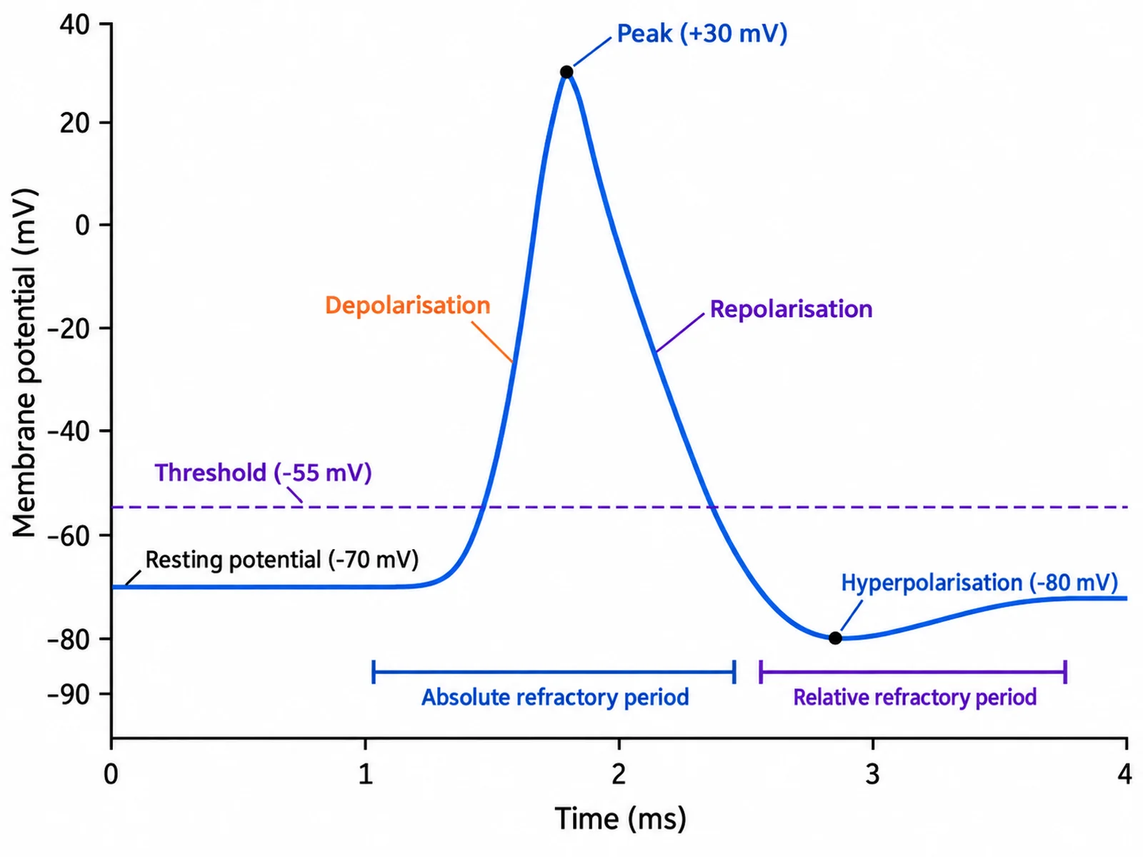
Synaptic transmission turns an electrical signal into a chemical one and back again. It starts when an action potential travels down the axon and reaches the axon terminal. The depolarisation opens voltage-gated calcium channels in the presynaptic membrane, and Ca²⁺ — which is far more concentrated outside the cell than inside — rushes into the terminal.
The rise in calcium is the trigger for release. Calcium-sensing proteins make synaptic vesicles docked at the membrane fuse with it, and each fused vesicle empties its neurotransmitter into the cleft by exocytosis. The neurotransmitter diffuses across the 20–40 nm gap in a fraction of a millisecond and binds to receptors on the postsynaptic membrane. If the receptor is an ion channel, it opens: the movement of ions through it changes the postsynaptic membrane potential, either toward threshold or away from it.
The signal has to end as quickly as it started, or the next one could not be read. The neurotransmitter is removed by being broken down by an enzyme, taken back up into the terminal or into glial cells, or simply diffusing away. The vesicle membrane that fused with the presynaptic membrane is pulled back in by endocytosis and refilled, ready for the next action potential. The whole sequence, from the action potential arriving to the postsynaptic channels opening, takes about one millisecond — the synaptic delay.
The synaptic cleft: how wide it is and what happens there
The synaptic cleft is the gap between the presynaptic and postsynaptic membranes, usually given as about 20–40 nanometres at a synapse between neurons, although different textbooks quote slightly different figures. It is not empty: it is filled with extracellular fluid and a scaffold of proteins that hold the two membranes in register, so that release sites on one side line up with receptors on the other. The gap is narrow enough for neurotransmitter to cross by diffusion almost instantly, but it is wide enough that the action potential itself cannot jump across. That is the single most important thing a synapse diagram has to show, and the reason a drawing in which the axon terminal touches the next cell is simply wrong. At cholinergic synapses the cleft also holds acetylcholinesterase, the enzyme that ends the signal.
Chemical vs electrical synapse diagram
Most synapses in the nervous system are chemical, but electrical synapses exist too, and comparison diagrams of the two are common in exams. At an electrical synapse the membranes of the two cells come within about 2–4 nm of each other and are joined by gap junctions. Each gap junction channel is made of two connexons, one contributed by each cell, and each connexon is a ring of six connexin proteins. Ions and small molecules pass straight through, so current flows from one cell into the next.
The differences follow from that structure. An electrical synapse has no vesicles and no neurotransmitter, it works almost instantly, and current can usually flow in both directions. It is ideal for keeping groups of cells in step — heart muscle cells, smooth muscle, and some groups of neurons that must fire together. A chemical synapse is slower, about a millisecond, and works in one direction only, but it can excite or inhibit, can amplify a signal, and can strengthen or weaken with use, which is the basis of learning. A comparison diagram should show the narrow gap and channels on one side and the cleft, vesicles, and receptors on the other.
Neuromuscular junction diagram
The neuromuscular junction is the chemical synapse between a motor neuron and a skeletal muscle fibre, and it is the synapse most anatomy courses draw in the most detail. The motor neuron’s axon branches near the muscle, and each branch ends in an axon terminal sitting in a shallow trough on the muscle fibre. The muscle membrane under the terminal, the motor end plate, is folded into junctional folds that increase the area available for receptors. When an action potential arrives, calcium enters the terminal and acetylcholine is released; it crosses the cleft and binds nicotinic acetylcholine receptors, which are ligand-gated channels. Na⁺ enters, the end plate depolarises, and a muscle action potential spreads along the fibre and triggers contraction. Acetylcholinesterase in the cleft splits acetylcholine into acetate and choline within milliseconds, and the choline is taken back into the terminal to make more. Unlike synapses between neurons, the neuromuscular junction is always excitatory.
Excitatory and inhibitory synapses, and how a neuron adds them up
Whether a synapse excites or inhibits depends on the receptor, not on the act of release. At an excitatory synapse — glutamate is the main excitatory transmitter in the brain — the receptor channels let Na⁺ into the postsynaptic cell. The membrane depolarises a little, producing an excitatory postsynaptic potential, or EPSP, that moves it closer to the threshold for firing. At an inhibitory synapse — GABA in the brain, glycine in the spinal cord — the channels let Cl⁻ in, or K⁺ out, and the membrane hyperpolarises, producing an inhibitory postsynaptic potential, or IPSP, that moves it further from threshold.
A single EPSP is far too small to fire a neuron on its own. A typical neuron receives thousands of synapses on its dendrites and cell body, and it adds their effects together at the axon hillock. Spatial summation adds inputs arriving at the same time from different synapses; temporal summation adds inputs arriving in quick succession at the same synapse. If the total depolarisation at the axon hillock reaches threshold, the neuron fires an action potential of its own. Synapses are also classified by where they land: axodendritic on a dendrite, axosomatic on the cell body, and axoaxonic on another axon.
How the signal is switched off
Every synapse diagram that shows release should, ideally, show removal too, because a synapse that cannot clear its transmitter cannot carry the next signal. There are three routes. Enzymatic breakdown: acetylcholinesterase in the cleft destroys acetylcholine almost as soon as it is released. Reuptake: transporter proteins in the presynaptic membrane, or in the membranes of nearby astrocytes, carry the transmitter back into the cell — this is how serotonin, dopamine, noradrenaline, and glutamate are cleared. Diffusion: some transmitter simply drifts out of the cleft. Many drugs work at exactly this step. SSRI antidepressants block the serotonin transporter so serotonin stays in the cleft longer, and some drugs for Alzheimer’s disease inhibit acetylcholinesterase so acetylcholine lasts longer. On a diagram, draw reuptake transporters on the presynaptic side with arrows pointing back in, and draw acetylcholinesterase in the cleft.
Synapse model labeled: building one for class
Many biology classes ask students to build a synapse model from clay, foam, beads, or craft materials and to label it. The labels are the same as on a printed synapse diagram, and they should follow the path of the signal: the action potential arriving, the axon terminal, voltage-gated calcium channels, synaptic vesicles, neurotransmitter, the presynaptic membrane, the synaptic cleft, receptors, the postsynaptic membrane, and the postsynaptic neuron. If the brief includes signal termination, add a reuptake transporter or an enzyme. Two practical tips: keep a clear physical gap between the two halves of the model so the cleft is obvious, and make the receptors a shape that the neurotransmitter beads visibly fit into, which captures the lock-and-key idea. A generated labeled synapse diagram works well as the reference picture and as the key card beside the model.
Serotonin and dopamine synapse diagrams
The basic layout of a chemical synapse stays the same whatever the transmitter, so a serotonin or dopamine synapse diagram is the standard drawing with the transmitter, its receptors, and its transporter named. At a serotonin synapse the reuptake protein is the serotonin transporter, SERT, in the presynaptic membrane; at a dopamine synapse it is the dopamine transporter, DAT. Dopamine receptors and almost all serotonin receptors are metabotropic: rather than opening a channel directly, they activate G proteins inside the postsynaptic cell, so their effects are slower and longer-lasting than the fast ionotropic glutamate and GABA synapses. These synapses are the ones psychology and pharmacology courses draw most often, because so many drugs act on them. SSRIs block SERT and leave more serotonin in the cleft; cocaine blocks DAT and the other monoamine transporters. If you want a drug on the figure, name it and say where it acts — the generator will not add one on its own.
Simple synapse drawing for younger students
A middle school synapse diagram does not need calcium channels or receptor subtypes. The idea it has to carry is simpler: one neuron does not touch the next, so it sends its message across a tiny gap as chemicals. Five or six labels are enough — the sending neuron, the bubbles of chemical messenger, the gap, the receiving neuron, the receptors the messengers fit into, and an arrow showing which way the message goes. Even a simple drawing should keep the gap open, keep the bubbles on the sending side, and point the arrow one way. To draw it by hand, sketch a rounded knob at the top with a few circles inside it, leave a clear space beneath it, draw a curved line for the next cell with small cups on it, and scatter dots crossing the space toward the cups.
What to check before you use a synapse diagram
Is there a visible gap? The axon terminal and the postsynaptic cell must never touch at a chemical synapse.
Are the vesicles on the sending side only? Vesicles and calcium channels belong in the presynaptic terminal.
Do the Ca²⁺ arrows point in? Calcium enters the terminal; it never leaves to trigger release.
Do the arrows across the cleft point one way? From presynaptic to postsynaptic, never both ways.
Are the receptors on the receiving side? Receptors for the transmitter sit on the postsynaptic membrane.
Is removal drawn in the right place? Reuptake transporters on the presynaptic side or on astrocytes, acetylcholinesterase in the cleft.
Do the labels match the type? An electrical synapse has gap junctions and no vesicles; a neuromuscular junction uses acetylcholine and has junctional folds.
Are the ions right on the receiving side? Na⁺ flows in at an excitatory synapse; Cl⁻ flows in, or K⁺ flows out, at an inhibitory one.
Is the scale honest? If the figure states a cleft width, it should say about 20–40 nm for a neuron-to-neuron synapse, and not a figure in micrometres.
Most common synapse diagram mistakes
These errors turn up again and again in student drawings, in synapse images found online, and in AI output. Check for each one before a diagram goes on a worksheet.
MistakeCorrect
MistakeThe axon terminal touches the next cell.
CorrectA chemical synapse always has an open synaptic cleft, about 20–40 nm wide.
MistakeSynaptic vesicles drawn in the postsynaptic cell.
CorrectVesicles and the transmitter store are only in the presynaptic axon terminal.
MistakeCa²⁺ arrows point out of the terminal.
CorrectCa²⁺ flows into the terminal through voltage-gated channels, and that influx triggers release.
MistakeSodium channels are labeled as the trigger for release.
CorrectCalcium entry triggers vesicle fusion. Na⁺ moves at the postsynaptic membrane at excitatory synapses.
MistakeThe electrical impulse is drawn jumping the cleft.
CorrectAt a chemical synapse the signal crosses as neurotransmitter molecules, not as current.
MistakeArrows point both ways across a chemical synapse.
CorrectChemical transmission is one-way, presynaptic to postsynaptic. Only electrical synapses are often two-way.
MistakeA reuptake transporter is drawn as a calcium channel, or on the postsynaptic side.
CorrectReuptake transporters carry transmitter back into the presynaptic terminal or into astrocytes.
MistakeAcetylcholinesterase is drawn inside a cell.
CorrectIt sits in the synaptic cleft and breaks acetylcholine into acetate and choline.
Chemical vs electrical synapse
Most synapses in the nervous system are chemical. Electrical synapses trade flexibility for speed.
Chemical synapse
Electrical synapse
Gap between the cells
Synaptic cleft, about 20–40 nm
About 2–4 nm, bridged by gap junctions
What carries the signal
Neurotransmitter released from vesicles
Ions flowing directly through connexon channels
Speed
About 1 ms synaptic delay
Virtually instantaneous
Direction
One way only
Often both ways
Can it inhibit?
Yes — EPSPs or IPSPs depending on the receptor
No — current passes through as it is
Can it change with use?
Yes, it can strengthen or weaken
Little
Where it is found
Most synapses between neurons; neuromuscular junction
Cardiac and smooth muscle; groups of neurons that fire together
Structures to draw
Vesicles, Ca²⁺ channels, cleft, receptors
Two membranes joined by gap junction channels
Synapse Diagram FAQ
A chemical synapse has three parts: the presynaptic neuron, the synaptic cleft, and the postsynaptic cell. The presynaptic side is the swollen axon terminal (also called the synaptic knob or bouton), which holds synaptic vesicles full of neurotransmitter, mitochondria to power it, voltage-gated Ca²⁺ channels in its membrane, and reuptake transporters. The synaptic cleft is the narrow fluid-filled gap between the two membranes. The postsynaptic side is the membrane of the next neuron or muscle fibre, studded with receptors that the neurotransmitter binds to.