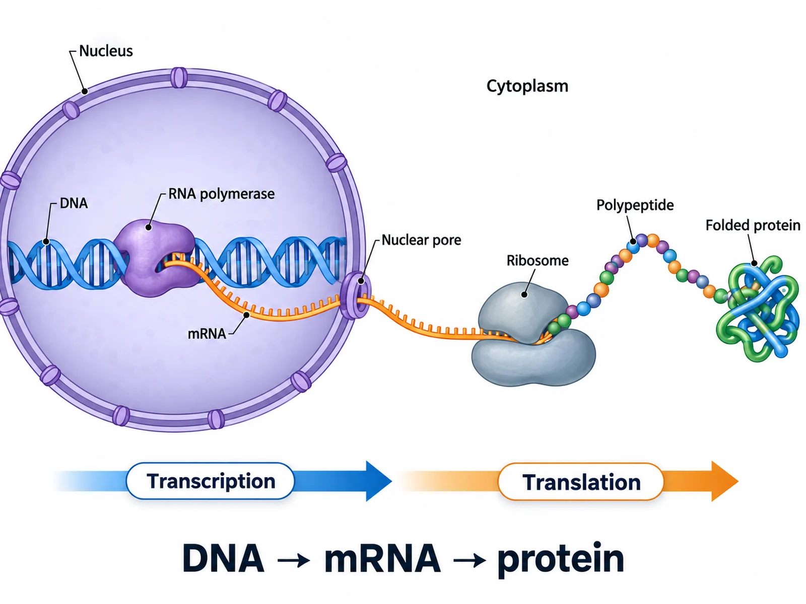
Draw the whole path from gene to protein on one page: DNA transcribed into mRNA in the nucleus, the mRNA leaving through a nuclear pore, and a ribosome in the cytoplasm where tRNA anticodons meet mRNA codons and the polypeptide grows. Make it fully labeled, simple for younger classes, or a blank worksheet with a matching answer key.
Describe what to show and label, or pick a simple, step-by-step, or blank version from the menus.
711 / 2000 characters
Generate a publication-ready PNG from your description.
Preview
Protein Synthesis Diagram Example Previews
Target aspect ratio: 4:3
Sample previews. Describe your diagram to generate your own protein synthesis figure.
Labeled, Simple and Blank Protein Synthesis Diagram Examples
The whole process labeled from DNA to polypeptide, a blank worksheet with its matching answer key, a step-by-step version with codons and anticodons, a DNA-to-protein decoding sheet, and a simple version for younger classes.
Prompt
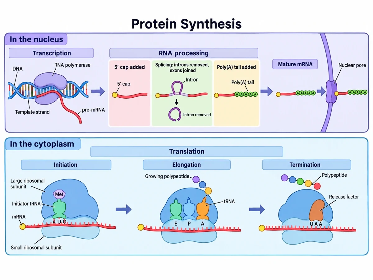
Create a fully labeled protein synthesis diagram on a white background in two stacked panels. Top panel, headed "In the nucleus": transcription, with RNA polymerase on an unwound DNA double helix building pre-mRNA from the template strand; then RNA processing in three boxes, "5′ cap added", "Splicing: introns removed, exons joined" with the intron loop cut out, and "Poly(A) tail added"; then the mature mRNA leaving through a nuclear pore. Bottom panel, headed "In the cytoplasm": translation in three steps. Initiation: the small and large ribosomal subunits and the initiator tRNA carrying Met paired with the AUG start codon. Elongation: tRNAs in the E, P and A sites from the 5′ cap end of the mRNA, with the growing polypeptide on the A-site tRNA just after a peptide bond forms. Termination: a release factor at the UAA stop codon and the finished polypeptide released. Name every structure with a thin leader line.
AP / intro college
Use this example
Prompt
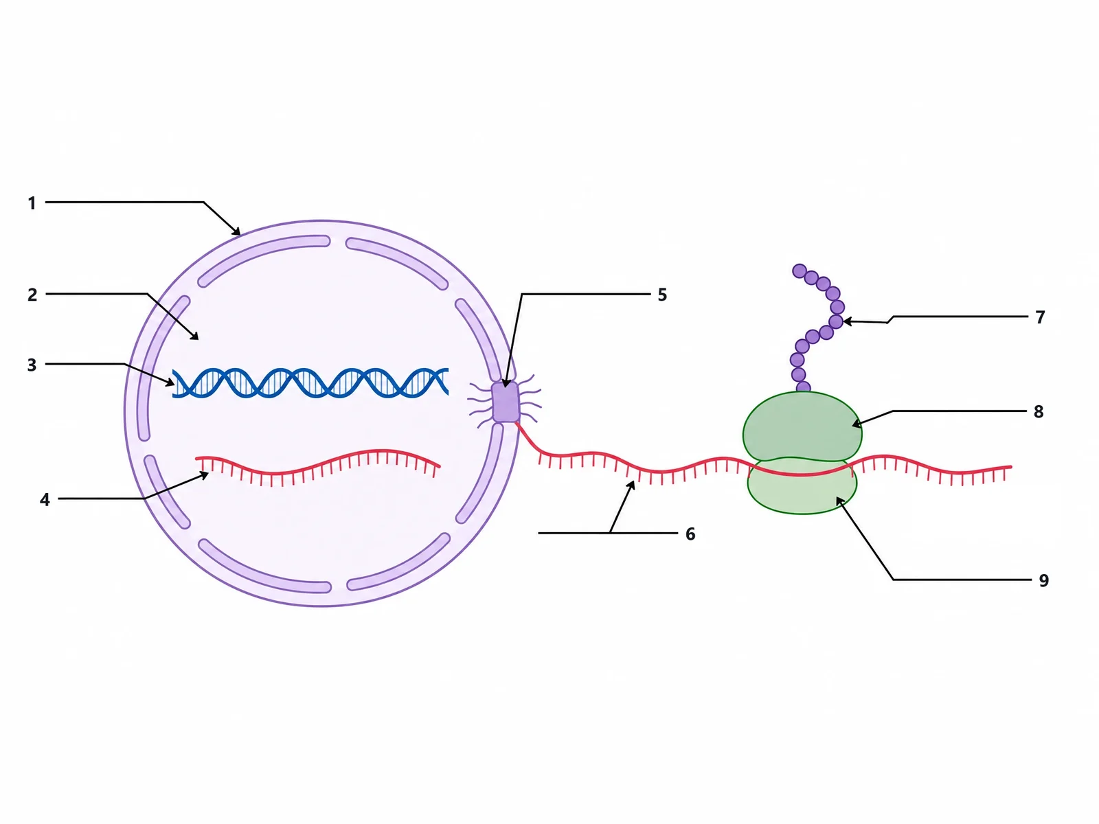
Create a blank protein synthesis worksheet for students to label. Draw a simple protein synthesis scene on a white background: on the left, a large round nucleus whose double envelope is broken by pores, with a DNA double helix and a separate single mRNA strand inside it; the mRNA leaving through a nuclear pore on the right of the nucleus and running across the cytoplasm between the large and small subunits of one ribosome; and a chain of amino acid beads rising from the top of the ribosome. Lines 1 to 4 sit on the left, numbered from the top down, and lines 5 to 9 on the right. Draw exactly 9 thin black leader lines with small arrowheads, each ending at a short horizontal answer line with its number at the outer end and no text, pointing at these targets in this order: 1 = the nuclear envelope, 2 = the inside of the nucleus, 3 = the DNA double helix, 4 = the mRNA strand inside the nucleus, 5 = the nuclear pore, 6 = the mRNA strand in the cytoplasm, 7 = the chain of amino acids, 8 = the large ribosomal subunit, 9 = the small ribosomal subunit. Put no structure names, no key and no word bank anywhere on the image.
Blank worksheetSimple overview
Use this example
Prompt
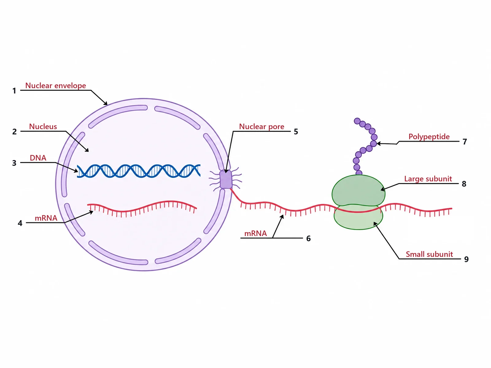
Create the answer key for a blank protein synthesis worksheet. Draw a simple protein synthesis scene on a white background: on the left, a large round nucleus whose double envelope is broken by pores, with a DNA double helix and a separate single mRNA strand inside it; the mRNA leaving through a nuclear pore on the right of the nucleus and running across the cytoplasm between the large and small subunits of one ribosome; and a chain of amino acid beads rising from the top of the ribosome. Lines 1 to 4 sit on the left, numbered from the top down, and lines 5 to 9 on the right. Draw exactly 9 thin black leader lines with small arrowheads, each ending at a short horizontal answer line with its number at the outer end, pointing at the same targets in the same order as the worksheet: 1 = the nuclear envelope, 2 = the inside of the nucleus, 3 = the DNA double helix, 4 = the mRNA strand inside the nucleus, 5 = the nuclear pore, 6 = the mRNA strand in the cytoplasm, 7 = the chain of amino acids, 8 = the large ribosomal subunit, 9 = the small ribosomal subunit. Write the answer on each line in dark red: 1 "Nuclear envelope", 2 "Nucleus", 3 "DNA", 4 "mRNA", 5 "Nuclear pore", 6 "mRNA", 7 "Polypeptide", 8 "Large subunit", 9 "Small subunit". Keep the numbering in this order so it matches the worksheet line for line.
Simple overview
Use this example
Prompt
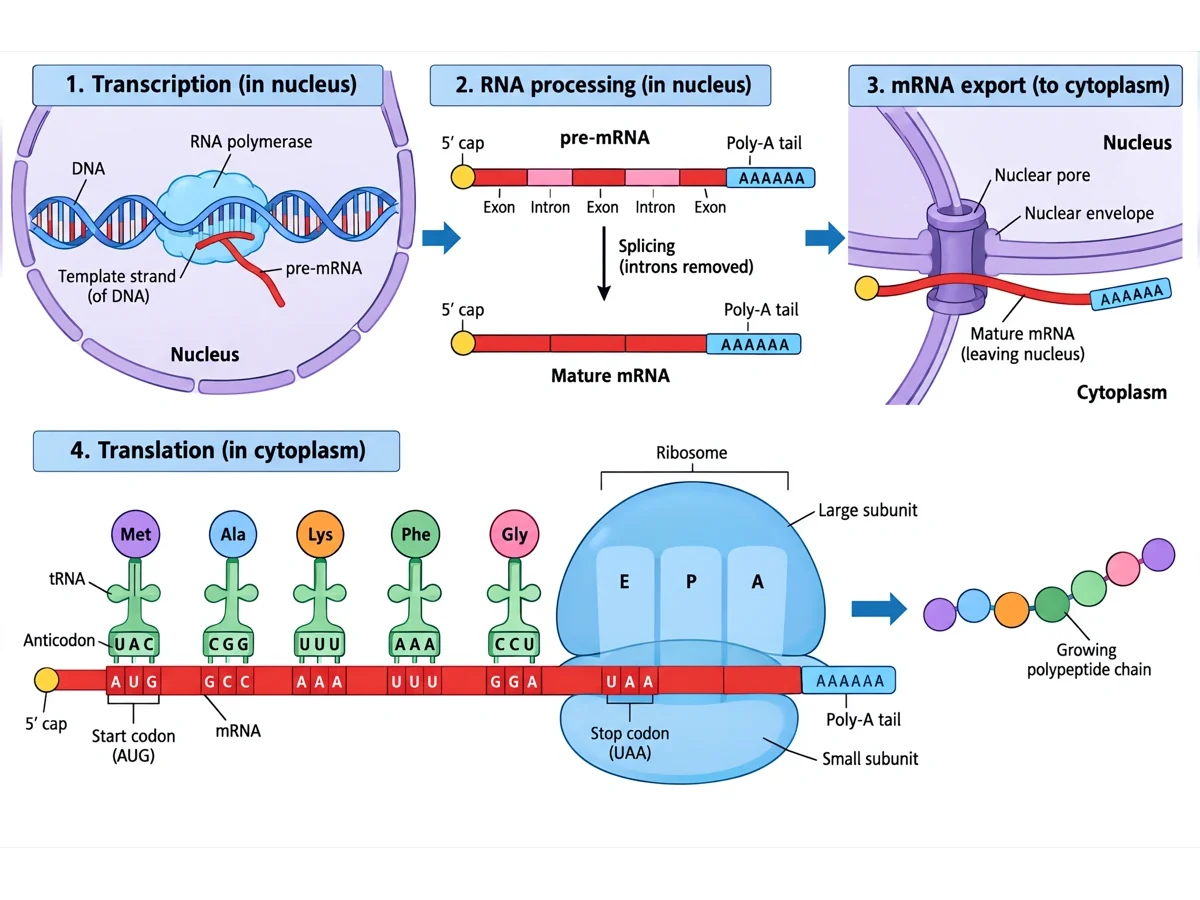
Create a four-step protein synthesis diagram on a white background with numbered headings. 1 "Transcription (in nucleus)": RNA polymerase on the DNA building pre-mRNA from the template strand. 2 "RNA processing (in nucleus)": a pre-mRNA with a 5′ cap, three exons, two introns and a poly-A tail, spliced into a mature mRNA with the introns removed. 3 "mRNA export (to cytoplasm)": the mature mRNA passing through a nuclear pore in the nuclear envelope. 4 "Translation (in cytoplasm)": a long mRNA from its 5′ cap to its poly-A tail with the codons AUG (marked "Start codon"), GCC, AAA, UUU and GGA, and above them, lined up as a codon key, tRNAs with the anticodons UAC, CGG, UUU, AAA and CCU carrying Met, Ala, Lys, Phe and Gly; then a ribosome with large and small subunits and E, P and A sites over the UAA stop codon, and the growing polypeptide chain leaving to the right.
Numbered stepsAP / intro college
Use this example
Prompt
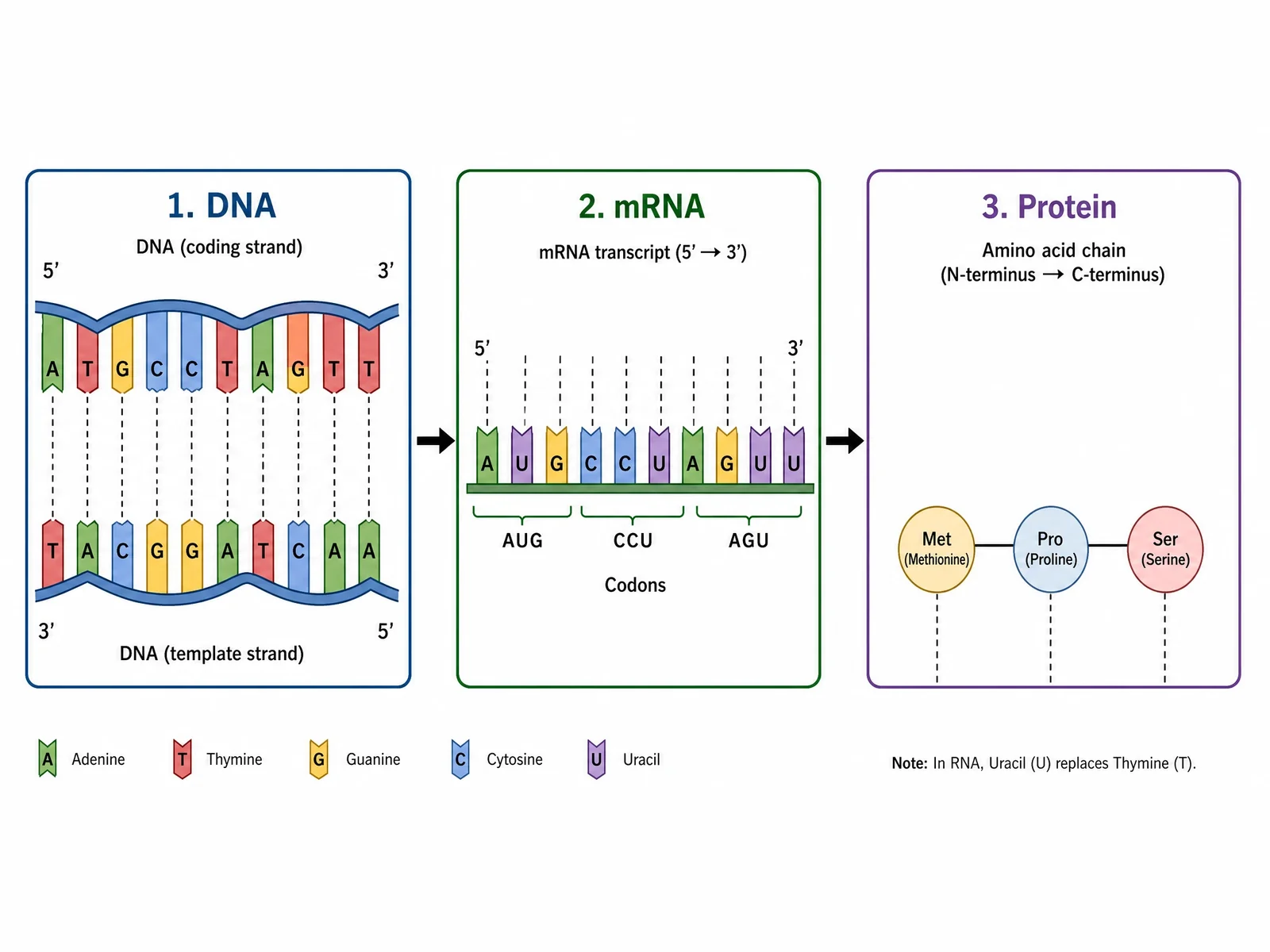
Create a three-panel decoding diagram on a white background, with a colour key for adenine, thymine, guanine, cytosine and uracil along the bottom. Panel 1 "DNA": a short double strand, the coding strand 5′-ATGCCTAGTT-3′ on top and the template strand 3′-TACGGATCAA-5′ below, each base pair joined by a dashed line. Panel 2 "mRNA": the transcript 5′-AUGCCUAGUU-3′ with brackets grouping the codons AUG, CCU and AGU. Panel 3 "Protein": three linked circles, Met (methionine), Pro (proline) and Ser (serine), headed "Amino acid chain (N-terminus → C-terminus)". Add the note "In RNA, uracil (U) replaces thymine (T)."
Codon decoding
Use this example
Prompt
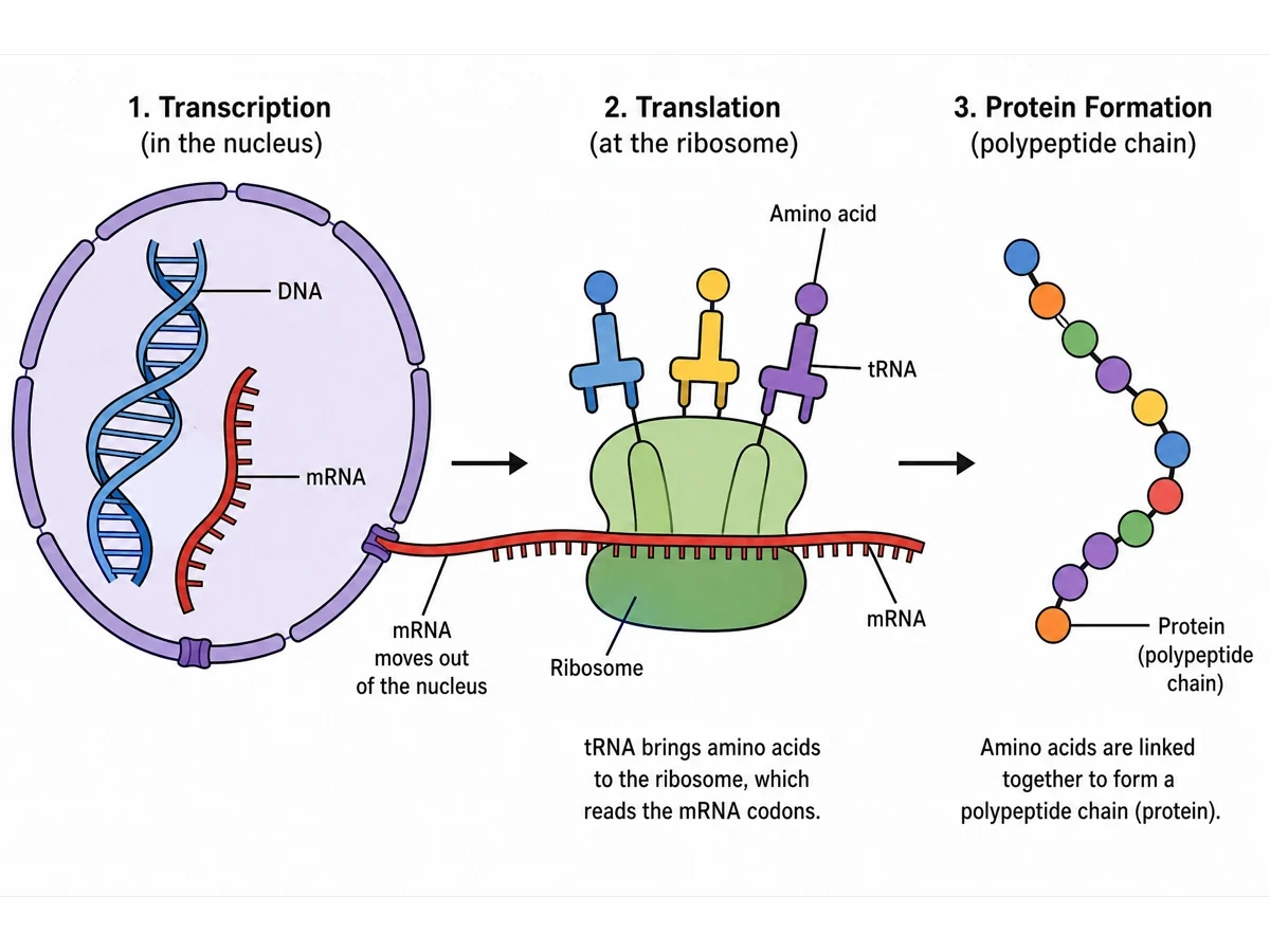
Create a simple protein synthesis diagram for younger students on a white background, in three steps from left to right with arrows between them. 1 "Transcription (in the nucleus)": a round nucleus with pores in its envelope, a DNA double helix and a single mRNA strand inside, and the mRNA leaving through a pore. 2 "Translation (at the ribosome)": the mRNA running through a green ribosome with three tRNAs above it, each carrying one amino acid bead, and the caption "tRNA brings amino acids to the ribosome, which reads the mRNA codons." 3 "Protein Formation (polypeptide chain)": a chain of coloured amino acid beads and the caption "Amino acids are linked together to form a polypeptide chain (protein)." Label DNA, mRNA, "mRNA moves out of the nucleus", ribosome, tRNA, amino acid and protein (polypeptide chain).
Simple overviewMiddle school
Use this example
How to make a labeled or blank protein synthesis diagram
1
Choose the diagram, level, and labels
Pick the full process, a simple overview, numbered steps, a codon decoding sheet, a ribosome close-up, or the bacterial version. Set the level from middle school to university, then choose labeled, numbered, a blank worksheet, or a worksheet with a word bank.
2
Generate the figure
The generator keeps transcription in the nucleus and translation in the cytoplasm, writes U in the mRNA, pairs every anticodon with its codon, and puts the tRNAs in the E, P and A sites in the right order, then adds names, numbers, or empty blanks.
3
Make the answer key, check, and export
For a worksheet, make the answer key from the same result so the numbers match. Check the enzyme name, the bases, and each codon and anticodon pair against the list below, then download the PNG.
A protein synthesis diagram follows one gene until it becomes a chain of amino acids. It has two halves. In the first, transcription, RNA polymerase copies the gene from DNA into messenger RNA. In the second, translation, a ribosome reads that mRNA three bases at a time while transfer RNAs bring the amino acids each codon calls for. The single mRNA strand is the thread that ties the two halves together.
In a eukaryotic cell the halves are drawn in different places, and that is the point of the figure: transcription inside the nucleus, translation on ribosomes in the cytoplasm, with the mRNA crossing the nuclear envelope through a pore in between. A labeled protein synthesis diagram names every part, which makes it a reference sheet or an answer key. A blank one keeps the drawing and the leader lines but leaves the names out, so students write them in.
Labels on a diagram of protein synthesis, by level
How many labels a diagram needs depends on the course. These are the sets the Level menu uses when your description does not list its own.
Middle school: DNA, nucleus, mRNA, ribosome, tRNA, and amino acids forming a protein chain. Six labels are enough to tell the story.
High school: add the template strand, RNA polymerase, the nuclear pore, the cytoplasm, codon, anticodon, peptide bond and polypeptide, and mark the AUG start codon.
AP and introductory college: add the promoter, pre-mRNA, the 5′ cap, the poly-A tail, introns and exons, the spliceosome, the two ribosomal subunits, the A, P and E sites, the initiator tRNA, a stop codon and the release factor.
University: add the TATA box, RNA polymerase II and transcription factors, the 7-methylguanosine cap, the 40S and 60S subunits, peptidyl transferase, and the N-terminus of the chain.
UK courses often call the same figure a protein synthesis labelled diagram; the labels are the same.
Step 1: transcription in the nucleus
Transcription makes an RNA copy of one gene. It has three phases, and a full diagram usually shows the middle one.
Initiation: in eukaryotes, transcription factors bind the promoter first, often at a TATA box about 25 to 35 bases upstream of the start. RNA polymerase II then joins them and opens a short stretch of the double helix, the transcription bubble.
Elongation: the polymerase reads the template strand from 3′ to 5′ and adds RNA nucleotides to the 3′ end of the growing chain, so the RNA is built from 5′ to 3′. Each base pairs with the template: A with U, T with A, G with C, and C with G. Behind the enzyme the DNA closes up again.
Termination: the polymerase reaches the end of the gene and the RNA is released. Eukaryotic RNA polymerase II runs on for a thousand or more bases before letting go, and the extra length is trimmed off.
The coding strand is the one not read. Its sequence matches the mRNA letter for letter, with T where the mRNA has U.
Step 2: mRNA processing and export
A eukaryotic transcript is not ready to translate the moment it is made. The first copy, the pre-mRNA, is modified in the nucleus, which is why AP and college diagrams draw a processing step between the two stages.
5′ cap: a modified guanine nucleotide, 7-methylguanosine, is added to the front end. It protects the mRNA and helps the ribosome find it.
Poly-A tail: an enzyme adds a run of about 200 adenine nucleotides to the 3′ end, which also protects the message and helps export it.
Splicing: the introns, stretches that do not code for the protein, are cut out by the spliceosome, a complex of proteins and small nuclear RNAs, and the exons are joined end to end. Alternative splicing joins different exons, which lets one gene make more than one protein.
Export: the mature mRNA leaves the nucleus through a nuclear pore. On a diagram this is the arrow that links the two halves.
Step 3: translation begins at the start codon
Initiation puts the ribosome and the first tRNA in place. The small ribosomal subunit binds the mRNA near its 5′ cap and scans along it until it finds the first AUG. An initiator tRNA carrying methionine pairs with that AUG, and the large subunit then joins to complete the ribosome, with the initiator tRNA sitting in the P site. Bacteria do the same job differently: their small subunit lines up on a short Shine–Dalgarno sequence just upstream of the start codon, and their first amino acid is formyl-methionine. Either way, every polypeptide starts with methionine, which is why a decoding exercise always begins at AUG and never at the first base of the message.
Step 4: elongation, codon by codon
Elongation is the repeating cycle that most diagrams freeze in mid-step.
A tRNA whose anticodon pairs with the codon in the A site enters, carrying its amino acid.
Peptidyl transferase, an activity of the ribosomal RNA in the large subunit, moves the growing chain from the P-site tRNA onto the amino acid in the A site, forming a new peptide bond.
The ribosome shifts one codon towards the 3′ end of the mRNA. The tRNA that held the chain moves to the E site and leaves empty; the tRNA now holding the chain sits in the P site; the A site is open for the next tRNA.
Each tRNA is recharged with a fresh amino acid in the cytoplasm and used again.
Several ribosomes can read one mRNA at the same time, a structure called a polysome, so one message makes many copies of its protein.
Step 5: termination and what happens to the new chain
When a stop codon (UAA, UAG or UGA) enters the A site, no tRNA can pair with it. A release factor binds instead, the bond between the chain and the last tRNA is broken, and the polypeptide is freed. The two ribosomal subunits separate from the mRNA and from each other, ready to start again. The chain then folds into its working shape, often with help from chaperone proteins. Proteins made on the rough endoplasmic reticulum go on through the Golgi apparatus to membranes, lysosomes or secretion; proteins made on free ribosomes stay in the cytoplasm or go to organelles such as the mitochondria.
Codons, anticodons, and reading the genetic code
The genetic code is the table that links each three-base codon to an amino acid. There are 64 codons: 61 code for the 20 amino acids and 3 are stop signals, so most amino acids have more than one codon. The code is nearly the same in every organism, which is why a bacterium can make a human protein from a human gene. To decode a sequence on a worksheet, work in this order.
Write the mRNA from the template strand by pairing each base: A gives U, T gives A, C gives G, G gives C. Template 3′-TAC CGT ACC TTT ACT-5′ gives mRNA 5′-AUG GCA UGG AAA UGA-3′.
Split the mRNA into codons starting at the AUG, with no gaps and no overlaps: AUG, GCA, UGG, AAA, UGA.
Look each codon up in a codon chart: methionine, alanine, tryptophan, lysine, then stop. The polypeptide is Met–Ala–Trp–Lys.
Write each anticodon as the complement of its codon: UAC, CGU, ACC, UUU. The stop codon has no anticodon, because no tRNA reads it.
Check the coding strand: 5′-ATG GCA TGG AAA TGA-3′. It matches the mRNA with T in place of U, which is a quick test of the whole answer.
DNA, mRNA, tRNA and rRNA: who does what
DNA holds the gene. It stays in the nucleus and is read, never used up, so the same gene can be transcribed again and again.
Messenger RNA (mRNA) is the working copy of one gene. It carries the codons from the nucleus to the ribosome and is broken down after it has been translated many times.
Transfer RNA (tRNA) is the adaptor. Each one is folded into a cloverleaf, or an L shape in three dimensions, with an anticodon loop at one end and its amino acid attached at the other.
Ribosomal RNA (rRNA) and proteins make up the ribosome. The rRNA does the chemistry: it forms the peptide bonds, which makes the ribosome an RNA enzyme.
RNA polymerase is the enzyme of transcription, and release factors are proteins that end translation. Neither is an RNA, which is a common worksheet trap.
Simple protein synthesis diagram for younger students
A simple protein synthesis diagram should leave things out, not draw them wrong. For a first lesson, keep a nucleus with a short piece of DNA, one mRNA strand leaving through the nuclear envelope, one ribosome in the cytoplasm with two or three tRNAs, and a short chain of amino acid beads. Six labels are enough: DNA, nucleus, mRNA, ribosome, tRNA and amino acids. Leave out RNA processing, the A, P and E sites and the 5′ and 3′ ends. Keep the facts that cannot be simplified: the DNA stays in the nucleus, the protein is made outside it, and RNA uses U instead of T. A useful extension is to print a three-codon message on the mRNA and let students decode it with a codon chart.
Using a blank protein synthesis worksheet in class
Start blank, end labeled: hand out the blank worksheet, then show the answer key made from the same result, so every number matches.
Match the labels to the lesson: six for a starter, about fourteen for an end-of-topic check, processing and ribosome sites for AP or college.
Word bank or not: include one for a scaffolded lesson, leave it out for a test. The word bank is alphabetical, so its order gives nothing away.
Pair the picture with a decoding row: a codon decoding sheet asks students to fill in the mRNA, anticodons and amino acids for a printed DNA sequence.
Steps for revision: the numbered-steps version turns into a six-box flow chart that fits on a revision card.
Protein synthesis in bacteria
Bacteria follow the same code with a simpler layout. With no nucleus, transcription and translation share the cytoplasm, and ribosomes start translating the free 5′ end of an mRNA while RNA polymerase is still making the rest of it, so one diagram can show a strand of DNA, a growing mRNA and a row of ribosomes all at once. Bacterial mRNA gets no cap, no poly-A tail and no splicing, bacterial ribosomes are 70S (30S and 50S subunits) rather than 80S, and one mRNA often carries several genes. For a side-by-side prokaryote and eukaryote panel, use the transcription and translation diagram page.
Showing mutations on a protein synthesis diagram
Once students can decode a sequence, the next exam question changes one base and asks what happens. A diagram with a short printed sequence makes this easy to show.
Silent mutation: the codon changes but the amino acid does not, because the code has several codons for most amino acids. GCA to GCG still codes for alanine.
Missense mutation: one amino acid is replaced by another. GCA (alanine) to ACA (threonine).
Nonsense mutation: a codon becomes a stop codon and the chain is cut short. UGG (tryptophan) to UGA (stop).
Frameshift: inserting or deleting bases in a number that is not a multiple of three shifts the reading frame, so every codon after the change is read differently. The rest of the chain is usually wrong, and a stop codon often turns up early.
Most common mistakes on a protein synthesis diagram
These are the errors image models, and students, make most often on this figure. Check each one before a diagram goes into graded work.
MistakeCorrect
MistakeT (thymine) written in the mRNA or tRNA
CorrectRNA uses U in place of T; only the DNA has T
MistakeA ribosome translating inside the nucleus
CorrectTranscription in the nucleus; translation on ribosomes in the cytoplasm or on rough ER
MistakeThe enzyme labeled DNA polymerase or helicase
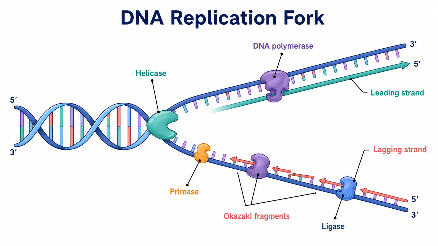
CorrectRNA polymerase transcribes; DNA polymerase and helicase belong to replication
MistakeAn anticodon that does not pair with its codon
CorrectBase by base complementary: codon AUG, anticodon UAC
MistakeThe amino acid drawn on the anticodon end of the tRNA
CorrectAmino acid at the top (3′ acceptor end); anticodon in the bottom loop, on the mRNA
MistakeFour or more tRNAs inside one ribosome
CorrectAt most three: E, P, A from the mRNA’s 5′ end; the chain sits on the P-site tRNA (briefly on the A-site tRNA after each new bond)
MistakeA tRNA drawn pairing with a stop codon
CorrectNo tRNA reads UAA, UAG or UGA; a release factor binds and frees the chain
MistakeBoth DNA strands copied, or the mRNA drawn double-stranded
CorrectOnly the template strand is read; the mRNA is a single strand matching the coding strand
MistakeAn arrow from protein back to DNA
CorrectInformation flows DNA → RNA → protein; protein is never copied back into nucleic acid
Transcription vs translation
A labeled protein synthesis diagram is really two diagrams joined by one mRNA. This is what changes between the two halves.
Transcription
Translation
Where (eukaryotes)
Nucleus
Cytoplasm: free ribosomes or rough ER
What is read
The DNA template strand, 3′ → 5′
The mRNA, 5′ → 3′, one codon at a time
What is made
mRNA (pre-mRNA in eukaryotes), built 5′ → 3′
A polypeptide, built from the N-terminus
Main machinery
RNA polymerase, transcription factors
Ribosome, tRNAs, amino acids, initiation and release factors
Base pairing
DNA to RNA: A–U, T–A, G–C, C–G
Codon to anticodon: A–U, G–C
Start signal
The promoter, upstream of the gene
The AUG start codon
Stop signal
A termination sequence at the end of the gene
A stop codon: UAA, UAG or UGA
Before the next stage
5′ cap, poly-A tail and splicing; export through a nuclear pore
Folding, and for some proteins transport through the ER and Golgi
Protein Synthesis Diagram FAQ
It shows a gene becoming a protein in one picture. In the nucleus, RNA polymerase copies the template strand of DNA into messenger RNA, which leaves through a nuclear pore. In the cytoplasm, a ribosome reads the mRNA one codon at a time, transfer RNAs with matching anticodons bring amino acids, and peptide bonds join them into a polypeptide. The usual labels are DNA, RNA polymerase, mRNA, nucleus, nuclear pore, ribosome, tRNA, codon, anticodon, amino acid, and polypeptide, plus the start and stop codons.
Generate DNA, replication, transcription, and gene transfer diagrams, blank labeling worksheets with matching answer keys, and revision-ready biology figures with the same SciFigure AI workflow.