DNA Diagram AI

DNA Diagram Generator

Draw a labeled DNA double helix with the sugar-phosphate backbone, correct A-T and G-C base pairing, and the parts of a nucleotide named. Ask for a simple twisted-ladder version or a blank one for students to label.

Correct base pairingSimple or detailedLabeled or blank

AI DNA Diagram Generator

Describe the level of detail and which parts to label.

Costs 5 credits492 / 2000 characters

Generate a publication-ready PNG from your description.

Preview

DNA Diagram Example Previews

Target aspect ratio: 3:4

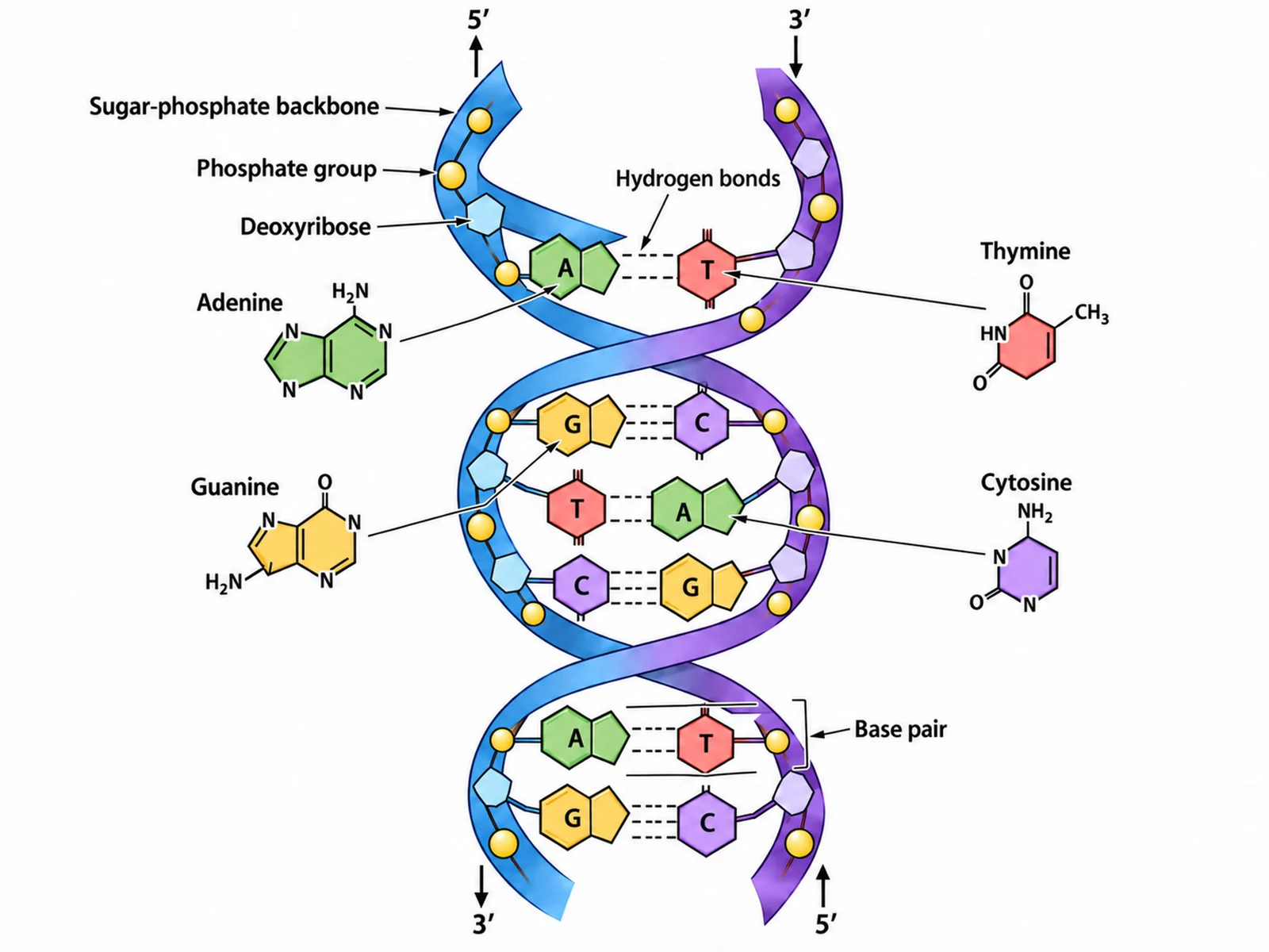
Labeled DNA double helix diagram naming the sugar-phosphate backbone, hydrogen bonds, the four bases, and the antiparallel 5 prime and 3 prime ends
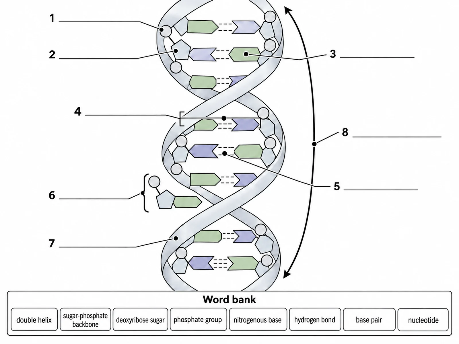
Blank unlabeled DNA double helix diagram with numbered empty label lines for students to complete
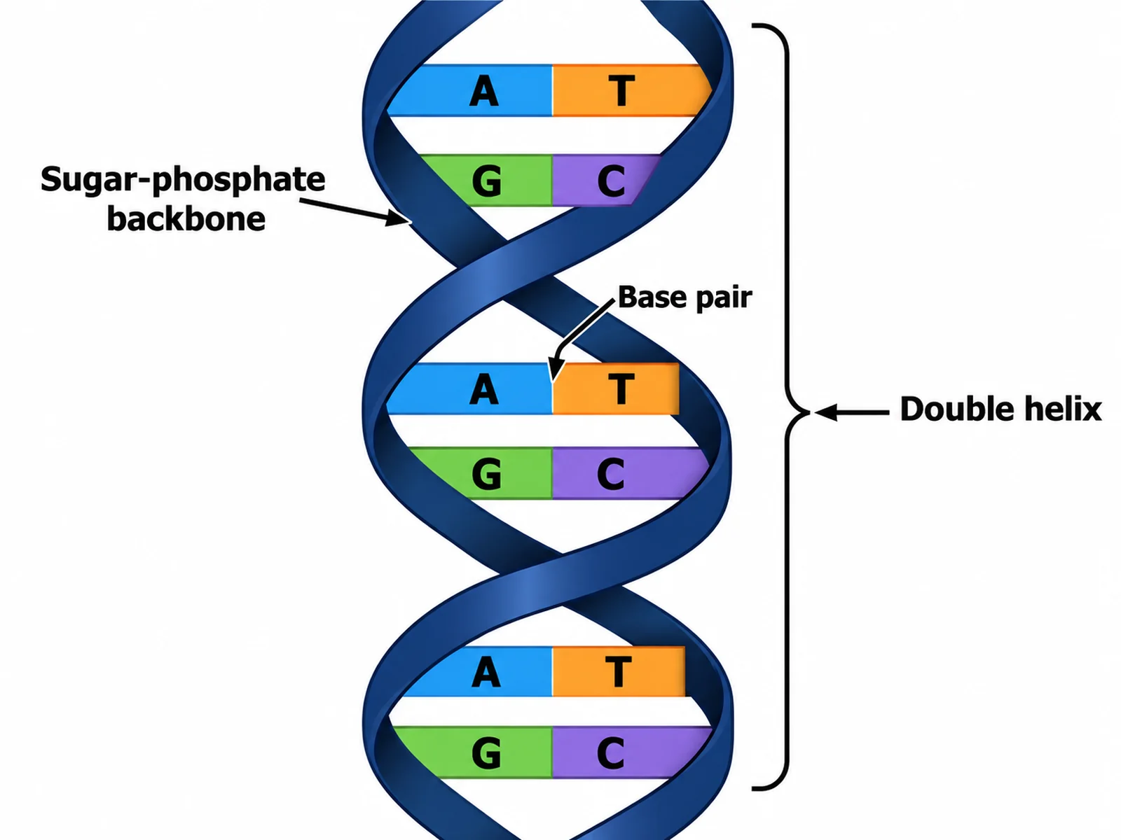
Simple easy-to-draw DNA twisted ladder with coloured base pairs and three basic labels

Sample previews. Describe your diagram to generate your own DNA figure.

DNA Diagram Examples

Use these as visual directions for labeled helices, simple twisted-ladder drawings, blank worksheets, base pairing charts, and nucleotide close-ups.

Labeled DNA double helix diagram naming the sugar-phosphate backbone, hydrogen bonds, the four bases, and the antiparallel 5 prime and 3 prime ends

Prompt

Create a fully labeled DNA double helix on a white background, drawn vertically as a right-handed spiral that twists clockwise as it recedes from the viewer. Draw the two sugar-phosphate backbones as ribbons on the outside, alternating pentagon deoxyribose sugars with circular phosphate groups, and point every base inward so the pairs meet in the middle. Colour adenine, thymine, guanine, and cytosine differently and pair A with T using two short hydrogen bond dashes and G with C using three. Label "Sugar-phosphate backbone", "Deoxyribose", "Phosphate group", "Hydrogen bonds", "Base pair", and each base by name. Mark 5′ and 3′ at opposite ends of each strand with small arrows showing they run antiparallel. Thin dark leader lines, readable sans-serif text.
Labeled DNA Double Helix

Blank unlabeled DNA double helix diagram with numbered empty label lines for students to complete

Prompt

Create a blank DNA worksheet. Draw the same right-handed double helix with backbones on the outside and paired bases meeting in the middle, in clean line art with pale uniform fills and no text anywhere on the figure. Keep the pairing structurally correct — two hydrogen bonds on one pair type and three on the other — but leave every base unlettered. Draw thin leader lines from each part out to a short horizontal ruled blank line, numbered 1 to 8. Add a word bank box below reading "double helix, sugar-phosphate backbone, deoxyribose, phosphate group, nitrogenous base, hydrogen bond, base pair, nucleotide". Generous white space, no answer key.

Blank DNA Worksheet

Simple easy-to-draw DNA twisted ladder with coloured base pairs and three basic labels

Prompt

Create a simple, easy-to-draw DNA diagram for younger students, styled as a twisted ladder. Draw two thick dark uprights spiralling gently to the right in a single flat colour, joined by straight rungs. Split every rung in the middle and colour the halves so that blue always faces orange and green always faces purple, with the letters A, T, G, and C printed large on the rungs and A always opposite T, G always opposite C. Label only three things in big readable text: "Sugar-phosphate backbone", "Base pair", and "Double helix". Bold flat colours, plenty of white space, no hydrogen bond detail.

Simple DNA Drawing

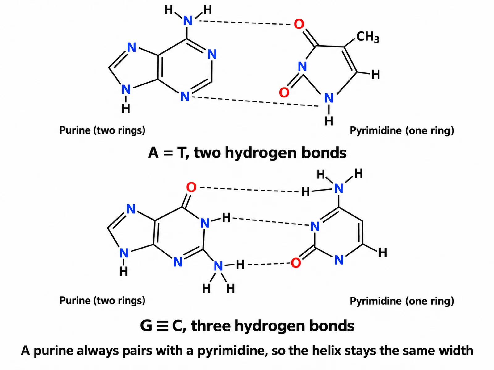
DNA base pairing diagram showing adenine with thymine joined by two hydrogen bonds and guanine with cytosine joined by three

Prompt

Create a base pairing rules diagram on a white background, drawn as a flat untwisted ladder so the chemistry is clear. Show two stacked panels. The top panel pairs adenine with thymine, joined by exactly TWO dashed hydrogen bonds, captioned "A = T, two hydrogen bonds". The bottom panel pairs guanine with cytosine, joined by exactly THREE dashed hydrogen bonds, captioned "G ≡ C, three hydrogen bonds". Draw adenine and guanine as two fused rings labeled "Purine (two rings)" and thymine and cytosine as single rings labeled "Pyrimidine (one ring)". Add a caption reading "A purine always pairs with a pyrimidine, so the helix stays the same width". Show no uracil anywhere.

DNA Base Pairing Rules

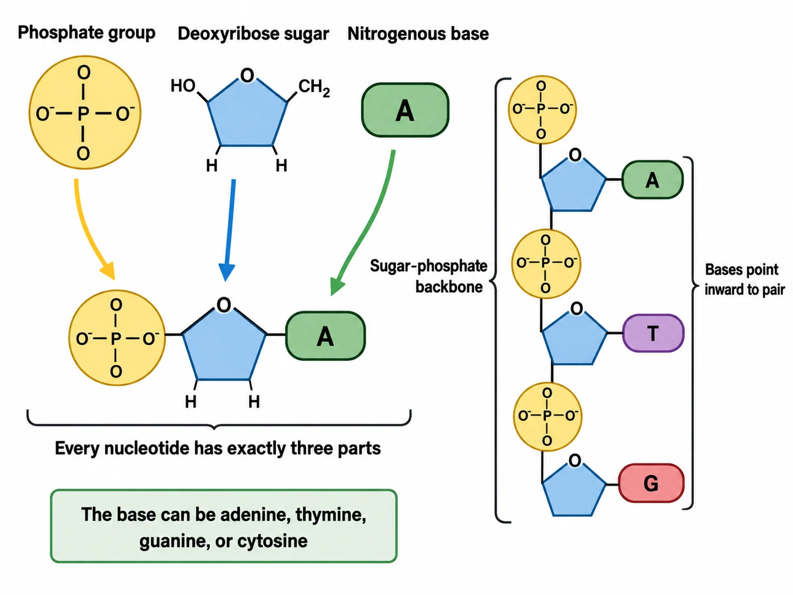
Diagram of a single DNA nucleotide showing the phosphate group, deoxyribose sugar, and nitrogenous base joined together

Prompt

Create a labeled diagram of a single DNA nucleotide on a white background. Show the three parts separately along the top — a circular "Phosphate group", a pentagon "Deoxyribose sugar", and a rounded "Nitrogenous base" — then show them joined below into one nucleotide with arrows from each part to its place in the assembled unit. Add a caption reading "Every nucleotide has exactly three parts". Beside it, draw three nucleotides linked into a short strand so the phosphate of one connects to the sugar of the next, labeled "Sugar-phosphate backbone", with the bases projecting sideways labeled "Bases point inward to pair". Note that the base can be adenine, thymine, guanine, or cytosine.

Structure of a Nucleotide

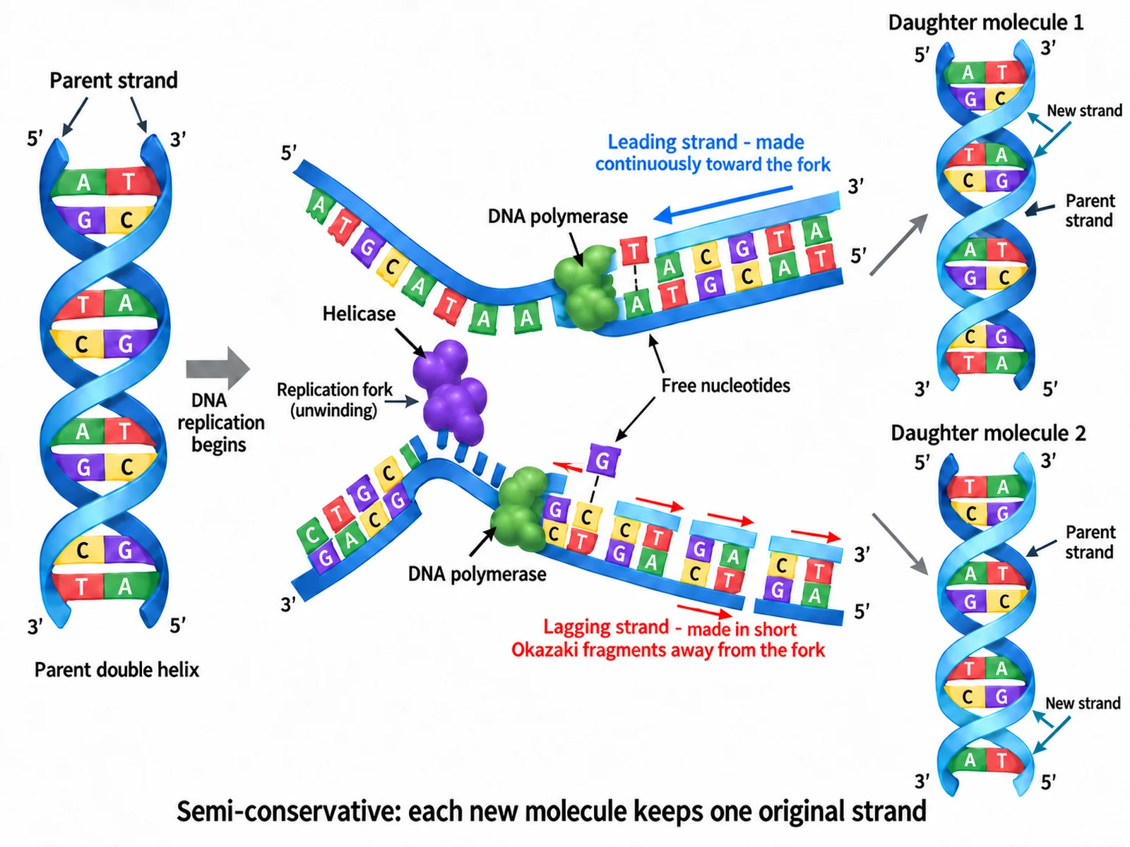
Semi-conservative DNA replication diagram with helicase unzipping the strands and DNA polymerase building the new strands

Prompt

Create a labeled DNA replication diagram on a white background. Draw the parent double helix at the left opening into a Y-shaped replication fork, with "Helicase" labeled at the fork unzipping the two strands by breaking the hydrogen bonds. Label "DNA polymerase" building each new strand by adding free nucleotides, keeping A opposite T and G opposite C on every rung. At the right, show two complete daughter molecules, each drawn with one dark original strand and one bright new strand, captioned "Semi-conservative: each new molecule keeps one original strand". Label "Parent strand", "New strand", and "Free nucleotides", and mark the 5′ and 3′ ends on the fork.

DNA Replication Diagram

How to draw a labeled DNA diagram

1

Choose the detail level

A simple twisted ladder for younger classes, a full labeled helix with backbone and hydrogen bonds, a flat base pairing chart, or a single nucleotide close-up.

2

Generate the diagram

The generator keeps the helix right-handed, the backbones on the outside, and the pairing fixed: A with T on two hydrogen bonds, G with C on three.

3

Check the pairing, then export

Confirm every rung reads A-T or G-C, that no uracil appears, and that the 5′ and 3′ ends sit at opposite ends of the two strands, then download the PNG.

What is DNA?

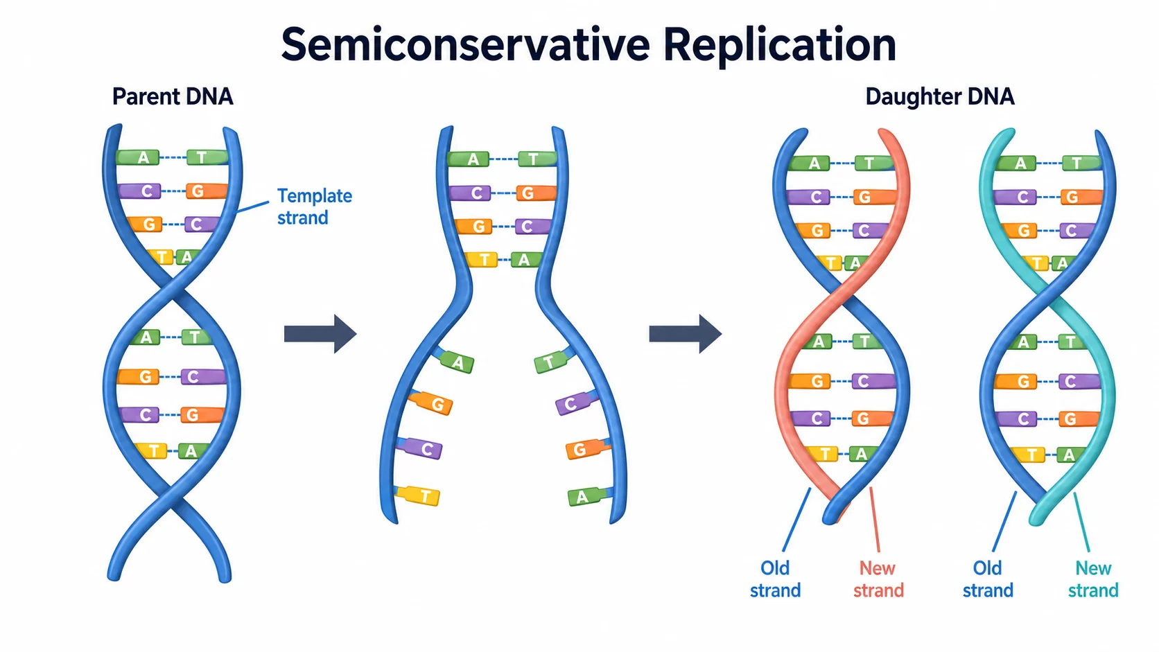
DNA is a long molecule built from two strands wound around each other into a double helix. Each strand is a chain of nucleotides, and the order of their bases spells out the instructions a cell uses to build proteins. The two strands are complementary rather than identical: wherever one carries an adenine the other carries a thymine, and wherever one carries a guanine the other carries a cytosine. That is what lets the molecule be copied — separate the strands and each one is a template for rebuilding its partner.

Parts of a DNA molecule

  • Double helix: the two strands twisted around each other, right-handed in the B-form DNA found in cells.
  • Sugar-phosphate backbone: alternating deoxyribose sugars and phosphate groups running down the outside of each strand.
  • Nitrogenous bases: adenine, thymine, guanine, and cytosine, pointing inward from the backbone to meet in the middle.
  • Hydrogen bonds: the weak links holding each pair together — two for A-T, three for G-C.
  • Nucleotide: one repeating unit of exactly three parts — a phosphate group, a deoxyribose sugar, and a base.
  • The 5′ and 3′ ends: the two strands run in opposite directions, which is what antiparallel means.

The base pairing rules

  • Adenine pairs only with thymine, held by two hydrogen bonds.
  • Guanine pairs only with cytosine, held by three hydrogen bonds.
  • Purines and pyrimidines: A and G have two rings, C and T have one, so every rung joins a two-ring base to a one-ring base.
  • Constant width: because a purine always faces a pyrimidine, the helix is the same width from top to bottom.
  • No uracil: uracil replaces thymine in RNA only — a DNA diagram should never show it.
  • Complementary strands: read one strand and you can write the other, which is how replication and transcription both work.

DNA Diagram FAQ

The double helix itself, the two sugar-phosphate backbones running down the outside, the nitrogenous bases pointing inward, the hydrogen bonds holding each pair together, and the four bases named: adenine, thymine, guanine, and cytosine. A fuller diagram also labels one complete nucleotide, the deoxyribose sugar, the phosphate group, and the 5′ and 3′ ends that show the strands are antiparallel.

Need other biology figures?

Generate molecular structures, cell diagrams, genetics figures, and worksheet-ready illustrations with the same SciFigure AI workflow.