Generate a coagulation cascade diagram with the intrinsic pathway (XII → XI → IX, with VIII), the extrinsic pathway (tissue factor with VII), and the common pathway where Xa and Va turn prothrombin (II) into thrombin (IIa), thrombin turns fibrinogen (I) into fibrin (Ia), and XIIIa cross-links it. Choose the classic cascade, the cell-based model, or PT and aPTT brackets — or a blank worksheet with its answer key.
Intrinsic, extrinsic, commonEvery factor numbered rightLabeled or blank worksheet
Checked against OpenStax A&P 2e
Every factor numbered right
Labeled, numbered, or blank
Print-ready PNG
AI Coagulation Cascade Diagram Generator
Describe the model, the factors to label, and the level, or ask for a blank worksheet.
584 / 2000 characters
Generate a publication-ready PNG from your description.
Preview
Coagulation Cascade Diagram Example Previews
Target aspect ratio: 4:3
Sample previews. Describe your diagram to generate your own coagulation cascade figure.
Coagulation Cascade Diagram Examples
Use these as visual directions for a labeled coagulation cascade, a clotting cascade made easy chart, the cell-based model, PT and aPTT brackets, anticoagulant drug targets, and a blank worksheet.
Prompt
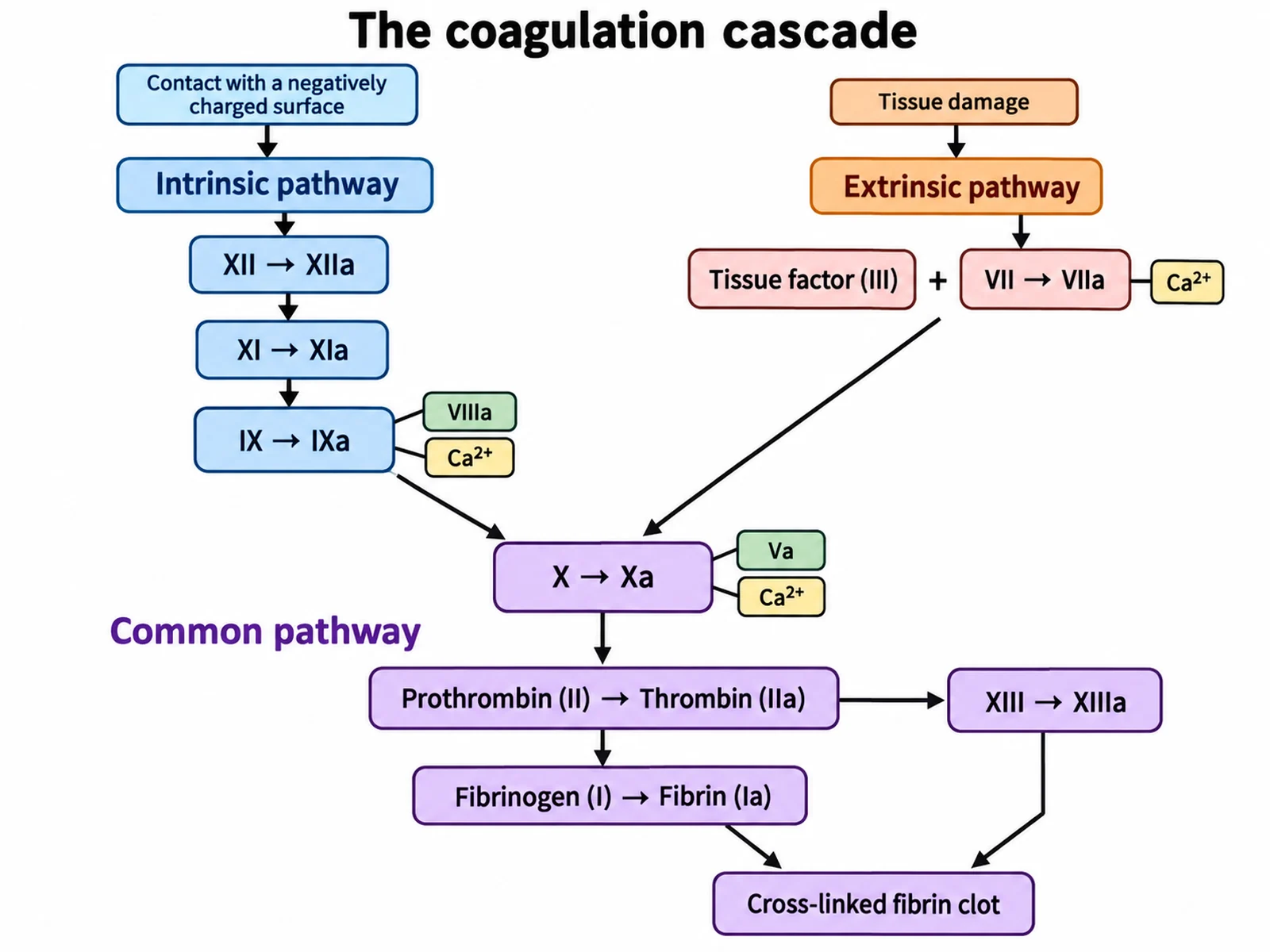
Create a labeled coagulation cascade diagram on a white background in clean medical textbook style, drawn as a Y. Upper left, headed "Intrinsic pathway" in blue, with a trigger label "Contact with a negatively charged surface": boxes "XII → XIIa", then "XI → XIa", then "IX → IXa", with small tags "VIIIa" and "Ca2+" attached to the IXa box. Upper right, headed "Extrinsic pathway" in orange, with a trigger label "Tissue damage": a box "Tissue factor (III)" joined by a plus sign to "VII → VIIa", with a tag "Ca2+". Both arrows meet at a central box "X → Xa". Below, headed "Common pathway" in purple: tags "Va" and "Ca2+" attached to Xa; arrow to "Prothrombin (II) → Thrombin (IIa)"; arrow from thrombin to "Fibrinogen (I) → Fibrin (Ia)"; a side arrow from thrombin to "XIII → XIIIa"; arrow from XIIIa to a final box "Cross-linked fibrin clot". Every arrow points downward toward the clot. No factor VI anywhere.
Use this example
Prompt
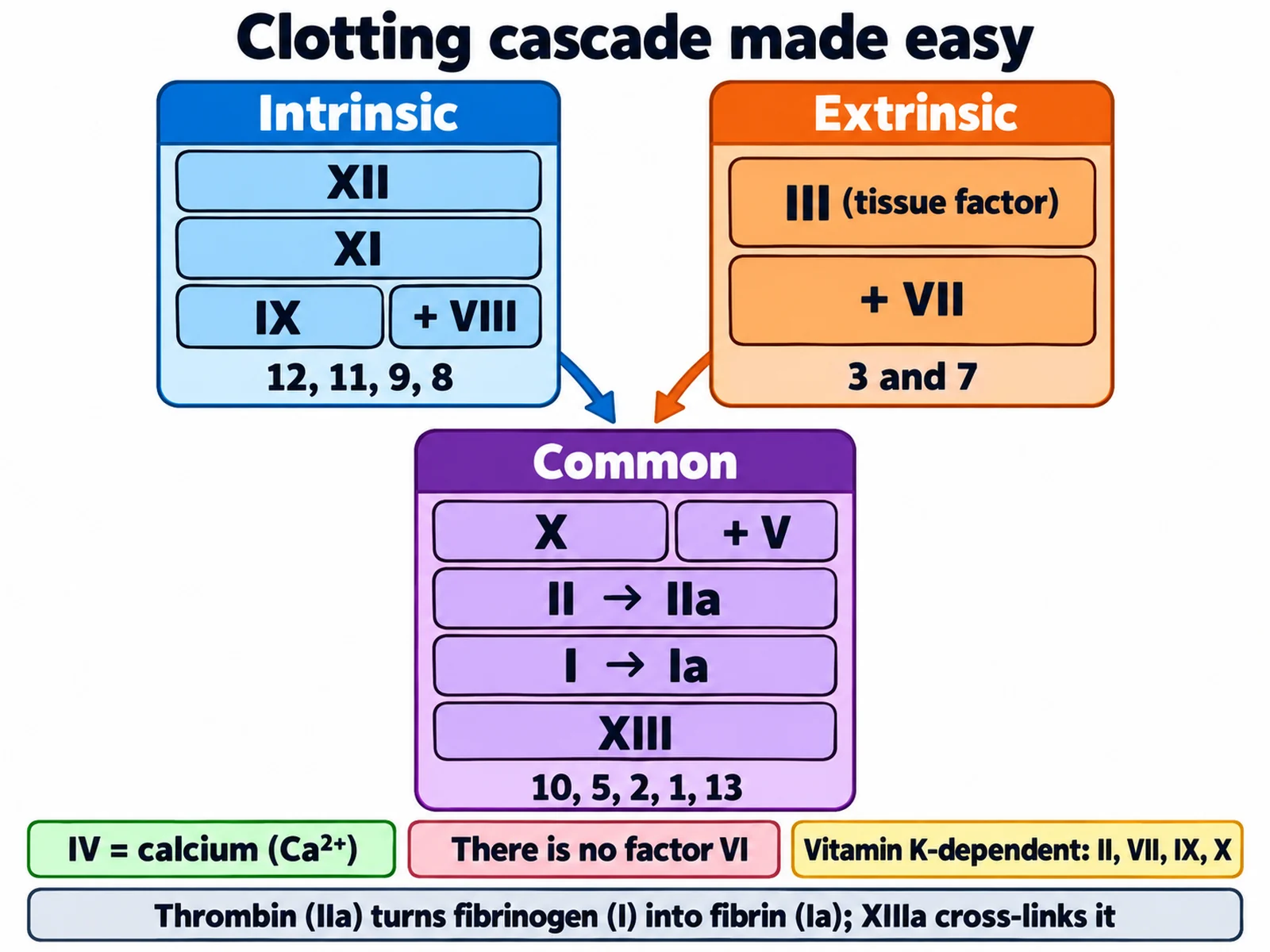
Create a "Clotting cascade made easy" study chart on a white background with bold coloured blocks and large Roman numerals. Three columns of stacked number blocks: blue column "Intrinsic: XII → XI → IX (+ VIII)" with the caption "12, 11, 9, 8"; orange column "Extrinsic: III (tissue factor) + VII" with the caption "3 and 7"; both columns point down into a purple column "Common: X (+ V) → II → I, then XIII" with the caption "10, 5, 2, 1, 13". Under the chart add three short rule cards: "IV = calcium (Ca2+)", "There is no factor VI", and "Vitamin K–dependent: II, VII, IX, X". Final caption at the bottom: "Thrombin (IIa) turns fibrinogen (I) into fibrin (Ia); XIIIa cross-links it".
Use this example
Prompt
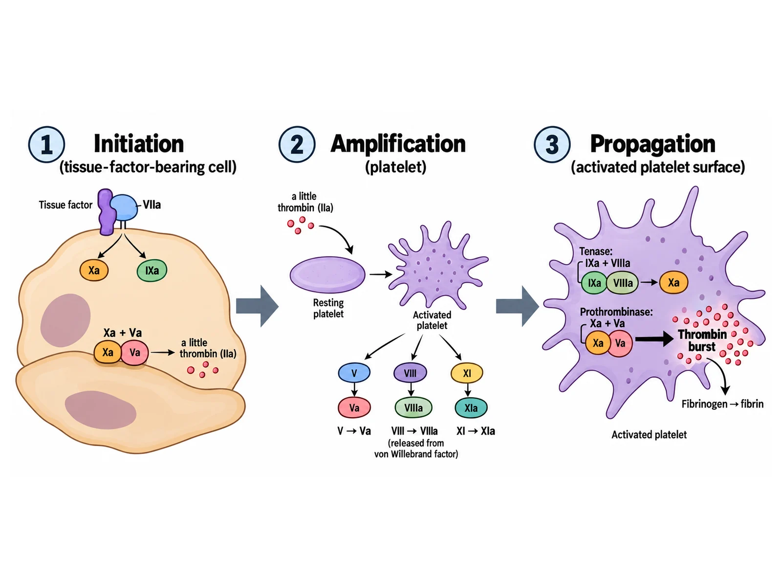
Create a three-panel diagram of the cell-based model of coagulation on a white background, panels left to right with arrows between them. Panel 1 "Initiation (tissue-factor-bearing cell)": a cell with "Tissue factor" on its surface bound to "VIIa", producing small amounts of "Xa" and "IXa", and "Xa + Va" making "a little thrombin (IIa)". Panel 2 "Amplification (platelet)": that thrombin arriving at a resting platelet and activating it, plus arrows "V → Va", "VIII → VIIIa (released from von Willebrand factor)", and "XI → XIa". Panel 3 "Propagation (activated platelet surface)": an activated platelet carrying the "Tenase: IXa + VIIIa" complex making Xa and the "Prothrombinase: Xa + Va" complex making a large "Thrombin burst", ending in "Fibrinogen → fibrin".
Use this example
Prompt
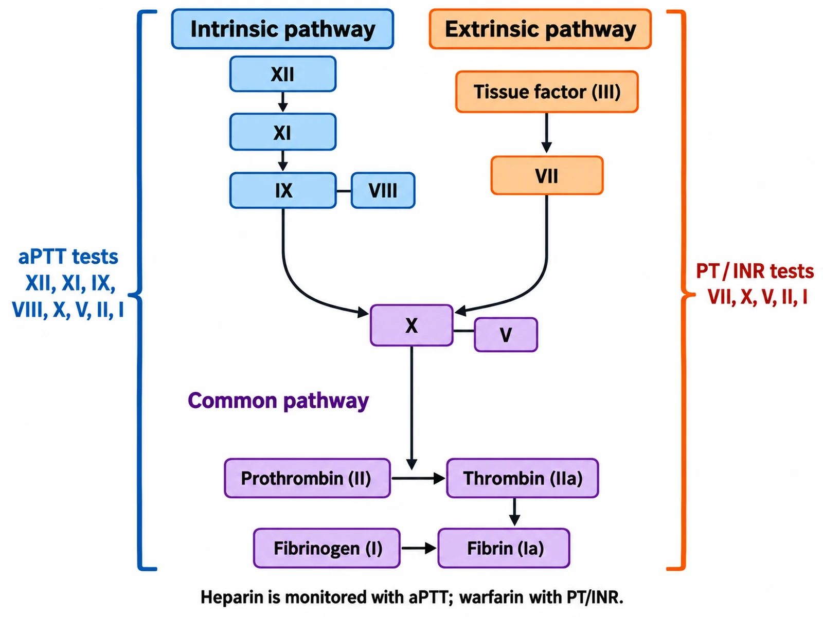
Create a classic coagulation cascade on a white background with lab test brackets. Left column "Intrinsic pathway": "XII", "XI", "IX", "VIII". Right column "Extrinsic pathway": "Tissue factor (III)", "VII". Both feed into "X", then the "Common pathway": "V", "Prothrombin (II) → Thrombin (IIa)", "Fibrinogen (I) → Fibrin (Ia)". Draw a large blue bracket on the left spanning the intrinsic column and the common pathway, labeled "aPTT tests XII, XI, IX, VIII, X, V, II, I". Draw a large orange bracket on the right spanning the extrinsic column and the common pathway, labeled "PT / INR tests VII, X, V, II, I". Add a footnote: "Heparin is monitored with aPTT; warfarin with PT/INR".
Use this example
Prompt
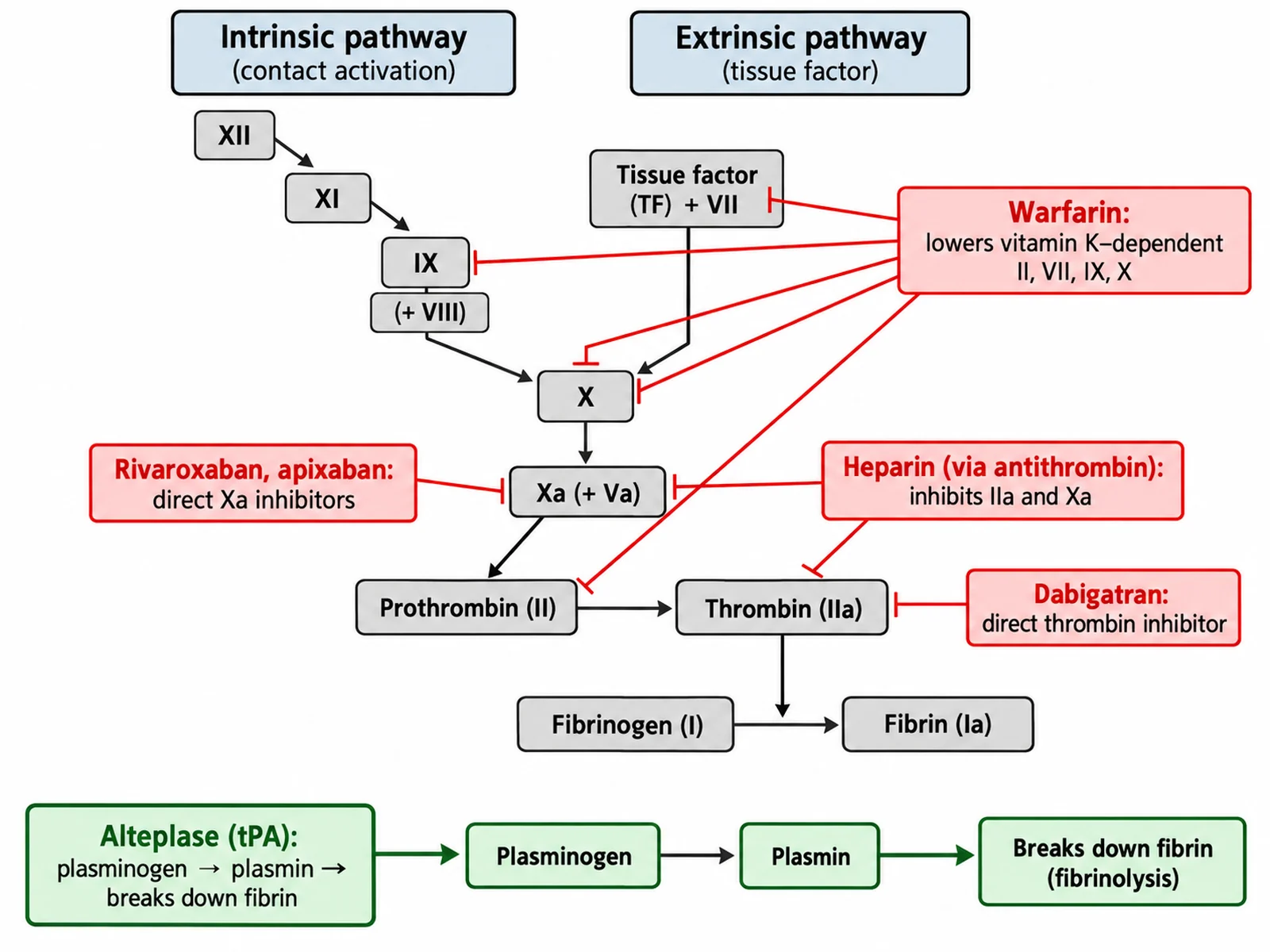
Create a coagulation cascade diagram for pharmacology on a white background showing where anticoagulant drugs act. Draw the classic cascade: intrinsic "XII → XI → IX (+ VIII)", extrinsic "Tissue factor + VII", both to "X", then "Xa (+ Va)" converting "Prothrombin (II)" to "Thrombin (IIa)", then "Fibrinogen (I) → Fibrin (Ia)". Add red drug tags with blunt-ended inhibition lines: "Warfarin: lowers vitamin K–dependent II, VII, IX, X" touching those four factors; "Heparin (via antithrombin): inhibits IIa and Xa"; "Rivaroxaban, apixaban: direct Xa inhibitors" at Xa; "Dabigatran: direct thrombin inhibitor" at IIa. Add a separate green branch below fibrin: "Alteplase (tPA): plasminogen → plasmin → breaks down fibrin".
Use this example
Prompt
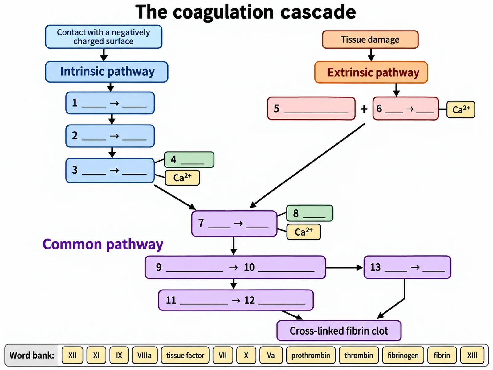
Create a blank coagulation cascade worksheet that is the same Y-shaped drawing as a labeled textbook cascade with the factor names removed. Keep the headings "Intrinsic pathway", "Extrinsic pathway", and "Common pathway", the trigger labels, the Ca2+ tags, the arrows, and the final box "Cross-linked fibrin clot" printed. Replace these thirteen with numbered empty boxes: intrinsic column top to bottom 1 (XII → XIIa), 2 (XI → XIa), 3 (IX → IXa), 4 the cofactor tag (VIIIa); extrinsic column 5 (tissue factor), 6 (VII → VIIa); centre 7 (X → Xa), 8 the cofactor tag (Va); common pathway 9 (prothrombin), 10 (thrombin), 11 (fibrinogen), 12 (fibrin), 13 (XIII → XIIIa). Add a word bank: "XII, XI, IX, VIIIa, tissue factor, VII, X, Va, prothrombin, thrombin, fibrinogen, fibrin, XIII".
Blank worksheet
Use this example
How to make a coagulation cascade diagram
1
Pick the model and the labels
Choose the classic intrinsic–extrinsic–common cascade, the cell-based model, or the classic cascade with PT and aPTT brackets, set the level, then choose labeled, numbered with a key, or a blank worksheet.
2
Generate the cascade
The generator joins both upstream pathways at factor X, attaches VIIIa and Va as cofactors, keeps calcium in the complexes, and ends with thrombin turning fibrinogen into fibrin and XIIIa cross-linking it.
3
Check the numerals, then export
Read every Roman numeral, confirm there is no factor VI, that VII is extrinsic and XIII is at the end, and that every arrow points toward the clot. Then download the PNG, or make the matching worksheet or answer key from the same image.
Coagulation cascade key facts
Pathways meet at
Factor X
Intrinsic factors
XII → XI → IX, with cofactor VIII
Extrinsic factors
Tissue factor (III) + VII
Common pathway
X (+ V) → II → I, then XIII
Factor IV
Calcium (Ca²⁺); there is no factor VI
Vitamin K–dependent
II, VII, IX, X (and proteins C, S)
PT / INR tests
Extrinsic + common: VII, X, V, II, I
aPTT tests
Intrinsic + common: XII, XI, IX, VIII, X, V, II, I
What is the coagulation cascade?
When a blood vessel is cut, the body stops the bleeding in overlapping stages called hemostasis. The vessel wall constricts (vascular spasm), platelets stick to the exposed collagen and to each other to form a soft plug (primary hemostasis), and then the coagulation cascade turns the liquid blood around the plug into a solid clot (secondary hemostasis). The cascade is a chain of plasma proteins called clotting factors, most made by the liver, that circulate in an inactive form. Each one, once activated, activates the next, so a tiny trigger is amplified at every step into a flood of thrombin.
Thrombin is the enzyme that makes the clot. It cuts soluble fibrinogen into fibrin, which polymerises into a mesh of strands that traps red cells and platelets, and it activates factor XIII, which cross-links those strands so the clot holds. A coagulation cascade diagram is the map of that chain. In its classic form it has three parts — the intrinsic, extrinsic, and common pathways — and its value as a teaching figure depends almost entirely on getting the Roman numerals in the right boxes and the arrows pointing the right way.
Primary vs secondary hemostasis: where the clotting cascade fits
A clotting cascade diagram shows only one part of hemostasis, and it helps to know which part. Primary hemostasis is the platelet plug: von Willebrand factor tethers platelets to exposed collagen through their GPIb receptors, the platelets activate, change shape, and release ADP and thromboxane A2, which recruit and activate more platelets until a soft plug seals the hole. Secondary hemostasis is the coagulation cascade, which builds a fibrin mesh through and around that plug so it can withstand arterial pressure. The two are not separate in time — activated platelets provide the phospholipid surface on which the tenase and prothrombinase complexes assemble, and thrombin activates more platelets — but they fail in different ways. Platelet and von Willebrand problems tend to cause bleeding from skin and mucous membranes, while clotting factor deficiencies such as hemophilia tend to cause deep bleeding into joints and muscles. Draw the platelet plug on a cascade diagram only if the request asks for it.
Coagulation cascade diagram: what to label
The three pathway names: intrinsic (contact activation), extrinsic (tissue factor), and common.
The intrinsic factors, top to bottom: XII → XIIa, XI → XIa, IX → IXa, with VIIIa as the cofactor of IXa.
The extrinsic factors: tissue factor (factor III) and VII → VIIa.
The meeting point: factor X → Xa, with cofactor Va.
The conversion steps of the common pathway: prothrombin (II) → thrombin (IIa), and fibrinogen (I) → fibrin (Ia).
Factor XIII → XIIIa, activated by thrombin, and the cross-linked fibrin clot it produces.
Calcium (Ca²⁺, factor IV) at the tenase and prothrombinase complexes.
Optional, if asked for: triggers (contact surface, tissue damage), positive feedback from thrombin, the PT and aPTT brackets, anticoagulant regulation, and fibrinolysis.
The intrinsic pathway step by step
The intrinsic pathway is also called the contact activation pathway, because it starts when factor XII (Hageman factor) touches a negatively charged surface — in the laboratory, glass or the activators added to an aPTT tube. Prekallikrein and high-molecular-weight kininogen help XII become XIIa. XIIa activates factor XI to XIa, and XIa, with calcium, activates factor IX to IXa. IXa on its own is a weak enzyme; it becomes a powerful one when it assembles with its cofactor VIIIa and calcium on a phospholipid membrane, mainly the surface of activated platelets. That assembly is called the intrinsic tenase complex, and it activates factor X. OpenStax describes the whole pathway as taking a few minutes. On a diagram, draw it as a column of four steps on one side of the Y, with VIIIa shown as a partner attached to IXa rather than as a fourth link in the chain.
The extrinsic pathway step by step
The extrinsic pathway, or tissue factor pathway, is short and fast. Damaged cells outside the vessel expose tissue factor, a membrane protein that is called factor III in the old numbering and thromboplastin in older texts. Tissue factor binds factor VII from the plasma, which becomes VIIa, and with calcium the pair forms the extrinsic tenase complex, which activates factor X directly. OpenStax describes these events as completed in a matter of seconds. In the body this is the pathway that actually starts clotting after an injury, which is why it is drawn as the trigger on the opposite side of the Y. In the classic diagram it joins the intrinsic pathway at factor X; the extra link by which tissue factor–VIIa also activates factor IX belongs on more detailed or cell-based diagrams.
The common pathway: from factor X to a cross-linked fibrin clot
Everything below factor X is the common pathway, shared by both upstream routes. Factor Xa assembles with its cofactor Va, calcium, and a phospholipid surface into the prothrombinase complex. Prothrombinase converts prothrombin, factor II, into thrombin, factor IIa — and because each Xa molecule makes many thrombin molecules, this is where the amplification of the cascade pays off.
Thrombin then does three jobs. It cleaves fibrinogen, factor I, into fibrin monomers, factor Ia, which line up and polymerise into strands. It activates factor XIII to XIIIa, which forms covalent cross-links between neighbouring fibrin strands and turns a loose mesh into a stable clot. And it feeds back up the cascade, activating factors V, VIII, and XI and activating platelets, which speeds its own production. A common-pathway diagram should therefore end fibrinogen → fibrin → cross-linked fibrin, with thrombin on the first arrow and XIIIa on the second.
Clotting cascade made easy: memory tricks for the factor numbers
Most students do not struggle with the idea of a cascade; they struggle with the numerals. The trick is to learn three short lists and a few rules. Intrinsic: 12, 11, 9, 8 — a popular way to remember it is that the intrinsic pathway "costs $12, or $11.98". Extrinsic: 3 and 7 — tissue factor plus factor VII. Common: 10, 5, 2, 1, then 13 — X with V makes thrombin from II, thrombin makes fibrin from I, and XIII stabilises the result.
Then add the rules that catch people out. IV is calcium, not a protein. There is no factor VI. II, VII, IX, and X are the vitamin K–dependent factors, which you can remember as "1972": ten, nine, seven, two. V and VIII are cofactors, not enzymes. And XIII is never upstream of fibrin. A coagulation cascade made easy diagram colour-codes the three lists — one colour for each pathway — so that a student can check any numeral against its column at a glance.
The cell-based model of coagulation
The classic cascade was built from test-tube experiments, and it explains clotting tests very well. It explains bleeding less well. If the extrinsic pathway can activate factor X on its own, why do people who lack factor VIII or IX — hemophilia A and B — bleed so badly? And why does a lack of factor XII, which prolongs the aPTT, cause no bleeding at all? In 2001 Hoffman and Monroe proposed a cell-based model that answers both questions by placing the reactions on cell surfaces in three overlapping phases.
In initiation, tissue factor–VIIa on a tissue-factor-bearing cell outside the vessel makes small amounts of Xa and IXa, and Xa with Va produces a little thrombin — not enough to clot, but enough to signal. In amplification, that thrombin activates nearby platelets and factors V, VIII, and XI, and releases VIII from its carrier, von Willebrand factor. In propagation, the activated platelet surface hosts the tenase complex (IXa–VIIIa) and the prothrombinase complex (Xa–Va), which together produce a burst of thrombin large enough to form a stable clot. Because the burst depends on IXa and VIIIa, a missing factor VIII or IX still causes bleeding, and because factor XII plays no part, its absence does not.
Coagulation tests on the diagram: PT/INR and aPTT
The two everyday clotting tests map neatly onto the classic diagram, which is why medical courses still teach it. The prothrombin time (PT), reported internationally as the INR, starts clotting with tissue factor, so it tests the extrinsic and common pathways: factors VII, X, V, II, and I. The activated partial thromboplastin time (aPTT) starts clotting with a contact activator, so it tests the intrinsic and common pathways: factors XII, XI, IX, VIII, X, V, II, and I. Reading them together localises a problem. A prolonged PT with a normal aPTT points at factor VII; a prolonged aPTT with a normal PT points at the intrinsic factors; both prolonged suggests the common pathway, several factors at once, or an anticoagulant. Warfarin is monitored with the PT/INR, and unfractionated heparin with the aPTT. Drawing the two tests as brackets on the cascade is the clearest way to show all of this on one figure.
How clotting is kept in check: natural anticoagulants and fibrinolysis
A cascade that amplifies itself needs brakes, or a small cut would clot the whole circulation. Antithrombin, a plasma protein, inactivates thrombin and factor Xa, along with IXa and XIa; heparin works by making antithrombin act far faster. Protein C, activated when thrombin binds thrombomodulin on healthy endothelium, works with its cofactor protein S to destroy the cofactors Va and VIIIa, shutting down the two big complexes. Tissue factor pathway inhibitor (TFPI) blocks tissue factor–VIIa once a little Xa has been made. Finally, once the vessel has healed, the clot is removed by fibrinolysis: tissue plasminogen activator (tPA) converts plasminogen into plasmin, and plasmin digests fibrin into fragments, among them D-dimer, the fragment measured when a clot is suspected. On a diagram, these are best drawn as red inhibition lines and a separate fibrinolysis branch — and only when the request asks for them.
Anticoagulant drug targets on a coagulation cascade diagram
Pharmacology courses use the cascade as a map of where drugs act, and each drug has one precise target. Warfarin blocks the recycling of vitamin K, so the liver makes less functional II, VII, IX, and X. Unfractionated heparin binds antithrombin and speeds its inhibition of thrombin and Xa; low-molecular-weight heparins such as enoxaparin act mainly on Xa, and fondaparinux on Xa only. The direct oral anticoagulants hit one enzyme each: rivaroxaban, apixaban, and edoxaban inhibit Xa, and dabigatran inhibits thrombin. Thrombolytics such as alteplase sit outside the cascade altogether: they are tPA, and they dissolve an existing clot by generating plasmin. A drug-target diagram draws each drug as a tag with an inhibition line ending on the right factor, which makes it obvious why, for example, warfarin raises the INR.
Bleeding disorders on the cascade
Hemophilia A: deficiency of factor VIII, about 80 percent of hemophilia; prolonged aPTT, normal PT.
Hemophilia B (Christmas disease): deficiency of factor IX; prolonged aPTT, normal PT.
Hemophilia C: deficiency of factor XI; rare and usually milder.
Factor XII deficiency: prolonged aPTT but no bleeding — evidence that contact activation is not how clotting starts in the body.
Vitamin K deficiency or warfarin: low II, VII, IX, and X; PT/INR prolonged first because factor VII has the shortest half-life.
Liver disease: most clotting factors are made in the liver, so several fall together.
How to draw a clotting cascade diagram by hand
If you need to sketch the cascade in an exam, draw a large Y. Put the extrinsic pathway on the upper right because it is short: write "tissue factor + VII". Put the intrinsic pathway on the upper left as a column: XII, XI, IX, and write VIII beside IX as its partner. Bring both arms down to a single box, X, at the fork, and write V beside it. Below that, write II → IIa (prothrombin to thrombin), then I → Ia (fibrinogen to fibrin), and finish with XIII cross-linking the fibrin. Add "Ca²⁺" next to the two complexes, and you have every factor in the classic picture in about a minute. If the question asks about tests, draw the aPTT bracket down the left arm and the common pathway, and the PT bracket down the right arm and the common pathway.
What to check before you use a coagulation cascade diagram
Read every numeral: XI and IX, and Ia and IIa, are the pairs most often swapped.
Look for a factor VI. There should never be one.
Check the columns: XII, XI, IX, VIII intrinsic; tissue factor and VII extrinsic; X, V, II, I, XIII common.
Check the meeting point: both pathways activate factor X in the classic diagram.
Check the cofactors: VIIIa partners IXa and Va partners Xa; neither is drawn as an enzyme step.
Check the ending: thrombin makes fibrin; XIIIa cross-links it.
Check the arrows: every one points from trigger toward the clot, except feedback arrows, which should be dashed.
Most common coagulation cascade diagram mistakes
Roman numerals make this the easiest diagram in physiology to get subtly wrong. Check each of these before a cascade goes on a handout.
MistakeCorrect
MistakeA box for factor VI.
CorrectThere is no factor VI. The number was retired when "VI" turned out to be activated factor V.
MistakeFactor VII drawn in the intrinsic pathway.
CorrectVII belongs to the extrinsic pathway with tissue factor. The intrinsic factors are XII, XI, IX, and VIII.
MistakeXIII shown as an intrinsic pathway factor.
CorrectXIII acts only at the end: thrombin activates it, and XIIIa cross-links fibrin.
MistakeThrombin cross-links the fibrin.
CorrectThrombin makes fibrin from fibrinogen and activates XIII; XIIIa does the cross-linking.
MistakeThe extrinsic pathway joins at factor IX in the classic picture.
CorrectIn the classic cascade both pathways meet at factor X. TF–VIIa activating IX belongs in the cell-based model.
MistakeV and VIII drawn as steps that activate the next factor.
CorrectVa and VIIIa are cofactors that partner Xa and IXa in the prothrombinase and tenase complexes.
MistakeIa and IIa swapped.
CorrectIa is fibrin; IIa is thrombin. I is fibrinogen; II is prothrombin.
MistakeCalcium left out.
CorrectCa²⁺, factor IV, is needed for the tenase and prothrombinase complexes to assemble.
Intrinsic vs extrinsic pathway
Both pathways end by activating factor X. They differ in what starts them, which factors they use, and which clotting test measures them.
Intrinsic pathway
Extrinsic pathway
Also called
Contact activation pathway
Tissue factor pathway
Trigger
Factor XII contacting a negatively charged surface
Tissue factor exposed by damaged tissue
Factors
XII, XI, IX, VIII (plus prekallikrein and HMWK)
Tissue factor (III), VII
Complex that activates X
Intrinsic tenase: IXa + VIIIa + Ca²⁺
Extrinsic tenase: tissue factor + VIIa + Ca²⁺
Speed (OpenStax)
Minutes
Seconds
Clotting test
aPTT
PT / INR
Deficiencies that cause bleeding
VIII (hemophilia A), IX (hemophilia B), XI (hemophilia C)
VII (rare)
Role in the body
Amplifies thrombin generation; XII itself is not needed to stop bleeding
Starts clotting after an injury
Coagulation Cascade Diagram FAQ
It is the chain of reactions in blood plasma that turns liquid blood into a solid clot after a vessel is injured. Each clotting factor circulates as an inactive precursor and, once activated, activates the next one, so a small trigger is amplified into a large burst of thrombin. Thrombin then converts soluble fibrinogen into strands of fibrin, and factor XIIIa cross-links the strands into a stable mesh that reinforces the platelet plug. The classic diagram splits the cascade into the intrinsic, extrinsic, and common pathways.