Generate a labeled skeletal system diagram with the major bones named, or a blank, unlabeled skeleton diagram with numbered lines for students to fill in, plus its answer key. Pick a front, back, or close-up view and a level, then download a clean figure for worksheets, revision, and slides.
Labeled, numbered or blank206 bones, 12 rib pairsMatching answer key
Checked against OpenStax A&P
Worksheet + answer key pairs
Print-ready PNG
AI Skeletal System Diagram Generator
Describe the view and the bones to label, or pick a blank worksheet from the Labels menu.
480 / 2000 characters
Generate a publication-ready PNG from your description.
Preview
Skeletal System Diagram Example Previews
Target aspect ratio: 3:4
Sample previews. Describe your diagram to generate your own skeleton figure.
Labeled and Blank Skeletal System Diagram Examples
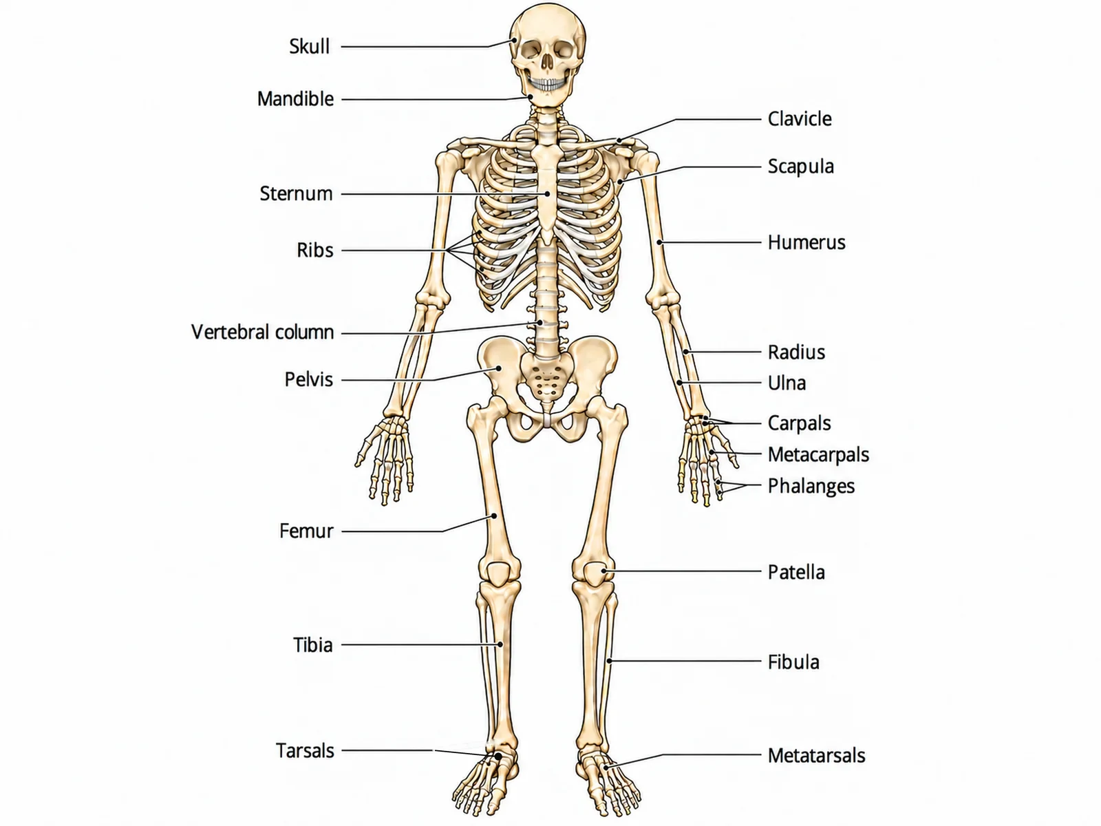
A labeled skeleton, a blank skeleton diagram worksheet with its matching answer key, the axial and appendicular divisions, and close-ups of the skull and the hand and foot bones.
Prompt
Create a fully labeled human skeletal system diagram on a white background: a complete adult skeleton seen from the front in anatomical position, palms facing forward, thumbs pointing away from the body, feet slightly apart, with the person’s right side on the viewer’s left. Draw exactly 12 pairs of ribs, five fingers on each hand and five toes on each foot, the radius and ulna side by side and uncrossed, the fibula on the outer side of each leg, and the patella over each knee. Label these bones with thin leader lines and readable dark sans-serif text: "Skull", "Mandible", "Clavicle", "Scapula", "Sternum", "Ribs", "Humerus", "Vertebral column", "Pelvis", "Radius", "Ulna", "Carpals", "Metacarpals", "Phalanges", "Femur", "Patella", "Tibia", "Fibula", "Tarsals", "Metatarsals".
Use this example
Prompt
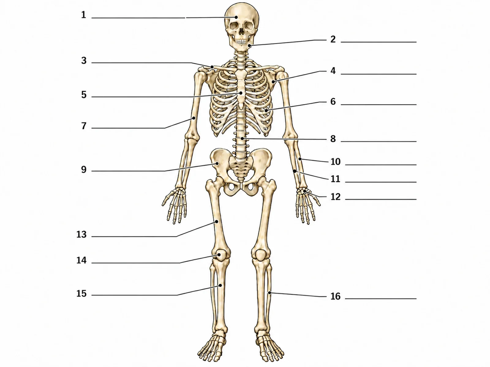
Create a blank skeleton diagram worksheet for students to label. Draw a complete adult human skeleton in anatomical position, seen from the front: standing upright, arms straight at the sides with the palms facing forward and the thumbs pointing away from the body, so the radius and ulna lie side by side and uncrossed, feet slightly apart. Draw exactly 12 pairs of ribs, five fingers on each hand and five toes on each foot, the scapulae showing only as edges behind the rib cage, the fibula on the outer side of each leg and the patella over each knee. Use clean dark outlines and a pale ivory fill on a white background. Draw exactly 16 thin leader lines, each ending at a short horizontal ruled answer line with a number beside it and no text, pointing at these targets in this order: 1 = Skull (cranium), 2 = Mandible, 3 = Clavicle, 4 = Scapula, 5 = Sternum, 6 = Ribs, 7 = Humerus, 8 = Vertebral column, 9 = Pelvis (hip bone), 10 = Radius, 11 = Ulna, 12 = Carpals, 13 = Femur, 14 = Patella, 15 = Tibia, 16 = Fibula. Put no bone names, no key and no word bank anywhere on the image. Leave generous white space.
Blank worksheet
Use this example
Prompt
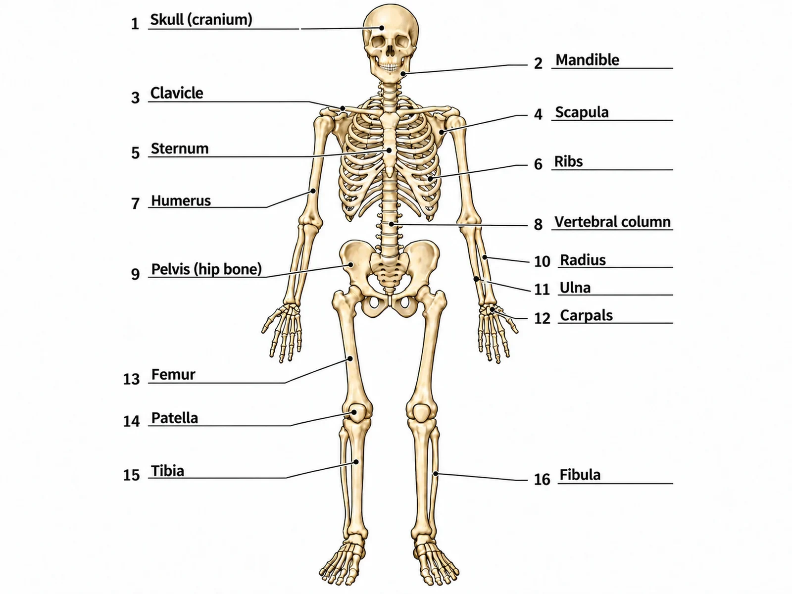
Create the answer key for a blank skeleton worksheet. Draw a complete adult human skeleton in anatomical position, seen from the front: standing upright, arms straight at the sides with the palms facing forward and the thumbs pointing away from the body, so the radius and ulna lie side by side and uncrossed, feet slightly apart. Draw exactly 12 pairs of ribs, five fingers on each hand and five toes on each foot, the scapulae showing only as edges behind the rib cage, the fibula on the outer side of each leg and the patella over each knee. Use clean dark outlines and a pale ivory fill on a white background. Draw exactly 16 thin leader lines, each ending at a short horizontal ruled line with its number, and write the answer on each line in dark sans-serif text: 1 = Skull (cranium), 2 = Mandible, 3 = Clavicle, 4 = Scapula, 5 = Sternum, 6 = Ribs, 7 = Humerus, 8 = Vertebral column, 9 = Pelvis (hip bone), 10 = Radius, 11 = Ulna, 12 = Carpals, 13 = Femur, 14 = Patella, 15 = Tibia, 16 = Fibula. Keep the numbering in this order so it matches the worksheet line for line.
Use this example
Prompt
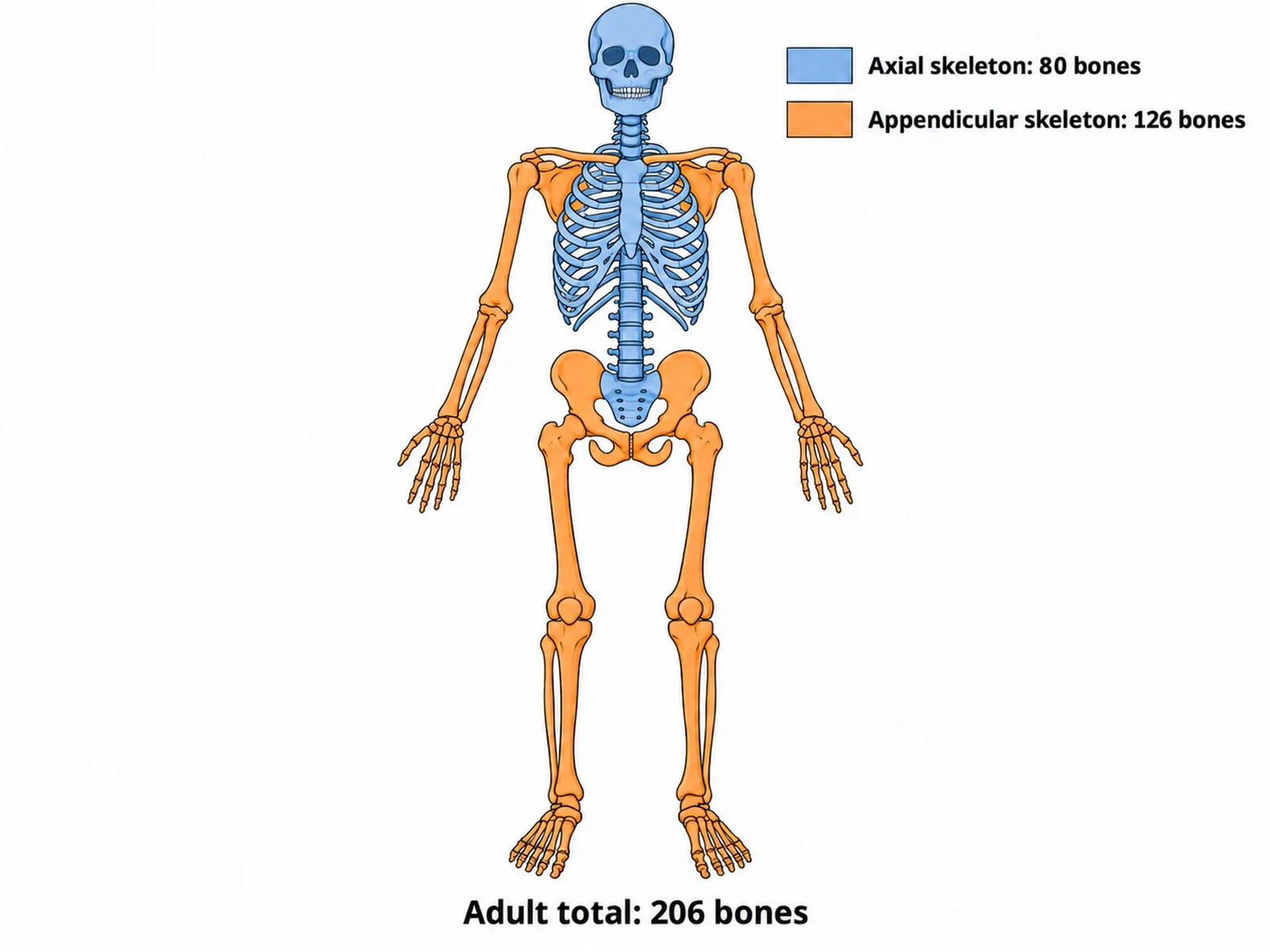
Create a skeletal system diagram that divides the adult skeleton into its two parts, drawn from the front in anatomical position on a white background. Shade the AXIAL skeleton (skull, vertebral column including the sacrum and coccyx, the 12 pairs of ribs and the sternum) in a soft blue, and the APPENDICULAR skeleton (clavicles, scapulae, arm and hand bones, the two hip bones, leg and foot bones) in a soft orange. Keep the sacrum blue, because it is axial even though it sits in the pelvis, and keep the hip bones orange. Add a legend reading "Axial skeleton: 80 bones" in blue and "Appendicular skeleton: 126 bones" in orange, and a caption reading "Adult total: 206 bones".
Use this example
Prompt
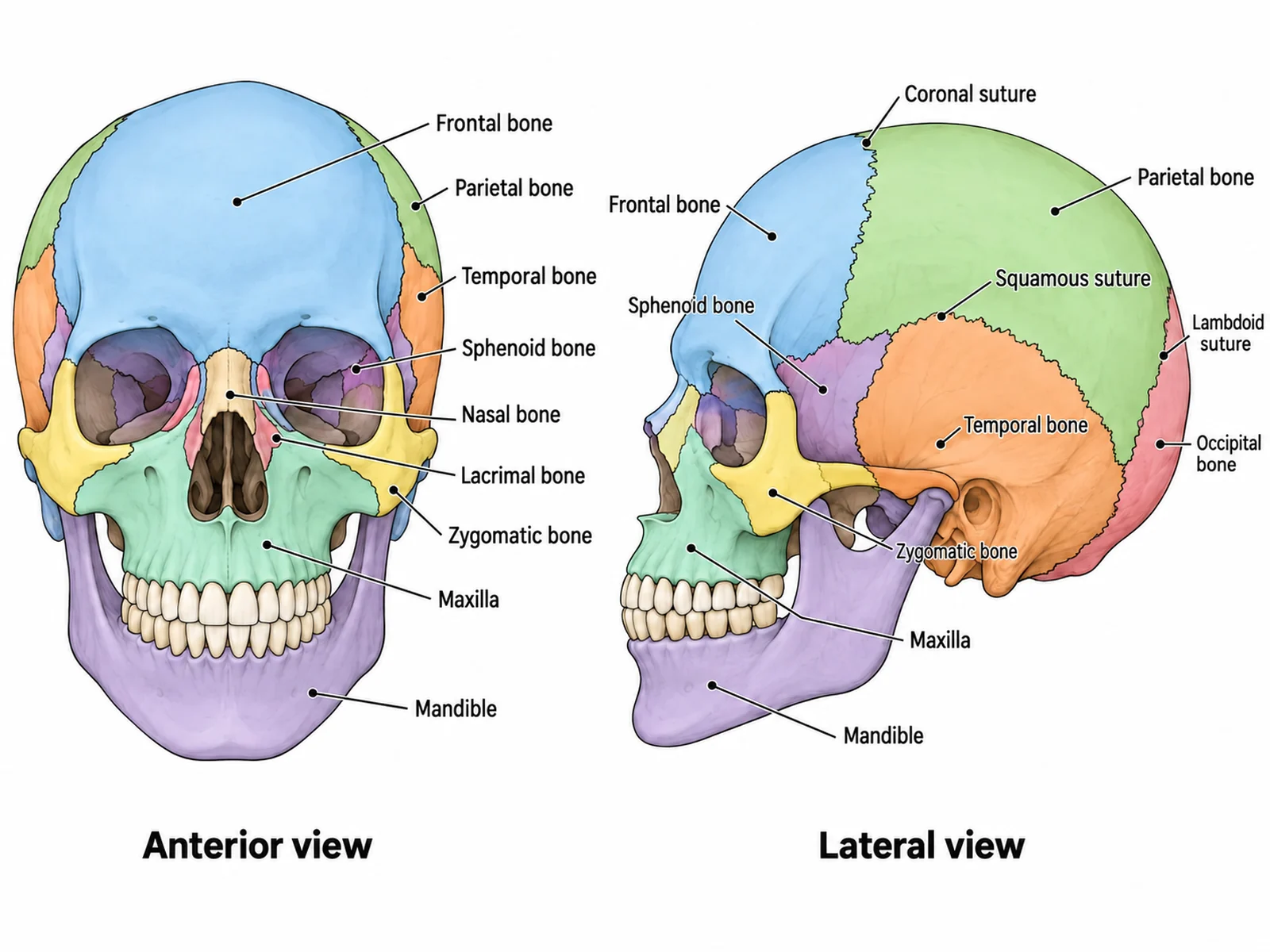
Create a labeled skull bones diagram on a white background with two views side by side: an anterior view on the left and a lateral view facing left on the right. In the anterior view label frontal bone, parietal bone, temporal bone, sphenoid bone, nasal bone, lacrimal bone, zygomatic bone, maxilla and mandible, and do not label the occipital bone, which cannot be seen from the front. In the lateral view label frontal bone, parietal bone, temporal bone, occipital bone, sphenoid bone, zygomatic bone, maxilla and mandible, and show the coronal, squamous and lambdoid sutures as thin lines. Tint each bone a different pale colour, the same colour in both views, and use thin leader lines.
Skull close-up
Use this example
Prompt
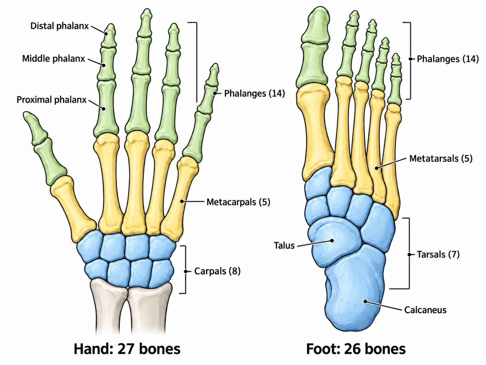
Create a labeled diagram of the bones of one hand and one foot, side by side on a white background. On the left, a hand seen from the palm side with the fingers pointing up: 8 carpals in two rows of four, 5 metacarpals, and 14 phalanges, the thumb with only two. Tint the carpals, metacarpals and phalanges three different pale colours and label "Carpals (8)", "Metacarpals (5)", "Phalanges (14)", "Proximal phalanx", "Middle phalanx" and "Distal phalanx". On the right, a foot seen from above with the toes pointing up: label "Calcaneus", "Talus", "Tarsals (7)", "Metatarsals (5)", "Phalanges (14)", with the big toe having only two phalanges. Exactly five digits on the hand and five toes on the foot.
Hand and foot close-up
Use this example
How to make a labeled or blank skeletal system diagram
1
Pick the view, level, and labels
Front, back, both, side, or a skull, hand, or foot close-up. Set the level from middle school to medical, then choose labeled, numbered with a key, a blank worksheet, or a worksheet with a word bank.
2
Generate the skeleton
The generator draws the skeleton in anatomical position with 12 pairs of ribs, the radius and ulna uncrossed and the fibula on the outside of the leg, then adds leader lines with names, numbers, or empty blanks.
3
Make the answer key, check, and export
For a worksheet, make the answer key from the same result so the numbers match. Count the ribs and fingers, check the forearm and leg bones, then download the PNG.
Skeletal system key facts
Adult bones
206
Axial skeleton
80 bones
Appendicular skeleton
126 bones
Ribs
12 pairs (7 true, 3 false, 2 floating)
Vertebrae above the sacrum
7 cervical, 12 thoracic, 5 lumbar
Skull
22 bones: 8 cranial, 14 facial
Hand / foot
27 / 26 bones each
Longest bone
Femur
Smallest bone
Stapes (middle ear)
Bony pieces at birth
About 270 to 300
What is a skeletal system diagram?
A skeletal system diagram is a labeled drawing of the human skeleton that names its bones. The standard version shows a whole adult skeleton from the front in anatomical position: standing, facing you, arms at the sides with the palms forward. It is one of the first figures anyone meets in biology or anatomy, because it ties the names of the bones to where they are in the body.
In class the figure appears in two forms. A labeled skeletal system diagram names every bone and works as a reference or an answer key. A blank skeleton diagram keeps the drawing and the leader lines but leaves the names off, so students write them in. This page makes both from one description, plus a numbered version with a key and a worksheet with a word bank.
The 206 bones: how the count breaks down
Textbooks give the adult total as 206, split into the axial skeleton (80 bones) and the appendicular skeleton (126). A whole-body diagram never labels all 206, because the small bones of the skull, wrist, and ankle would be unreadable at that scale. What it should get right is the structure behind the count.
Skull: 22 bones. Eight cranial bones (frontal, two parietal, two temporal, occipital, sphenoid, ethmoid) and 14 facial bones, including the mandible.
Ear ossicles and hyoid: three tiny bones in each middle ear (malleus, incus, stapes) and the U-shaped hyoid in the neck, 7 in all. They are rarely drawn on a whole skeleton.
Vertebral column: 26 bones. That is 7 cervical, 12 thoracic and 5 lumbar vertebrae, plus the sacrum and the coccyx.
Thoracic cage: 24 ribs in 12 pairs, plus the sternum.
Upper limbs and pectoral girdle: 64 bones. Each side has a clavicle, a scapula, a humerus, a radius, an ulna, 8 carpals, 5 metacarpals and 14 phalanges.
Lower limbs and pelvic girdle: 62 bones. Each side has a hip bone, a femur, a patella, a tibia, a fibula, 7 tarsals, 5 metatarsals and 14 phalanges.
The axial skeleton: 80 bones along the midline
The axial skeleton is the central column of the body. It protects the brain, spinal cord, heart and lungs, and it is the frame the limbs hang from. On a color-coded diagram it is the part left once the arms, legs and girdles are taken away.
Skull: the cranium encloses the brain; the facial bones shape the face and hold the teeth. The mandible is the only skull bone that moves.
Vertebral column: seen from the side it has four curves, cervical and lumbar curving forward, thoracic and sacral curving back. That S-shape is why a lateral view looks different from a front view.
Ribs: all 12 pairs join a thoracic vertebra at the back. Ribs 1 to 7 reach the sternum by their own costal cartilage, ribs 8 to 10 reach it through the cartilage above, and ribs 11 and 12 have free front ends.
Sternum: the manubrium at the top, where the clavicles attach, then the body, then the small xiphoid process at the bottom.
Sacrum and coccyx: five vertebrae fused into the sacrum, and usually four tiny ones into the coccyx. Both are axial even though they sit inside the pelvis.
The appendicular skeleton: 126 bones in the limbs and girdles
The appendicular skeleton is built for movement. Each limb hangs from a girdle that connects it to the axial skeleton. The two limbs follow the same plan: one bone in the upper segment, two in the lower segment, then a wrist or ankle, then five digits.
Pectoral girdle: the clavicle and the scapula on each side. The clavicle is the only bony link between the arm and the trunk, which is one reason it breaks so often in falls on an outstretched hand.
Arm and hand: humerus in the upper arm; radius (thumb side) and ulna (little-finger side) in the forearm; 8 carpals in the wrist; 5 metacarpals in the palm; 14 phalanges in the fingers.
Pelvic girdle: two hip bones, each made of the ilium, ischium and pubis fused together. They meet at the pubic symphysis in front and join the sacrum at the back.
Leg and foot: femur in the thigh, the patella in front of the knee, the tibia (inner, weight-bearing) and fibula (outer) in the leg, 7 tarsals including the calcaneus (heel) and talus, 5 metatarsals and 14 phalanges.
Bones to label on a blank skeleton diagram, by level
The right number of labels depends on the class. Too few and the worksheet is trivial; too many and the small bones crowd each other. These are the sets the level menu uses when your description does not list its own bones.
Middle school: about ten bones with everyday names, such as skull, jaw, collarbone, ribs, breastbone, backbone, pelvis, thigh bone (femur), kneecap and shin bone.
High school: the twenty standard bones, from the skull, mandible, clavicle, scapula and sternum down to the tarsals and metatarsals.
College: the major bones plus the regions of the vertebral column, the three parts of the sternum, the parts of the hip bone, and the calcaneus and talus.
Medical: bony landmarks as well as bones, such as the acromion, olecranon, iliac crest, greater trochanter, tibial tuberosity and the malleoli.
Any level: a clean unlabeled skeleton with no lines at all, for coloring, quizzing aloud, or as a slide background.
Reading a front-view skeleton: sides, pairs, and anatomical position
Most labeling mistakes on skeletons come from the view, not the bone names. In a front view the person faces you, so their right arm is on your left. Labels such as right humerus or left femur always mean the person’s side. A back view flips this: the person’s right is on your right.
Anatomical position also decides how the forearm looks. With the palms forward the radius and ulna lie parallel, the radius on the outside on the thumb side. A skeleton drawn with its palms against the thighs has the radius crossing the ulna. That is not wrong anatomy, but it confuses students labeling the two bones, so this generator keeps the palms forward unless you ask otherwise. The same habit applies to the leg: the tibia is always on the inner, big-toe side, whichever way the figure faces.
Skull, hand, and foot close-ups
Some of the most tested bones are too small for a whole-body figure, so they get their own close-up.
Skull, front view: frontal, parietal, temporal, sphenoid, ethmoid and lacrimal around the orbit, nasal, zygomatic (cheekbone), maxilla (upper jaw) and mandible (lower jaw). The occipital bone is at the back and cannot be seen from the front.
Skull, side view: adds the occipital bone and shows the sutures. The coronal suture runs between the frontal and parietal bones, the squamous suture above the temporal bone, and the lambdoid suture in front of the occipital bone.
Hand: the carpals sit in two rows of four. Students often learn them as scaphoid, lunate, triquetrum and pisiform in the near row, and trapezium, trapezoid, capitate and hamate in the far row. Then come 5 metacarpals and 14 phalanges.
Foot: the talus forms the ankle joint with the tibia and fibula, and the calcaneus forms the heel below it. The navicular, cuboid and three cuneiforms complete the 7 tarsals, followed by 5 metatarsals and 14 phalanges.
Bone shapes and the parts of a long bone
Anatomy courses often pair the whole skeleton with a cut-open long bone, and both ask students to sort bones by shape. Ask for "a labeled long bone section" and the generator draws a femur or humerus opened lengthwise.
Long bones are longer than they are wide: the femur, humerus, radius, ulna, tibia, fibula, the metacarpals and metatarsals, and even the small phalanges.
Short bones are roughly cube-shaped, like the carpals and tarsals. Flat bones are thin and often curved: the skull roof, sternum, ribs and scapula. Irregular bones, such as the vertebrae, fit none of these shapes.
Sesamoid bones grow inside a tendon. The patella is the largest; smaller ones sit in the hands and feet.
Diaphysis: the shaft, a tube of compact bone around the medullary cavity, which holds yellow marrow in adults.
Epiphyses: the two ends, made of spongy bone with red marrow in its spaces and capped with articular cartilage where they form a joint.
Epiphyseal plate: the band of cartilage between shaft and end where a child’s bone grows in length. In an adult it is only a thin epiphyseal line.
Periosteum and endosteum: the periosteum is the tough membrane over the outside of the bone, except on the joint surfaces; the endosteum lines the medullary cavity.
Joints a skeleton diagram can show
A skeleton is also a map of the joints between its bones, and joint labels are a common extension once the bones are known. The structural classes are fibrous, cartilaginous and synovial.
Fibrous joints barely move. The sutures between the skull bones are the standard example.
Cartilaginous joints allow a little movement: the pubic symphysis at the front of the pelvis, and the intervertebral discs between vertebral bodies.
Ball-and-socket synovial joints, at the shoulder and the hip, move in every direction. The hip socket is deeper, which makes it more stable and less mobile than the shoulder.
Hinge joints, at the elbow and the knee, bend in one plane. The pivot joint between the first two cervical vertebrae lets the head turn side to side.
Saddle and condyloid joints, at the base of the thumb and at the wrist, give two directions of movement.
Male and female skeletons
Men and women have the same 206 bones and the same 12 pairs of ribs. The idea that men have one rib fewer is a myth. The clearest difference is the pelvis. A female pelvis is wider and shallower, with a rounder inlet and a wider angle under the pubic arch, which leaves room for childbirth. A male pelvis is narrower and taller, with a heart-shaped inlet and a sharper pubic angle. Male bones are also on average heavier, with more pronounced ridges where muscles attach. On a standard diagram none of this matters, but if a worksheet compares the two pelves, say so in your description so the difference is drawn on purpose.
What the skeletal system does
Support: the skeleton holds the body’s shape and carries its weight down through the spine, pelvis and legs.
Protection: the skull surrounds the brain, the vertebrae the spinal cord, and the rib cage the heart and lungs.
Movement: bones are levers, joints are pivots, and skeletal muscles attached by tendons pull on them.
Mineral storage: bone holds most of the body’s calcium and phosphate and releases them into the blood when needed.
Blood cell formation: red bone marrow, mainly in the flat bones, the vertebrae and the ends of long bones in adults, makes red cells, white cells and platelets.
Fat storage: yellow marrow in the shafts of adult long bones stores fat.
Using an unlabeled skeleton diagram in class
Start blank, end labeled: hand out the blank skeleton worksheet, then show the answer key made from the same result so every number matches.
Match the labels to the lesson: ten bones for a starter, twenty for an end-of-unit check, landmarks for an anatomy practical.
Word bank or not: include one for a scaffolded lesson, leave it out for a test. The word bank is alphabetical, so its order gives nothing away.
One skeleton, two divisions: use the axial and appendicular version to sort bones before naming them.
Close-ups for detail: test the carpals, tarsals and skull bones on their own sheets instead of squeezing them onto the whole body.
How the skeleton changes from birth to adulthood
A newborn has around 270 to 300 separate bony pieces, many still joined by cartilage. As a child grows these fuse. The skull plates close at the sutures, the five sacral vertebrae fuse into one sacrum, and the ilium, ischium and pubis join into each hip bone during the teenage years. The growth plates at the ends of long bones turn to bone once growth stops, usually by the early twenties. That is why an adult skeleton has 206 bones and a child’s X-ray shows gaps at the ends of the long bones. A diagram of a child’s skeleton should show those growth plates; an adult one should not.
Most common mistakes on a skeletal system diagram
These are the errors image models make most often on skeletons. Check each one before a diagram goes into graded work.
Pectoral girdle, arms and hands, pelvic girdle, legs and feet
Girdle bones
None
Clavicles and scapulae; the two hip bones
Where it sits
The central axis of the body
The limbs and the bones that attach them to the axis
Main job
Protects the brain, spinal cord, heart, and lungs; holds the body upright
Movement and handling objects
Easy to misplace
The sacrum and coccyx (axial, although they sit in the pelvis)
The hip bones (appendicular, although they form most of the pelvis)
Skeletal System Diagram FAQ
A standard front-view diagram names the skull, mandible, clavicle, scapula, sternum, ribs, vertebral column, humerus, radius, ulna, carpals, metacarpals, phalanges, pelvis, femur, patella, tibia, fibula, tarsals, and metatarsals. That is enough for most school and introductory college courses. Anatomy courses add the parts of each bone and the individual skull, wrist, and ankle bones, which are clearer on a close-up than on a whole-body figure.
Generate body system diagrams, organ close-ups, blank labeling worksheets with matching answer keys, and revision-ready biology figures with the same SciFigure AI workflow.