Eye Diagram AI

Eye Diagram Generator

Generate a blank unlabeled eye diagram for students to complete, or a fully labeled cross-section with the cornea, iris, lens, retina, and optic nerve named.

Blank or labeledFull cross-sectionWorksheet-ready

AI Eye Diagram Generator

Describe which eye structures to label and the style you need.

Costs 5 credits347 / 2000 characters

Generate a publication-ready PNG from your description.

Preview

Eye Diagram Example Previews

Target aspect ratio: 4:3

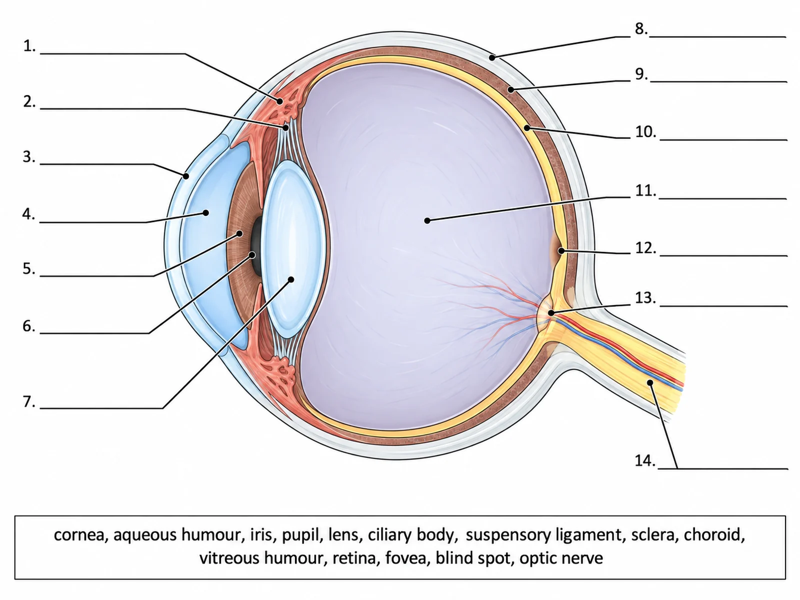
Blank unlabeled eye diagram cross-section with numbered empty label lines for students to complete
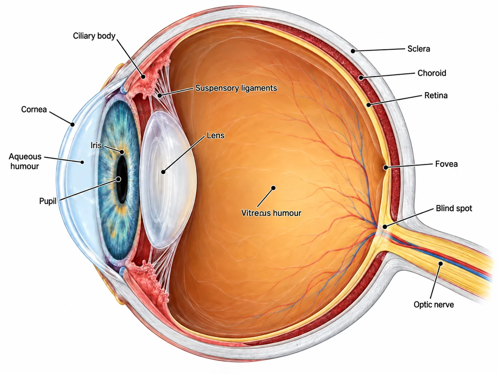
Labeled eye anatomy cross-section naming the cornea, aqueous humour, iris, pupil, lens, ciliary body, sclera, choroid, retina, fovea, blind spot, and optic nerve
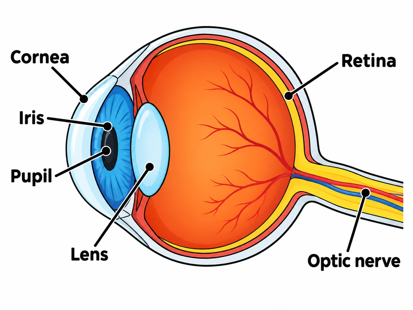
Simple eye diagram for younger students labeling only the cornea, iris, pupil, lens, retina, and optic nerve

Sample previews. Describe your diagram to generate your own eye figure.

Eye Diagram Examples

Use these as visual directions for blank worksheets, labeled cross-sections, light pathway figures, accommodation comparisons, and retina close-ups.

Blank unlabeled eye diagram cross-section with numbered empty label lines for students to complete

Prompt

Create a blank eye diagram worksheet on a white background. Draw a horizontal cross-section of the human eye in clean line art with pale flat fills and absolutely no text inside the drawing: a round eyeball with the transparent cornea bulging at the front left, the iris and pupil behind it, the lens, the three concentric layers at the back, and the optic nerve leaving at the lower right. Draw thin leader lines from fourteen structures out to short horizontal ruled blank lines, numbered 1 to 14. Add a word bank box below reading "cornea, aqueous humour, iris, pupil, lens, ciliary body, suspensory ligament, sclera, choroid, vitreous humour, retina, fovea, blind spot, optic nerve". Generous white space, no answer key.
Blank Eye Diagram

Labeled eye anatomy cross-section naming the cornea, aqueous humour, iris, pupil, lens, ciliary body, sclera, choroid, retina, fovea, blind spot, and optic nerve

Prompt

Create a fully labeled human eye diagram on a white background, drawn as a horizontal cross-section facing left. Show the transparent cornea bulging at the front, the aqueous humour chamber behind it, a coloured iris with a central pupil, a biconvex lens held by suspensory ligaments to the ciliary body, the large vitreous humour filling the main chamber, and three clearly distinct concentric layers at the back drawn in order from outside in: a thick white sclera, a dark red choroid, and a pale retina. Mark the fovea directly opposite the pupil on the visual axis and the blind spot beside it where the optic nerve exits at the lower right. Label every structure with a thin dark leader line and readable sans-serif text.

Labeled Eye Anatomy

Simple eye diagram for younger students labeling only the cornea, iris, pupil, lens, retina, and optic nerve

Prompt

Create a simple eye diagram for primary or lower-secondary students. Show only six labeled parts: cornea, iris, pupil, lens, retina, and optic nerve. Use bold flat colours, thick clean outlines, large readable text, and plenty of white space. Omit the aqueous and vitreous humour, choroid, ciliary body, fovea, and blind spot.

Simple Eye Diagram

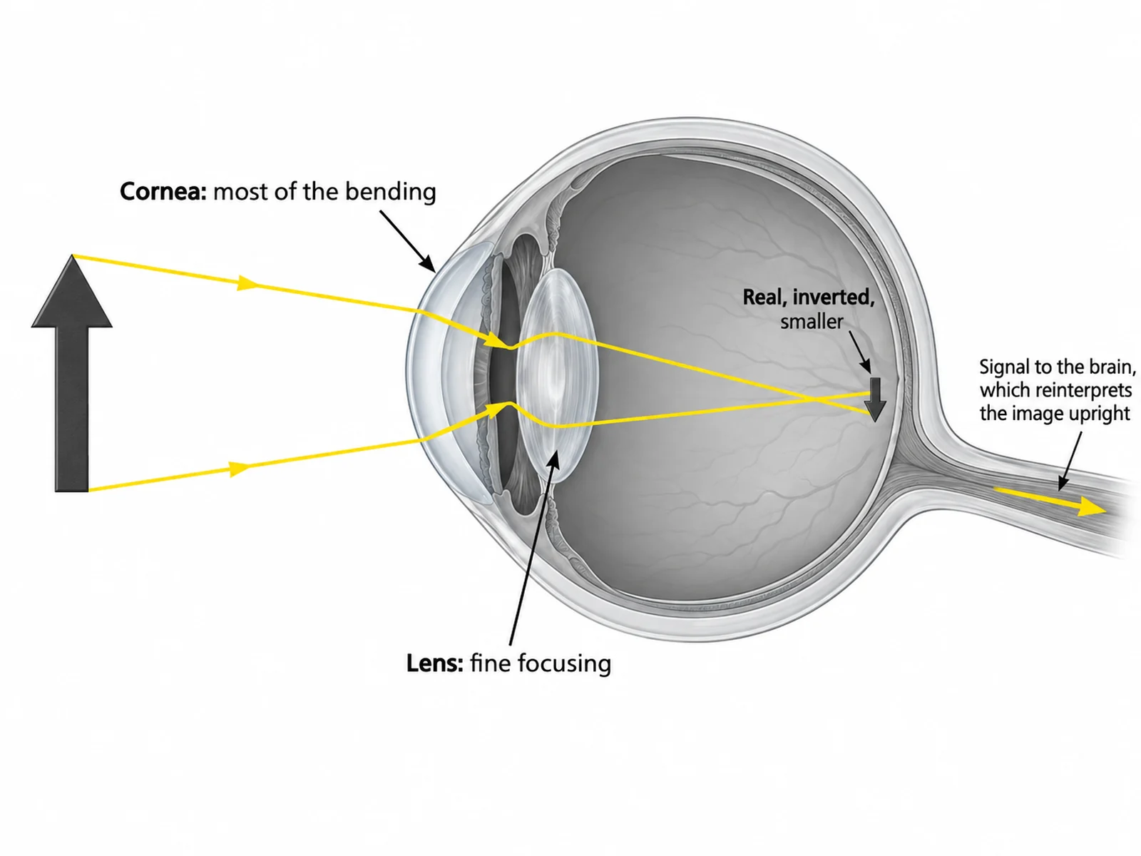
Eye diagram tracing light rays through the cornea, pupil, and lens to form an inverted image on the retina

Prompt

Create an eye diagram that traces the path of light. Draw the labeled eye cross-section in muted grey, then overlay bright yellow light rays entering from an upright arrow-shaped object on the left, bending sharply at the cornea, passing through the pupil, bending again at the lens, and converging to form a small INVERTED arrow image on the retina at the back. Label the refraction points "Cornea: most of the bending" and "Lens: fine focusing", and caption the image on the retina "Real, inverted, smaller". Add an arrow to the optic nerve labeled "Signal to the brain, which reinterprets the image upright".

How the Eye Focuses Light

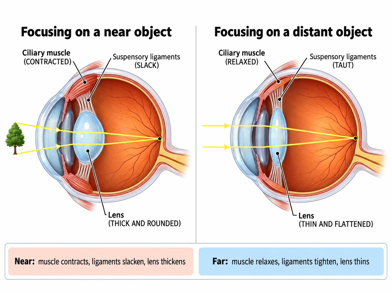
Two-panel diagram comparing lens shape and ciliary muscle state when focusing on near and distant objects

Prompt

Create a two-panel accommodation diagram on a white background. The left panel, headed "Focusing on a near object", shows the ciliary muscle contracted, the suspensory ligaments slack, and the lens thick and rounded, with light rays converging on the retina. The right panel, headed "Focusing on a distant object", shows the ciliary muscle relaxed, the suspensory ligaments taut, and the lens thin and flattened. Label "Ciliary muscle", "Suspensory ligaments", and "Lens" in both panels, and add a small comparison strip beneath reading "Near: muscle contracts, ligaments slacken, lens thickens" and "Far: muscle relaxes, ligaments tighten, lens thins".

Accommodation: Near and Far

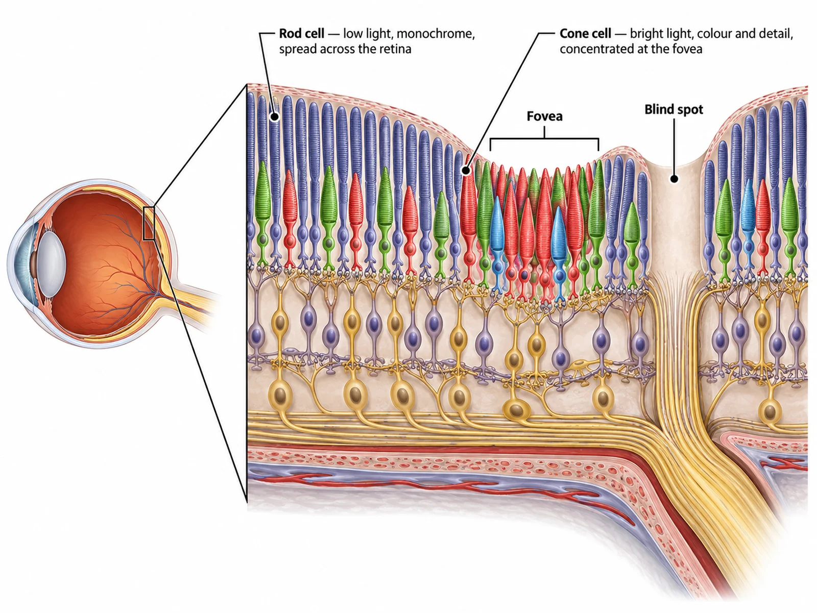
Magnified retina diagram showing rod and cone cells, the fovea, and the blind spot where the optic nerve leaves

Prompt

Create a magnified retina diagram on a white background. On the left show a small eye outline with a callout wedge pointing to the back wall. On the right show the magnified retina in cross-section with rod cells drawn as slender cylinders and cone cells as shorter tapered shapes, connected to a layer of nerve fibres that gather into the optic nerve. Label "Rod cell — low light, monochrome, spread across the retina" and "Cone cell — bright light, colour and detail, concentrated at the fovea". Mark the fovea as a dense patch of cones and the blind spot as a gap with no receptors where the nerve fibres exit.

Retina, Rods and Cones

How to make an eye diagram to label

1

Choose blank or labeled

A blank unlabeled cross-section with numbered lines for the worksheet, and the labeled version as the answer key. Say how many structures you need.

2

Generate the diagram

The generator keeps the sclera, choroid, and retina in the correct order and places the fovea and blind spot properly.

3

Check the anatomy, then export

Confirm the fovea sits on the visual axis and the blind spot is beside the optic nerve exit, then download the PNG for a worksheet, slide, or revision sheet.

What is an eye diagram?

An eye diagram is a horizontal cross-section through the eyeball, cut so the internal structures become visible. It is one of the most commonly set labelling exercises in school biology, which is why the blank version is requested more often than the labeled one: the unlabeled drawing is the worksheet, and the labeled drawing is the answer key.

Parts of the eye and what each one does

  • Cornea: the transparent front layer that does most of the light bending as it enters.
  • Aqueous humour: watery fluid in the front chamber that maintains shape and nourishes the cornea and lens.
  • Iris: the coloured muscular ring that adjusts pupil size to control light entry.
  • Pupil: the opening at the centre of the iris that light passes through.
  • Lens: a flexible biconvex structure that fine-tunes focus by changing shape.
  • Ciliary body and suspensory ligaments: the muscle and fibres that alter lens thickness during accommodation.
  • Sclera: the tough white outer coat that protects the eye and anchors the muscles.
  • Choroid: the dark pigmented middle layer that supplies blood and absorbs stray light.
  • Retina: the innermost light-sensitive layer containing rod and cone cells.
  • Fovea: the small retinal pit packed with cones, giving the sharpest colour vision.
  • Blind spot (optic disc): where the optic nerve leaves; no receptors, so no image is detected.
  • Optic nerve: carries impulses from the retina to the visual cortex of the brain.

Common errors to check before you print

  • Layers out of order. From outside in it is always sclera, then choroid, then retina.
  • Fovea off-axis. It sits directly opposite the pupil, where the sharpest image falls.
  • Blind spot drawn on the visual axis. It lies beside the fovea, where the optic nerve exits.
  • Lens drawn rigid. It is flexible, and accommodation depends on it changing shape.

Eye Diagram Generator FAQ

From front to back: cornea, aqueous humour, iris, pupil, lens, ciliary body and suspensory ligaments, sclera, choroid, vitreous humour, retina, fovea (macula), blind spot (optic disc), and optic nerve. Most school syllabuses require at least cornea, iris, pupil, lens, retina, and optic nerve.

Need other anatomy figures?

Generate sense organ diagrams, body system figures, cell diagrams, and worksheet-ready illustrations with the same SciFigure AI workflow.