Cell Membrane Diagram AI

Cell Membrane Diagram Generator

Generate a labeled cell membrane diagram of the fluid mosaic model, with the phospholipid bilayer, embedded proteins, cholesterol, and glycoproteins named. Ask for a blank version and students can label it themselves.

Fluid mosaic modelBilayer, proteins, cholesterolLabeled or blank

AI Cell Membrane Diagram Generator

Describe which membrane components to label and the style you need.

Costs 5 credits447 / 2000 characters

Generate a publication-ready PNG from your description.

Preview

Cell Membrane Diagram Example Previews

Target aspect ratio: 16:9

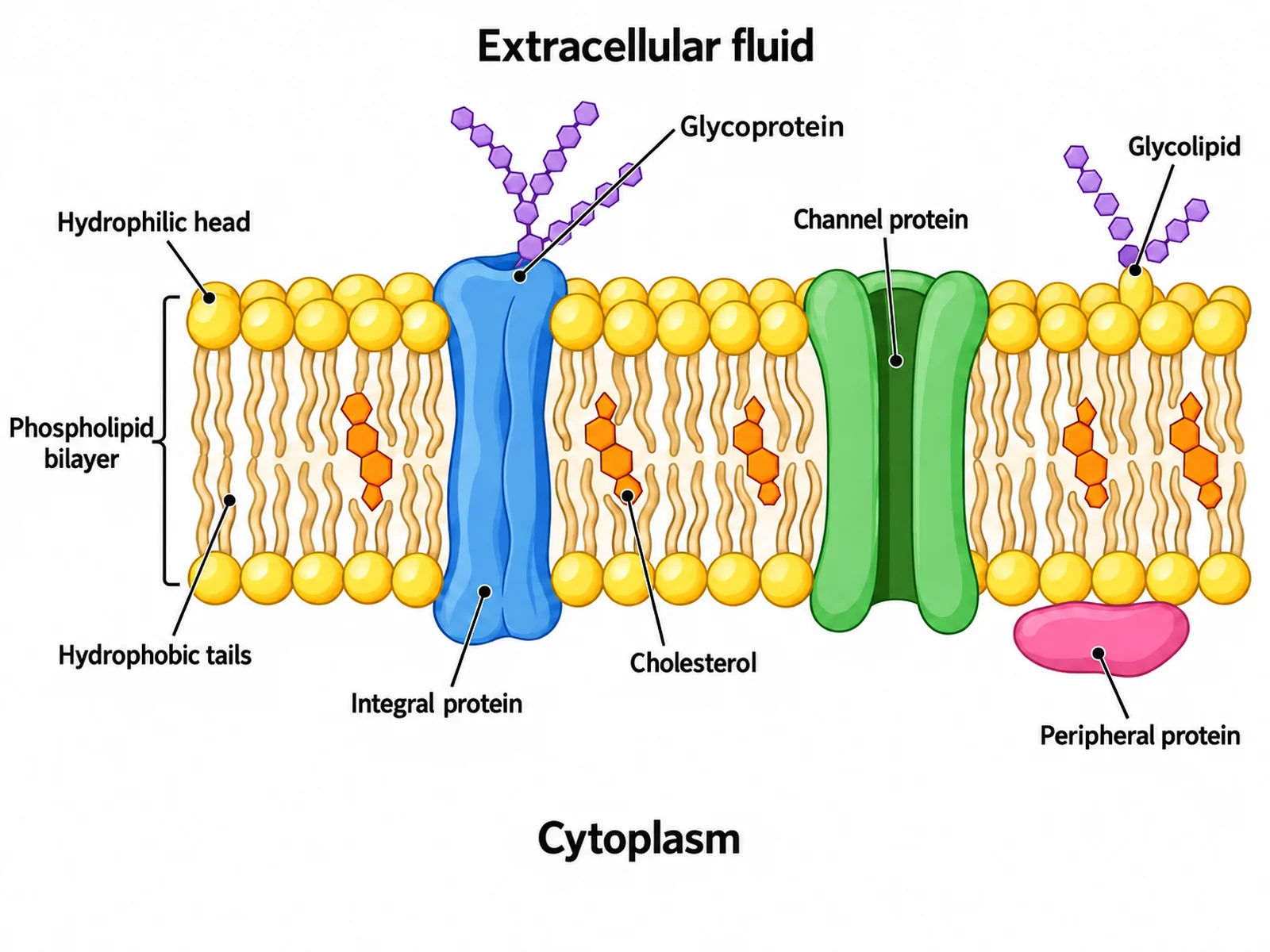
Labeled fluid mosaic model of the cell membrane showing the phospholipid bilayer, hydrophilic heads, hydrophobic tails, integral and peripheral proteins, channel protein, cholesterol, glycoprotein, and glycolipid
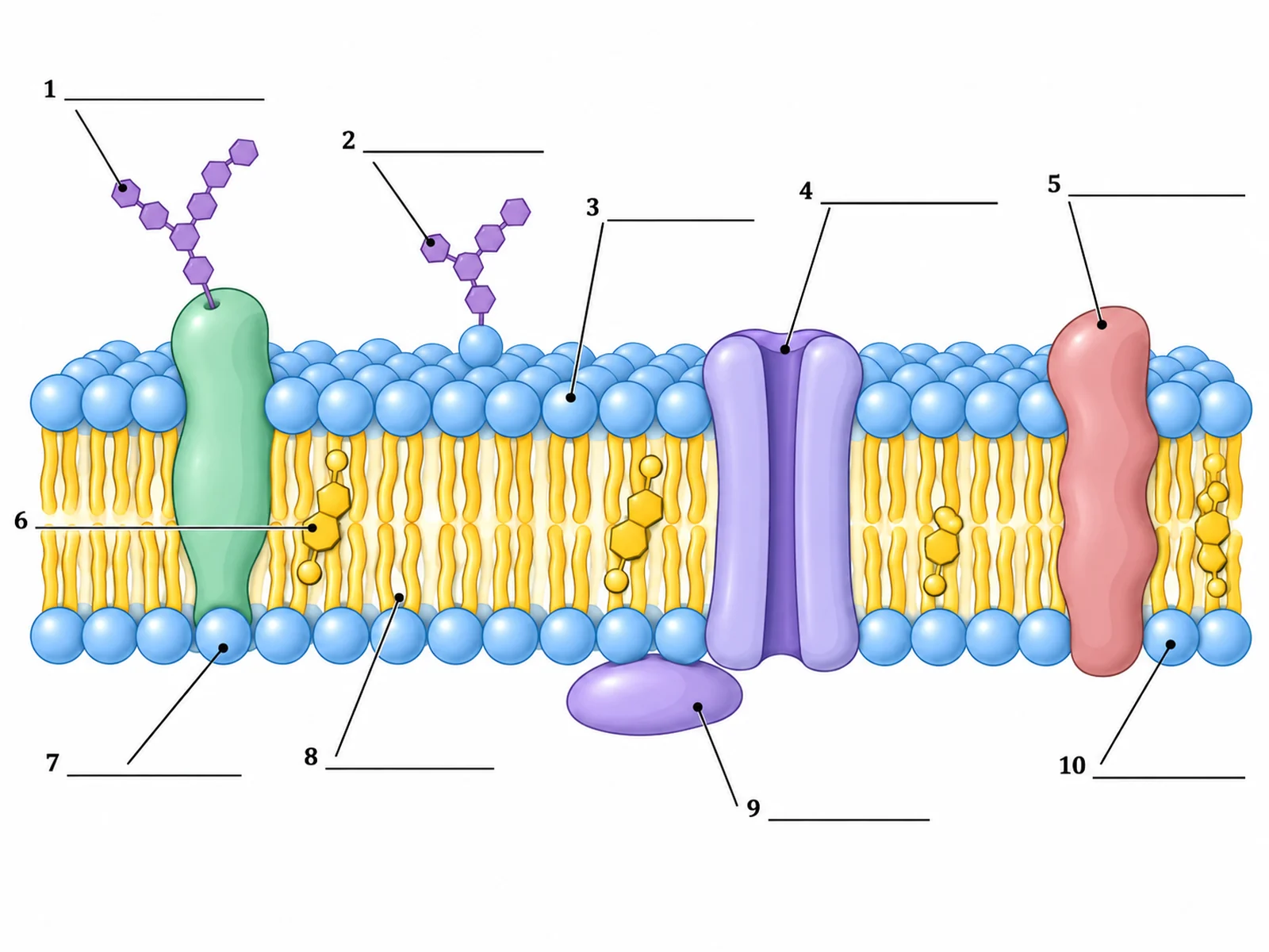
Blank unlabeled cell membrane diagram with numbered empty label lines for students to complete
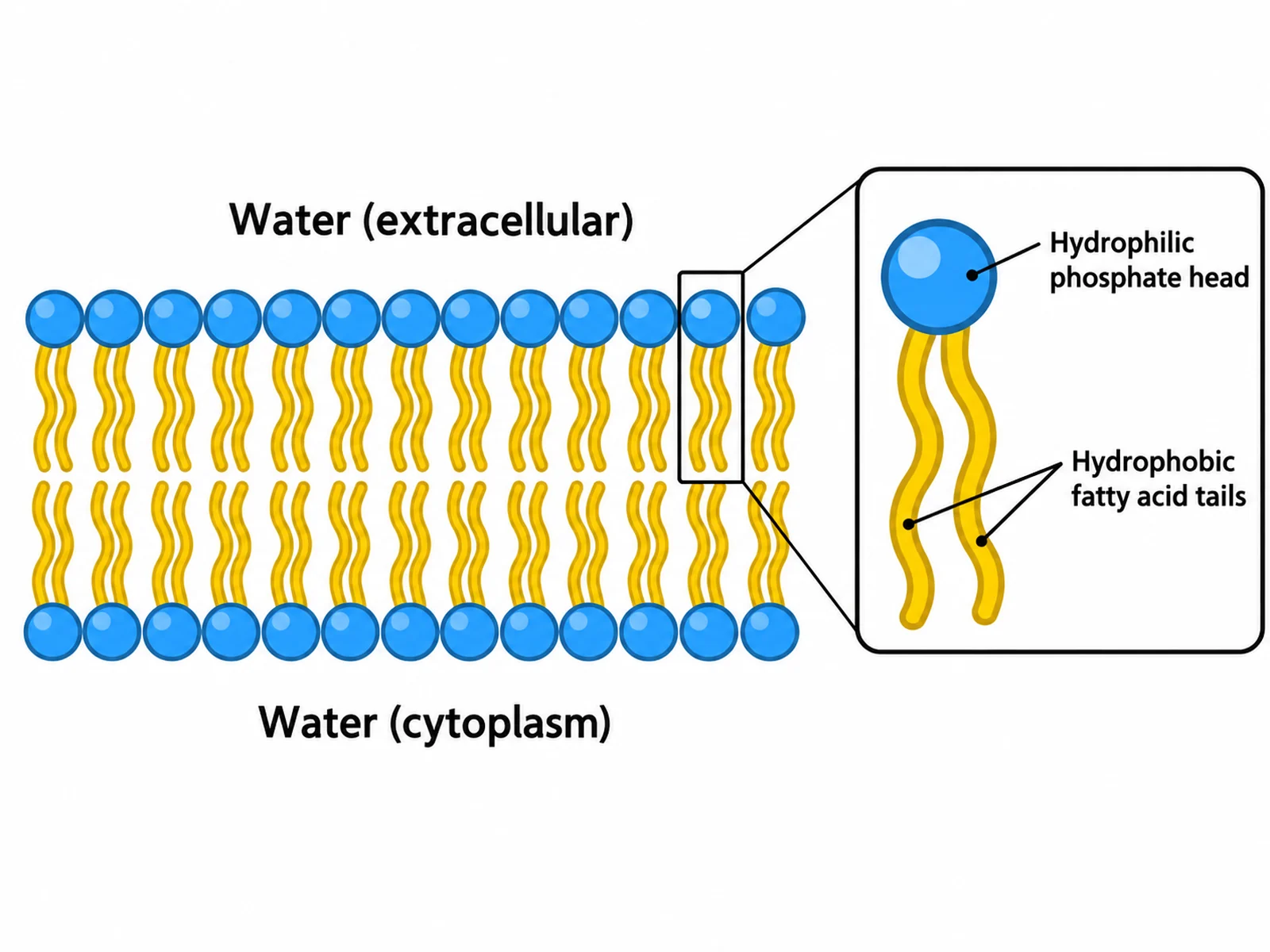
Simple phospholipid bilayer diagram labeling the hydrophilic phosphate head, the hydrophobic fatty acid tails, and the two sides of the membrane

Sample previews. Describe your diagram to generate your own membrane figure.

Labeled Cell Membrane Diagram Examples

Use these as visual directions for the fluid mosaic model, blank worksheets, simple bilayer drawings, transport routes, and membrane protein types.

Labeled fluid mosaic model of the cell membrane showing the phospholipid bilayer, hydrophilic heads, hydrophobic tails, integral and peripheral proteins, channel protein, cholesterol, glycoprotein, and glycolipid

Prompt

Create a fully labeled fluid mosaic model of the cell membrane on a white background, viewed in cross-section. Draw two rows of phospholipids: round yellow phosphate heads facing outward at the top and bottom, with paired wavy fatty acid tails pointing inward and meeting in the middle. Embed a blue integral protein spanning the full bilayer, a green channel protein with a visible central pore, a pink peripheral protein resting on the lower surface only, orange cholesterol molecules wedged between the tails, and branched purple carbohydrate chains attached to a glycoprotein and a glycolipid on the upper surface only. Label every component with a thin leader line, and add side labels reading "Extracellular fluid" above and "Cytoplasm" below.
Labeled Fluid Mosaic Model

Blank unlabeled cell membrane diagram with numbered empty label lines for students to complete

Prompt

Create a blank cell membrane worksheet. Draw the same fluid mosaic cross-section in clean line art with soft flat fills, but leave every label empty: draw thin leader lines from each component out to a short horizontal ruled blank line with no text on it. Number the blanks 1 to 10. Include generous white space and no answer key.

Blank Membrane Worksheet

Simple phospholipid bilayer diagram labeling the hydrophilic phosphate head, the hydrophobic fatty acid tails, and the two sides of the membrane

Prompt

Create a simple phospholipid bilayer diagram with no proteins. Show two rows of phospholipids, each drawn as one round head with two wavy tails, heads facing outward and tails meeting in the middle. Enlarge one phospholipid to the side as a callout and label its hydrophilic phosphate head and its two hydrophobic fatty acid tails. Add side labels reading "Water (extracellular)" above and "Water (cytoplasm)" below. Use bold flat colors and large readable text.

Simple Phospholipid Bilayer

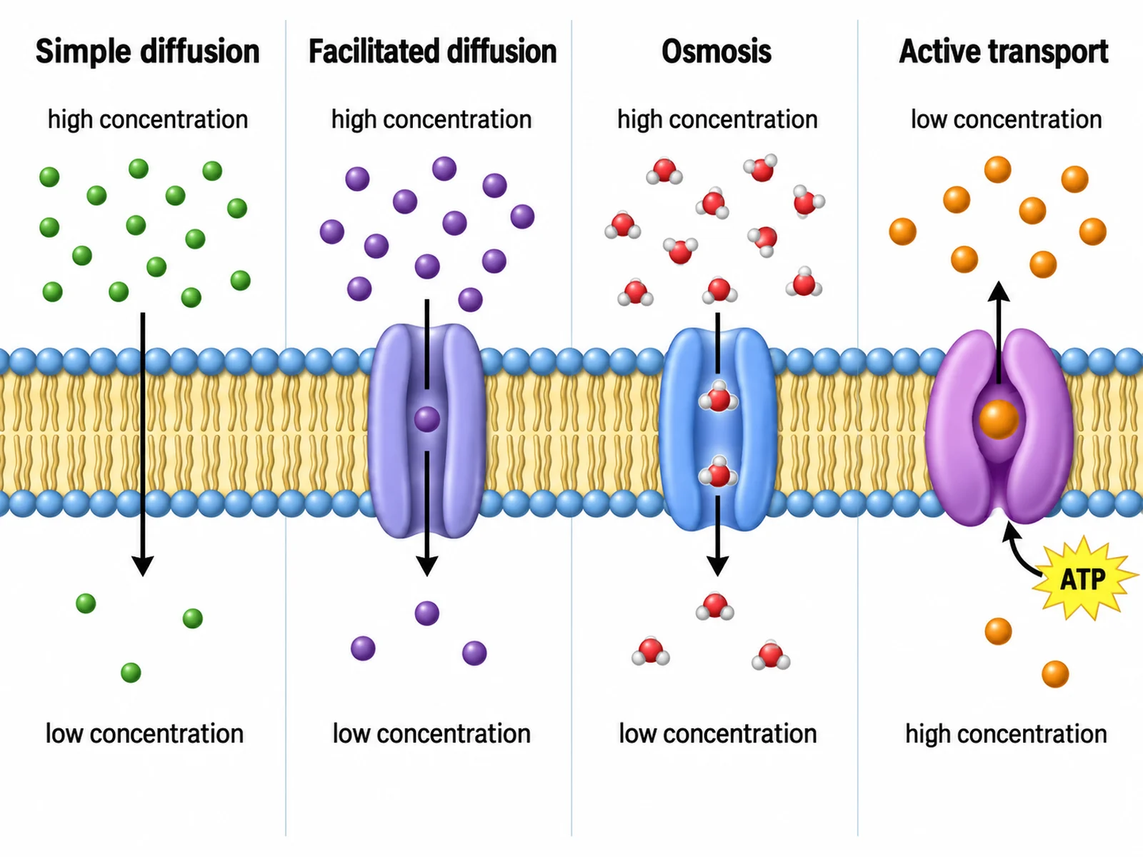
Cell membrane diagram comparing simple diffusion, facilitated diffusion, osmosis, and active transport with directional arrows

Prompt

Create a labeled membrane transport diagram. Draw one phospholipid bilayer across the width of the image and show four transport routes through it, left to right, each with a directional arrow and a heading: simple diffusion of small molecules straight through the bilayer, facilitated diffusion of a molecule through a channel protein, osmosis of water through an aquaporin, and active transport of a molecule through a carrier protein against the gradient with a small ATP symbol beside it. Add a subtle concentration gradient shading behind the membrane and label "high concentration" and "low concentration".

Membrane Transport

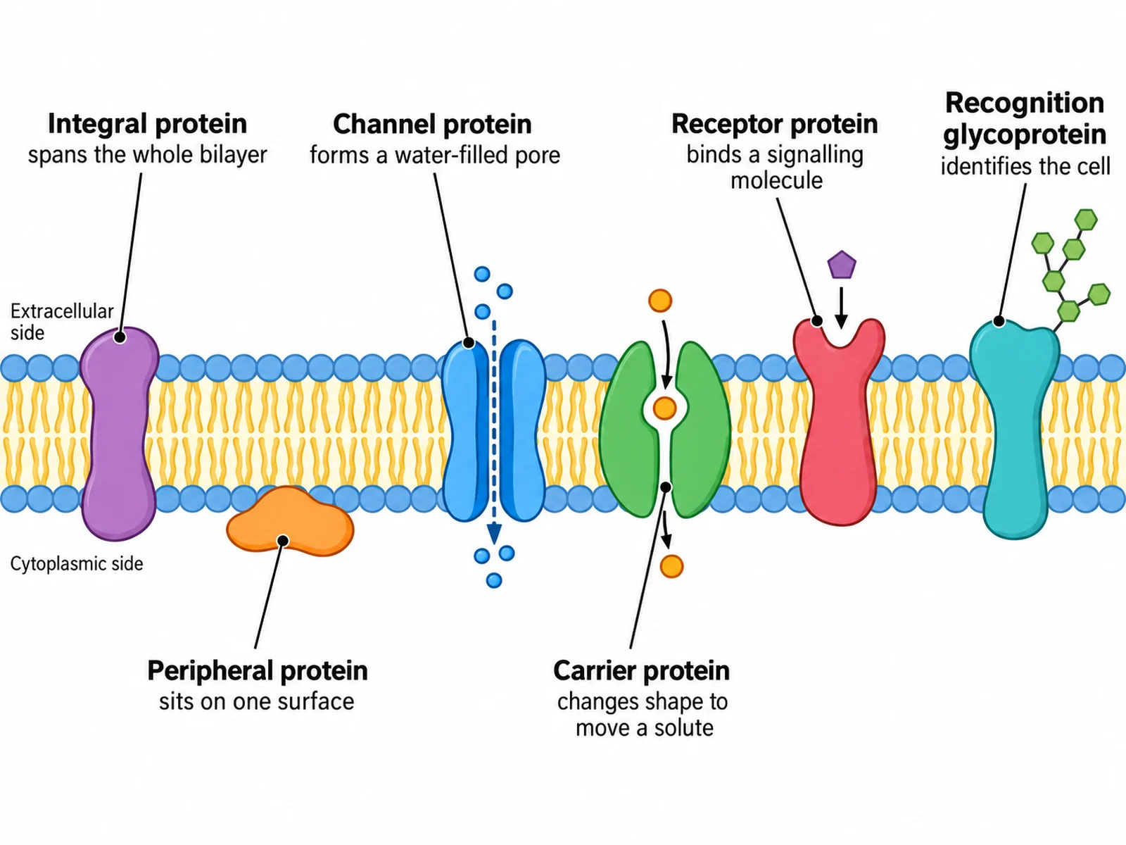
Cell membrane diagram labeling integral, peripheral, channel, carrier, receptor, and recognition proteins with their functions

Prompt

Create a labeled diagram of membrane protein types. Draw one phospholipid bilayer and embed six different proteins along it, each labeled with a name and a one-line function: integral protein "spans the whole bilayer", peripheral protein "sits on one surface", channel protein "forms a water-filled pore", carrier protein "changes shape to move a solute", receptor protein "binds a signalling molecule", and recognition glycoprotein "identifies the cell". Use a distinct color for each protein and keep the bilayer orientation correct with heads outward.

Membrane Proteins

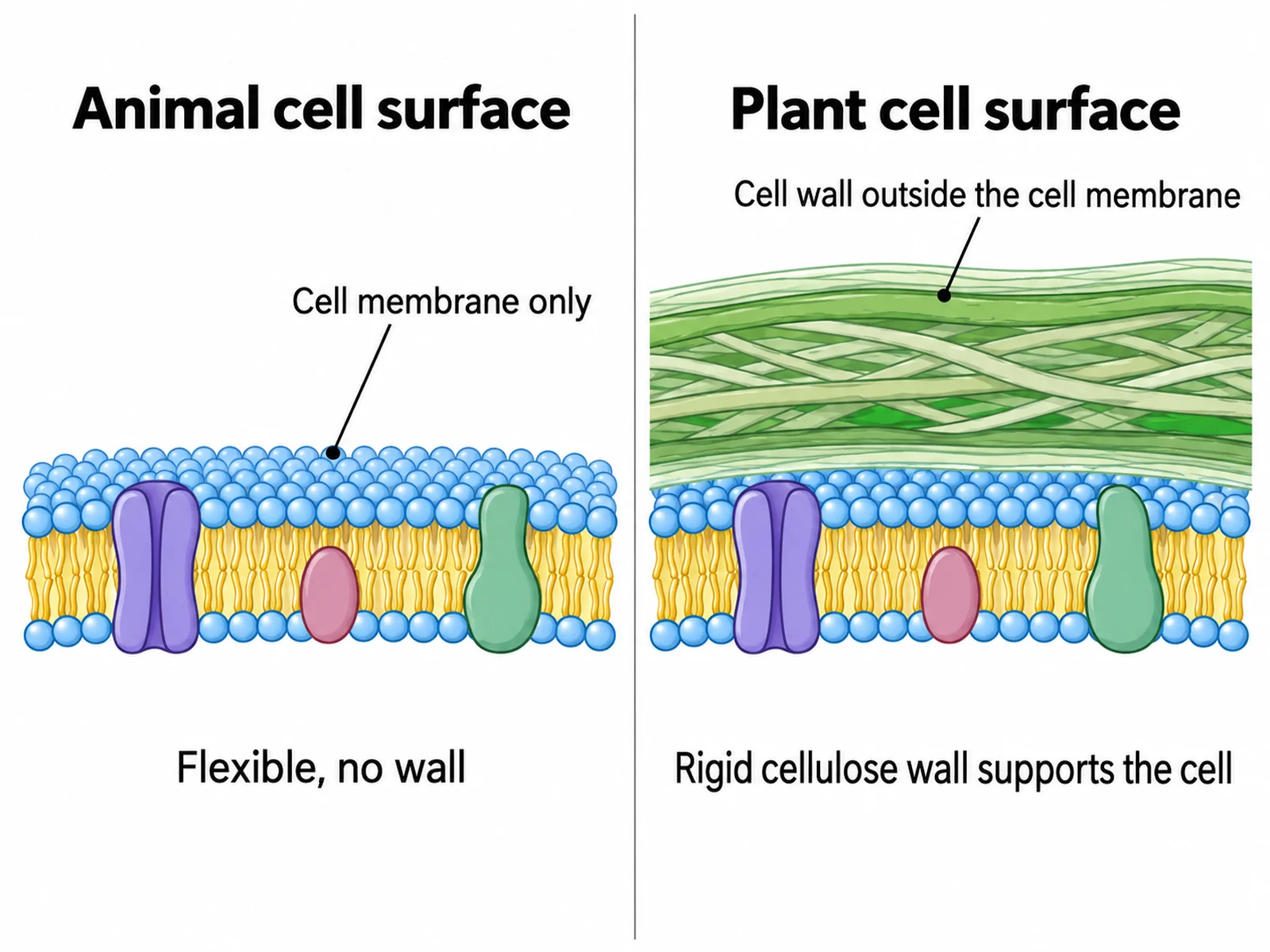
Comparison of an animal cell surface and a plant cell surface showing the cellulose cell wall outside the plant cell membrane

Prompt

Create a side-by-side comparison of the cell surface in an animal cell and a plant cell. On the left draw an animal cell surface: a single phospholipid bilayer with embedded proteins, labeled "Cell membrane only". On the right draw a plant cell surface: the same bilayer with a thick layered cellulose cell wall drawn outside it, labeled "Cell wall outside the cell membrane". Add a short note under each panel: "Flexible, no wall" and "Rigid cellulose wall supports the cell".

Plant vs Animal Cell Membrane

How to draw a labeled cell membrane diagram

1

Choose the level of detail

A simple bilayer for heads and tails only, the full fluid mosaic model with proteins and cholesterol, or a transport diagram showing molecules crossing the membrane.

2

Generate the diagram

The generator draws the bilayer with heads outward and tails inward, places proteins correctly as integral or peripheral, and adds readable leader lines.

3

Check the orientation, then export

Confirm the phosphate heads face the watery sides and the fatty acid tails meet in the middle, then download the PNG for a worksheet, slide, or revision sheet.

What is a cell membrane diagram?

A cell membrane diagram is a cross-section through the plasma membrane drawn as the fluid mosaic model. It shows a double layer of phospholipids with proteins scattered through it, and it explains how the membrane can be both a barrier and a controlled gateway. Because the membrane is only about 7 nanometres thick and invisible in a light microscope, this diagram is the only practical way to teach its structure.

Parts of the cell membrane and what each one does

  • Phospholipid bilayer: two layers of phospholipids forming the basic barrier that blocks large and charged molecules.
  • Hydrophilic phosphate head: water-attracting, faces the extracellular fluid above and the cytoplasm below.
  • Hydrophobic fatty acid tails: water-repelling, point inward and meet in the middle of the bilayer.
  • Integral (transmembrane) protein: spans the full width of the bilayer and often transports substances.
  • Peripheral protein: attached to one surface only, often acting in signalling or as an enzyme.
  • Channel protein: forms a water-filled pore for facilitated diffusion of ions and polar molecules.
  • Carrier protein: changes shape to move a specific solute, in facilitated diffusion or active transport.
  • Cholesterol: wedges between the fatty acid tails and stabilises membrane fluidity as temperature changes.
  • Glycoprotein and glycolipid: carbohydrate chains on the outer surface only, used for cell recognition.

The three mistakes to check before you print

  • Tails drawn outward. The hydrophilic heads must face the water on both sides; the tails always meet in the middle.
  • Carbohydrates on the wrong side. Glycoproteins and glycolipids appear on the extracellular surface only, never in the cytoplasm.
  • Cholesterol drawn on the surface. It sits between the fatty acid tails, inside the hydrophobic core.

Cell Membrane Diagram Generator FAQ

A complete fluid mosaic diagram labels the phospholipid bilayer with its hydrophilic phosphate heads facing outward and hydrophobic fatty acid tails facing inward, integral (transmembrane) proteins, peripheral proteins, channel and carrier proteins, cholesterol molecules wedged between the phospholipids, glycoproteins and glycolipids on the outer surface, and the extracellular and intracellular (cytoplasm) sides.

Need other biology figures?

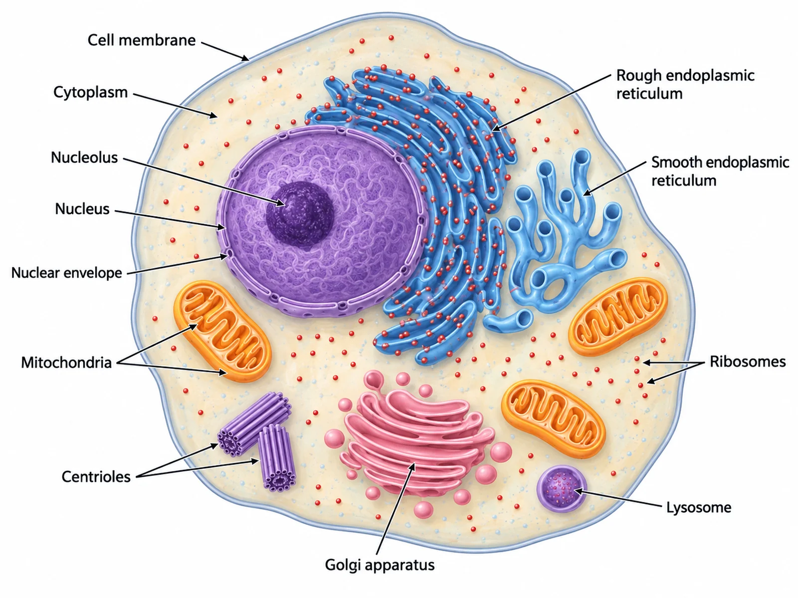
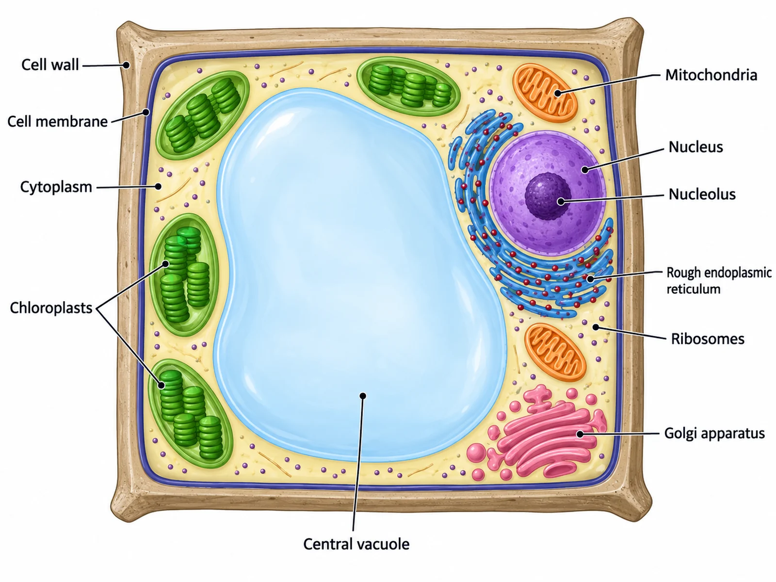
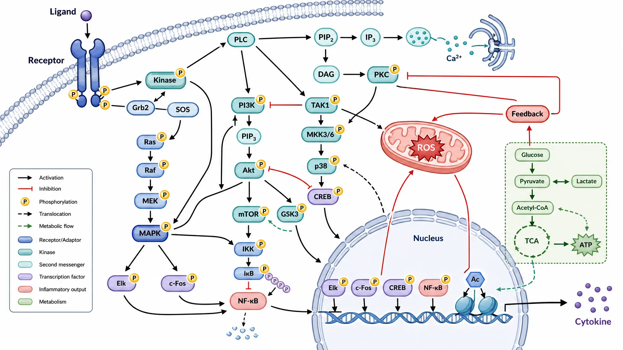
Generate animal and plant cell diagrams, pathway figures, mitosis sequences, and worksheet-ready biology illustrations with the same SciFigure AI workflow.