Plant vs Animal Cell AI

Plant and Animal Cell Comparison Maker

Compare plant and animal cells as a labeled Venn diagram, a side-by-side drawing, or a comparison table. Ask for a blank version and students can complete the comparison themselves.

Venn diagram or side by sideLabeled or blankShared and unique parts

AI Plant vs Animal Cell Diagram Maker

Describe the comparison format and which parts to label.

Costs 5 credits363 / 2000 characters

Generate a publication-ready PNG from your description.

Preview

Plant vs Animal Cell Example Previews

Target aspect ratio: 16:9

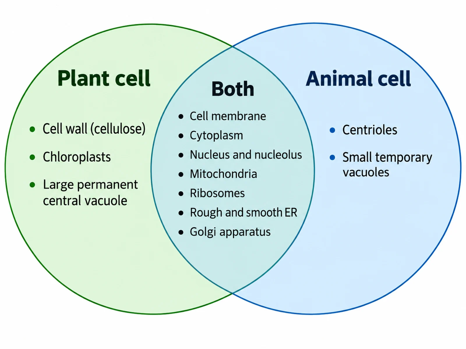
Labeled Venn diagram comparing plant and animal cells, with cell wall, chloroplasts, and central vacuole in the plant-only circle and shared organelles in the overlap
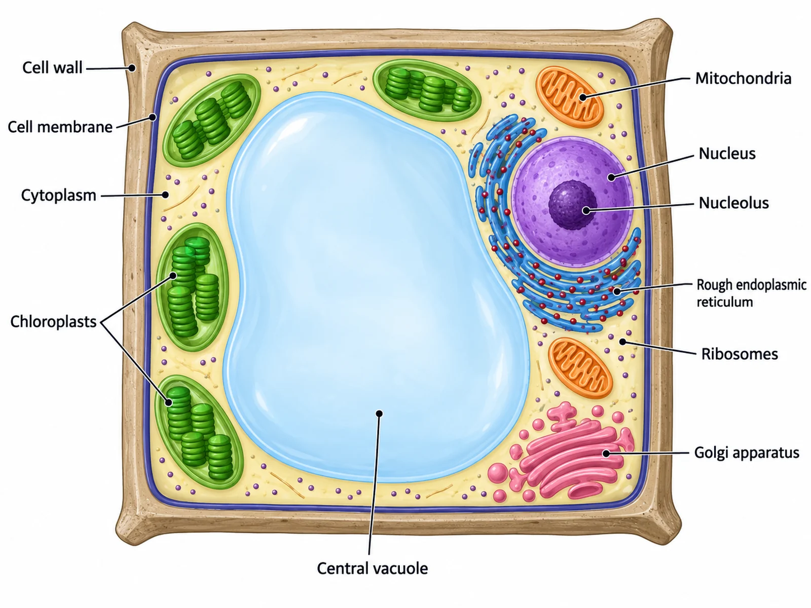
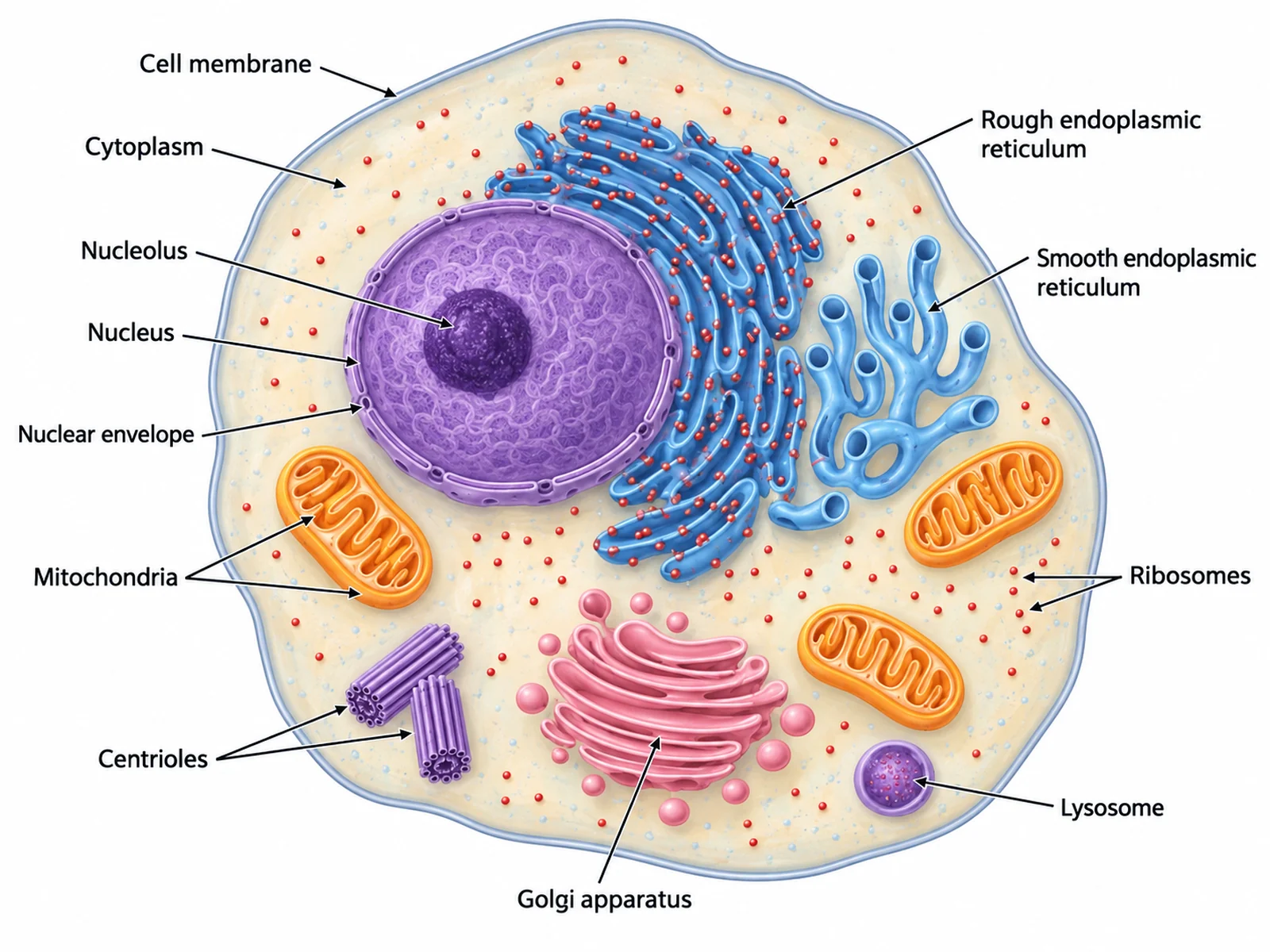
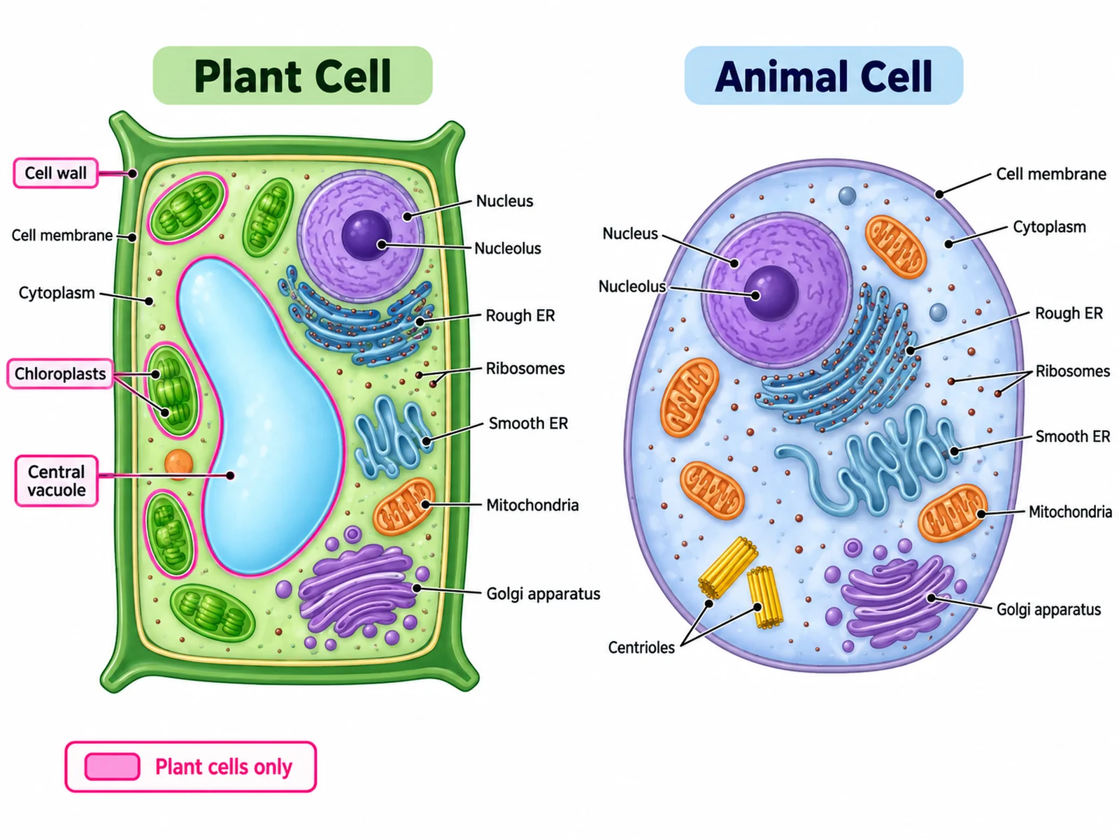
Side-by-side labeled plant cell and animal cell with the three plant-only structures highlighted
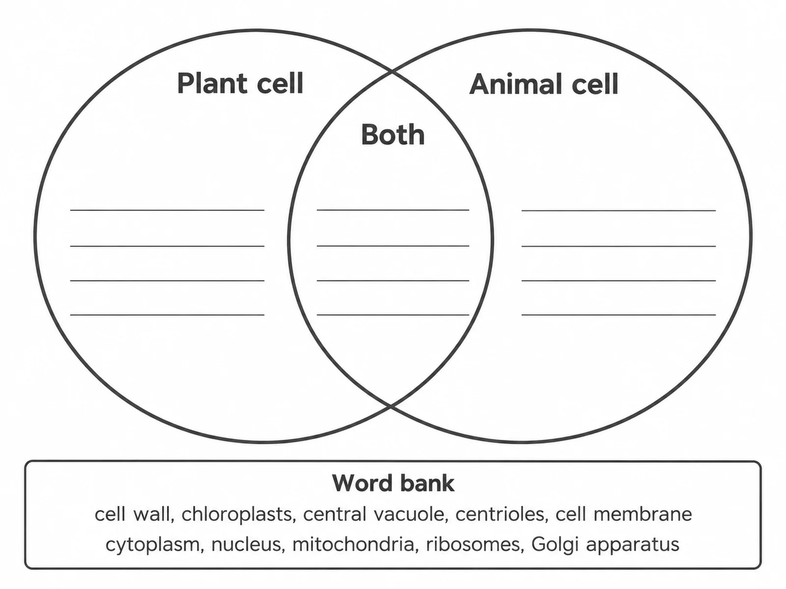
Blank plant and animal cell Venn diagram with empty circles and ruled lines for students to complete

Sample previews. Describe your comparison to generate your own figure.

Plant and Animal Cell Comparison Examples

Use these as visual directions for Venn diagrams, side-by-side labeled cells, comparison tables, and blank worksheets.

Labeled Venn diagram comparing plant and animal cells, with cell wall, chloroplasts, and central vacuole in the plant-only circle and shared organelles in the overlap

Prompt

Create a labeled plant and animal cell Venn diagram on a white background. Draw two large overlapping circles, the left one tinted pale green and headed "Plant cell", the right one tinted pale blue and headed "Animal cell". In the left crescent list "Cell wall (cellulose)", "Chloroplasts", and "Large permanent central vacuole". In the right crescent list "Centrioles" and "Small temporary vacuoles". In the central overlap, headed "Both", list "Cell membrane", "Cytoplasm", "Nucleus and nucleolus", "Mitochondria", "Ribosomes", "Rough and smooth ER", and "Golgi apparatus". Use clean dark sans-serif text and small bullet dots.
Plant vs Animal Venn Diagram

Side-by-side labeled plant cell and animal cell with the three plant-only structures highlighted

Prompt

Create a side-by-side comparison of a labeled plant cell and a labeled animal cell on a white background. Draw the boxy plant cell on the left with a thick outer cell wall, a large central vacuole, and green chloroplasts; draw the rounded animal cell on the right with a single thin membrane and a pair of centrioles. Label every organelle in both cells with thin leader lines. Outline the three plant-only structures — cell wall, chloroplasts, central vacuole — in a bright accent color and add a small legend reading "Plant cells only".

Side-by-Side Labeled Cells

Blank plant and animal cell Venn diagram with empty circles and ruled lines for students to complete

Prompt

Create a blank plant and animal cell Venn diagram worksheet. Draw two large empty overlapping circles with dark grey outlines, headed "Plant cell" on the left and "Animal cell" on the right, and "Both" over the overlap. Fill each region with three or four faint horizontal ruled writing lines and nothing else. Below the diagram, add a small word bank box headed "Word bank" containing, in two rows of plain text: "cell wall, chloroplasts, central vacuole, centrioles, cell membrane, cytoplasm, nucleus, mitochondria, ribosomes, Golgi apparatus". Generous white space, no answer key.

Blank Venn Worksheet

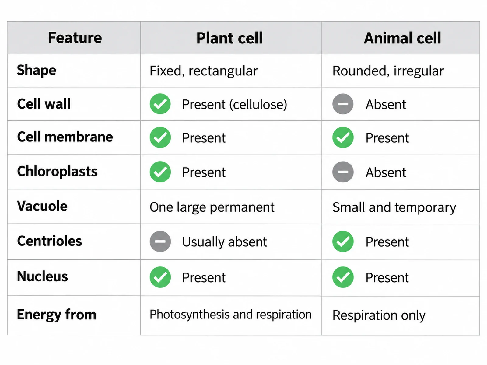
Comparison table of plant and animal cell features listing shape, cell wall, chloroplasts, vacuole, centrioles, and energy source

Prompt

Create a clean comparison table on a white background with three columns headed "Feature", "Plant cell", and "Animal cell", and a pale grey header row. Fill eight rows exactly: "Shape" — "Fixed, rectangular" / "Rounded, irregular"; "Cell wall" — "Present (cellulose)" / "Absent"; "Cell membrane" — "Present" / "Present"; "Chloroplasts" — "Present" / "Absent"; "Vacuole" — "One large permanent" / "Small and temporary"; "Centrioles" — "Usually absent" / "Present"; "Nucleus" — "Present" / "Present"; "Energy from" — "Photosynthesis and respiration" / "Respiration only". Use a green tick and a grey dash icon where a row is simply present or absent.

Comparison Table

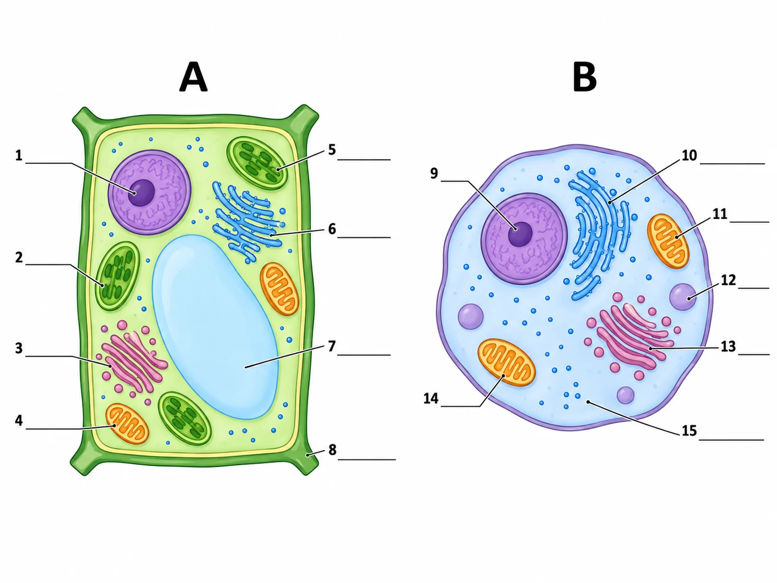
Blank unlabeled plant and animal cell drawings side by side with numbered empty label lines

Prompt

Create a blank side-by-side plant and animal cell worksheet. Draw both cells in clean line art with soft flat fills but no text: the boxy plant cell on the left, the rounded animal cell on the right. Draw thin leader lines from each organelle out to a short horizontal ruled blank line. Number the plant cell blanks 1 to 8 and the animal cell blanks 9 to 15. Head the left drawing "A" and the right drawing "B". Generous white space, no answer key.

Blank Side-by-Side Worksheet

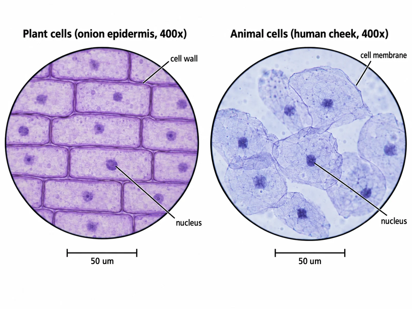
Microscope view comparing tessellating plant cells with thick walls and rounded overlapping animal cheek cells

Prompt

Create a two-panel microscope comparison on a white background in the style of a biology practical guide. The left panel, headed "Plant cells (onion epidermis, 400x)", shows a tessellating grid of rectangular cells with thick shared walls and one visible nucleus each. The right panel, headed "Animal cells (human cheek, 400x)", shows flat, irregular, overlapping cells with a thin membrane and one darkly stained nucleus each. Label "cell wall" and "nucleus" on the left and "cell membrane" and "nucleus" on the right. Add a scale bar reading "50 um" under each panel.

Both Cells Under the Microscope

How to compare plant and animal cells in a diagram

1

Pick the format

A Venn diagram for shared versus unique features, a side-by-side drawing for recognising organelles in place, or a table for revision notes.

2

Generate the comparison

The generator keeps the cell wall, chloroplasts, and central vacuole on the plant side and puts every shared organelle in the overlap.

3

Check the sorting, then export

Confirm no shared organelle has drifted into a single-cell region, then download the PNG for a worksheet, slide, or revision sheet.

What is a plant and animal cell Venn diagram?

A plant and animal cell Venn diagram sorts cell structures into three regions: features only plant cells have, features only animal cells have, and features both share. Because both are eukaryotic, the shared overlap is the largest region, which is the point of the exercise — the two cell types are far more alike than different, and only three structures truly separate them.

Plant cells only

  • Cell wall: a rigid cellulose layer outside the membrane that fixes the cell shape and resists turgor pressure.
  • Chloroplasts: green organelles holding chlorophyll, where photosynthesis converts light energy into glucose.
  • Large permanent central vacuole: a sap-filled sac that presses outward on the wall and keeps the cell turgid.

Animal cells only

  • Centrioles: paired cylinders that organise spindle fibres during cell division. Most plant cells divide without them.
  • Small temporary vacuoles: present in some animal cells for storage or transport, never one large permanent sac.
  • Rounded, flexible shape: with no wall, the membrane alone defines the outline, so the cell is irregular rather than boxy.

Shared by both

  • Cell membrane, cytoplasm, and cytoskeleton.
  • Nucleus with a nuclear envelope and nucleolus, holding the DNA.
  • Mitochondria, which release energy by aerobic respiration in both cell types.
  • Ribosomes, rough and smooth endoplasmic reticulum, and the Golgi apparatus for making and shipping proteins.

Plant vs Animal Cell Diagram FAQ

The plant-only circle holds the cell wall, chloroplasts, and the large permanent central vacuole. The animal-only circle holds centrioles and small temporary vacuoles, with a rounded shape rather than a fixed one. The shared overlap holds the cell membrane, cytoplasm, nucleus, nucleolus, mitochondria, ribosomes, rough and smooth endoplasmic reticulum, and the Golgi apparatus.

Need other biology figures?

Generate cell diagrams, membrane figures, mitosis and meiosis sequences, and worksheet-ready biology illustrations with the same SciFigure AI workflow.