Golgi Apparatus AI

Golgi Apparatus Diagram Generator

Draw a labeled Golgi apparatus with its cis and trans faces, stacked cisternae, and budding vesicles. Ask for a simple easy-to-draw version or a blank one for students to label.

Cis and trans facesSimple or detailedLabeled or blank

AI Golgi Apparatus Diagram Generator

Describe the level of detail and which parts to label.

Costs 5 credits390 / 2000 characters

Generate a publication-ready PNG from your description.

Preview

Golgi Apparatus Example Previews

Target aspect ratio: 4:3

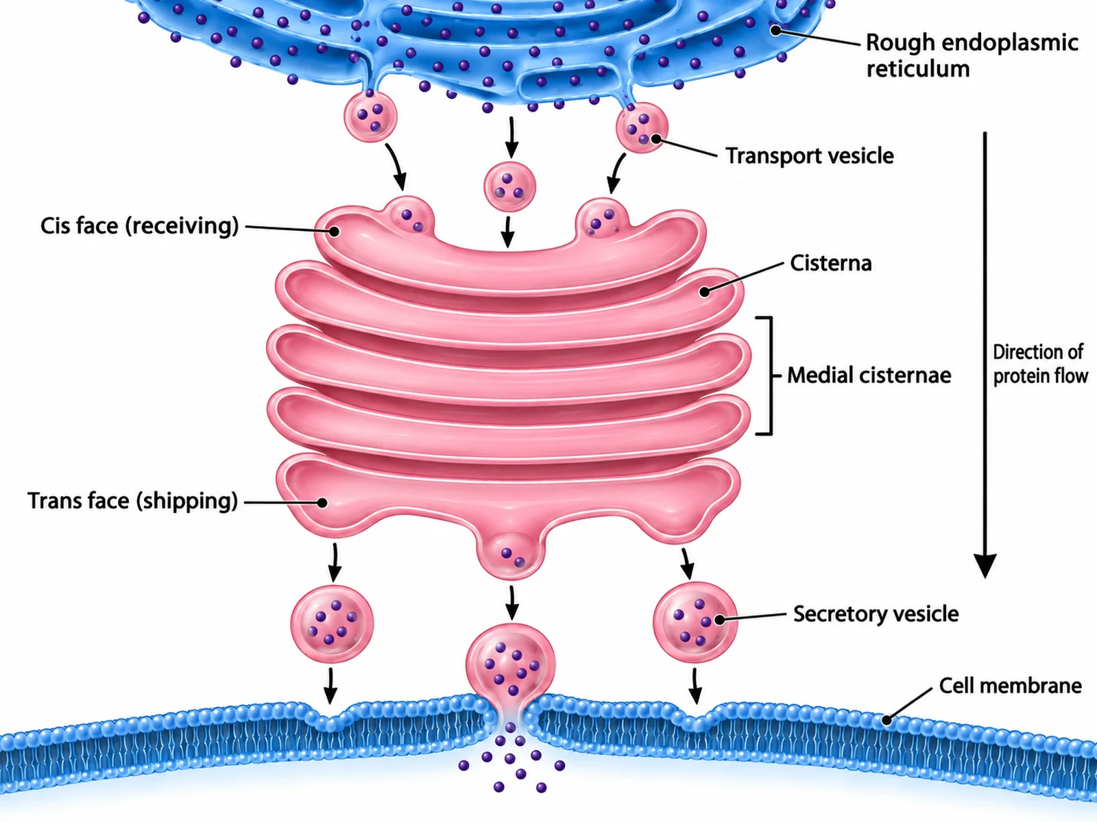
Labeled Golgi apparatus diagram naming the cis face, medial cisternae, trans face, cisternae, transport vesicles, and secretory vesicles
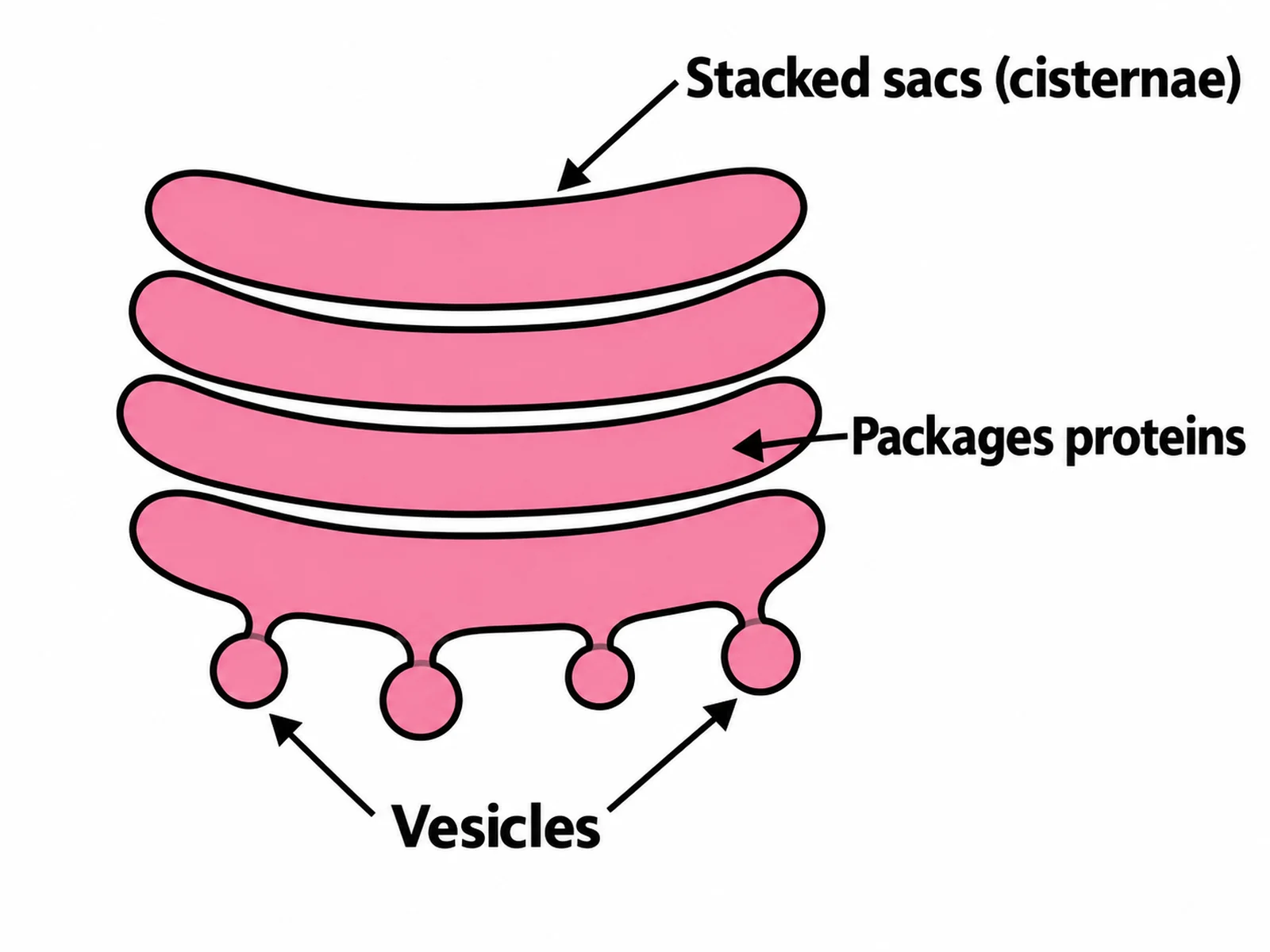
Simple easy-to-draw Golgi apparatus with stacked sacs and three basic labels
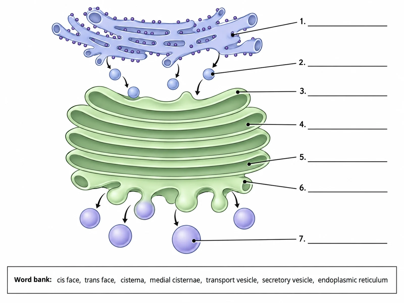
Blank unlabeled Golgi apparatus diagram with numbered empty label lines for students to complete

Sample previews. Describe your diagram to generate your own Golgi figure.

Golgi Apparatus Diagram Examples

Use these as visual directions for labeled stacks, simple drawings, blank worksheets, secretory pathway figures, and in-cell context views.

Labeled Golgi apparatus diagram naming the cis face, medial cisternae, trans face, cisternae, transport vesicles, and secretory vesicles

Prompt

Create a fully labeled Golgi apparatus diagram on a white background. Draw five flattened pink cisternae stacked in parallel, each slightly curved, all curving the same way so the stack has a convex upper side and a concave lower side. Label the convex upper side "Cis face (receiving)" with the rough endoplasmic reticulum drawn above it sending small "Transport vesicle" spheres toward it. Label the middle sacs "Medial cisternae" and one individual sac "Cisterna". Label the concave lower side "Trans face (shipping)" with larger "Secretory vesicle" spheres budding off and moving downward toward a cell membrane edge. Add a long thin arrow through the stack labeled "Direction of protein flow". Use thin dark leader lines and readable sans-serif text.
Labeled Golgi Apparatus

Simple easy-to-draw Golgi apparatus with stacked sacs and three basic labels

Prompt

Create a simple, easy-to-draw Golgi apparatus diagram for younger students. Draw four flattened sacs stacked like a pile of pita breads in a single flat colour with thick dark outlines, and three or four small circles budding off the bottom edge. Label only three things in large readable text: "Stacked sacs (cisternae)", "Vesicles", and "Packages proteins". Bold flat colours, plenty of white space, no ER, no cis or trans labels.

Simple Golgi Drawing

Blank unlabeled Golgi apparatus diagram with numbered empty label lines for students to complete

Prompt

Create a blank Golgi apparatus worksheet. Draw the same stack of five cisternae with the ER above and vesicles budding below, in clean line art with pale uniform fills and no text. Draw thin leader lines from each part out to a short horizontal ruled blank line, numbered 1 to 7. Add a word bank box below reading "cis face, trans face, cisterna, medial cisternae, transport vesicle, secretory vesicle, endoplasmic reticulum". Generous white space, no answer key.

Blank Golgi Worksheet

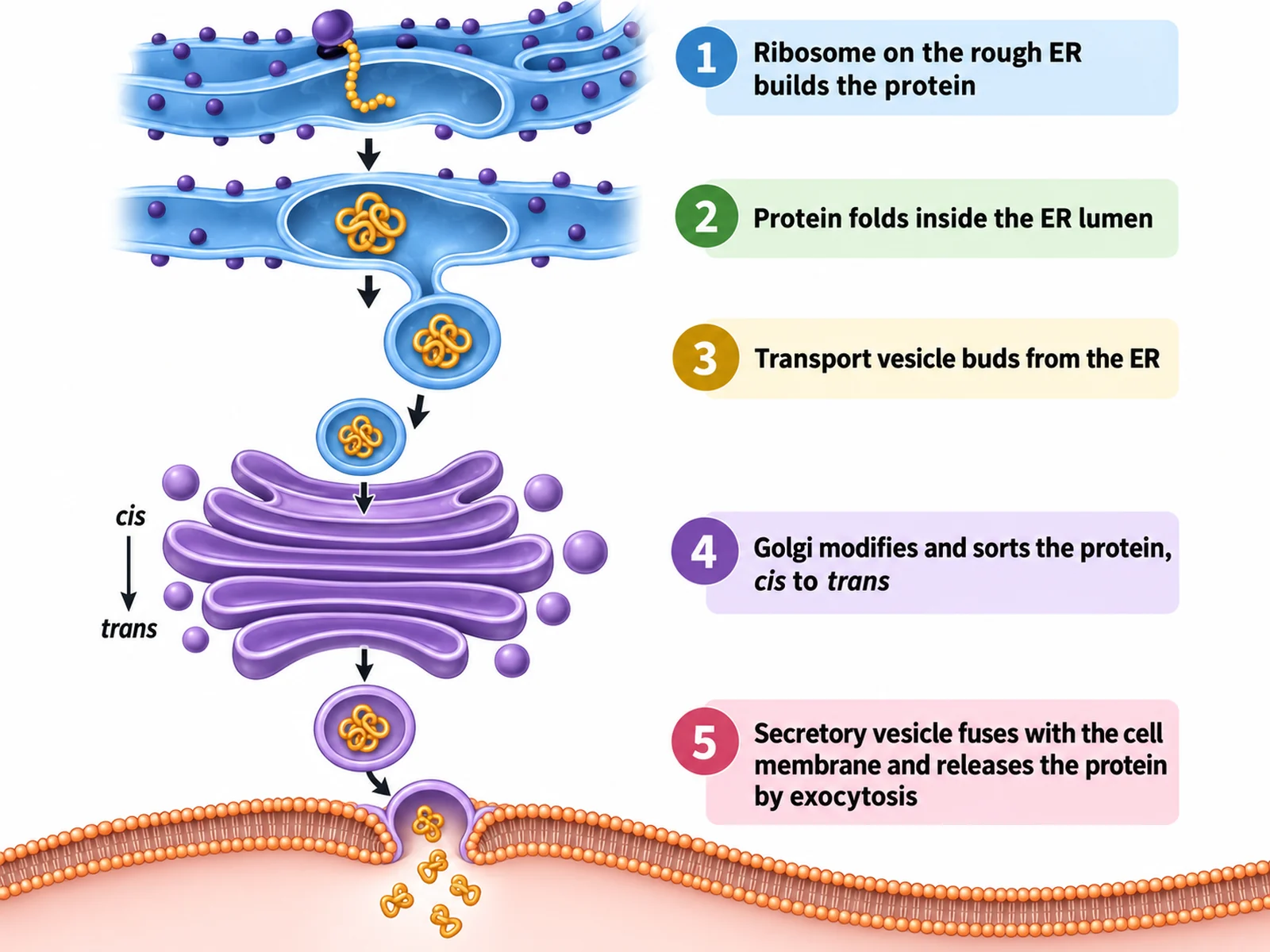
Diagram tracing a protein from the ribosome through the ER and Golgi to secretion at the cell membrane

Prompt

Create a labeled secretory pathway diagram on a white background, running top to bottom. Show five numbered stages with short captions: "1 Ribosome on the rough ER builds the protein", "2 Protein folds inside the ER lumen", "3 Transport vesicle buds from the ER", "4 Golgi modifies and sorts the protein, cis to trans", "5 Secretory vesicle fuses with the cell membrane and releases the protein by exocytosis". Draw the rough ER with ribosome dots at the top, the Golgi stack in the middle with an arrow through it, and the cell membrane at the bottom with a vesicle fusing and contents leaving the cell.

The Secretory Pathway

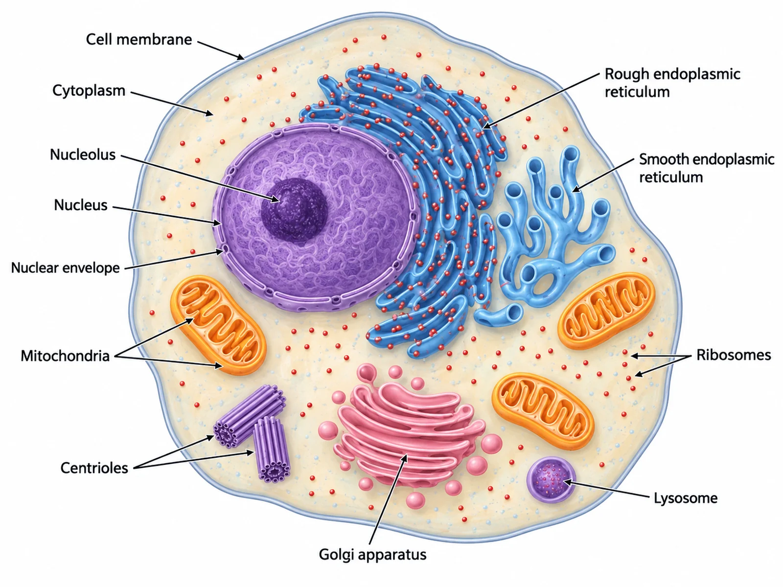
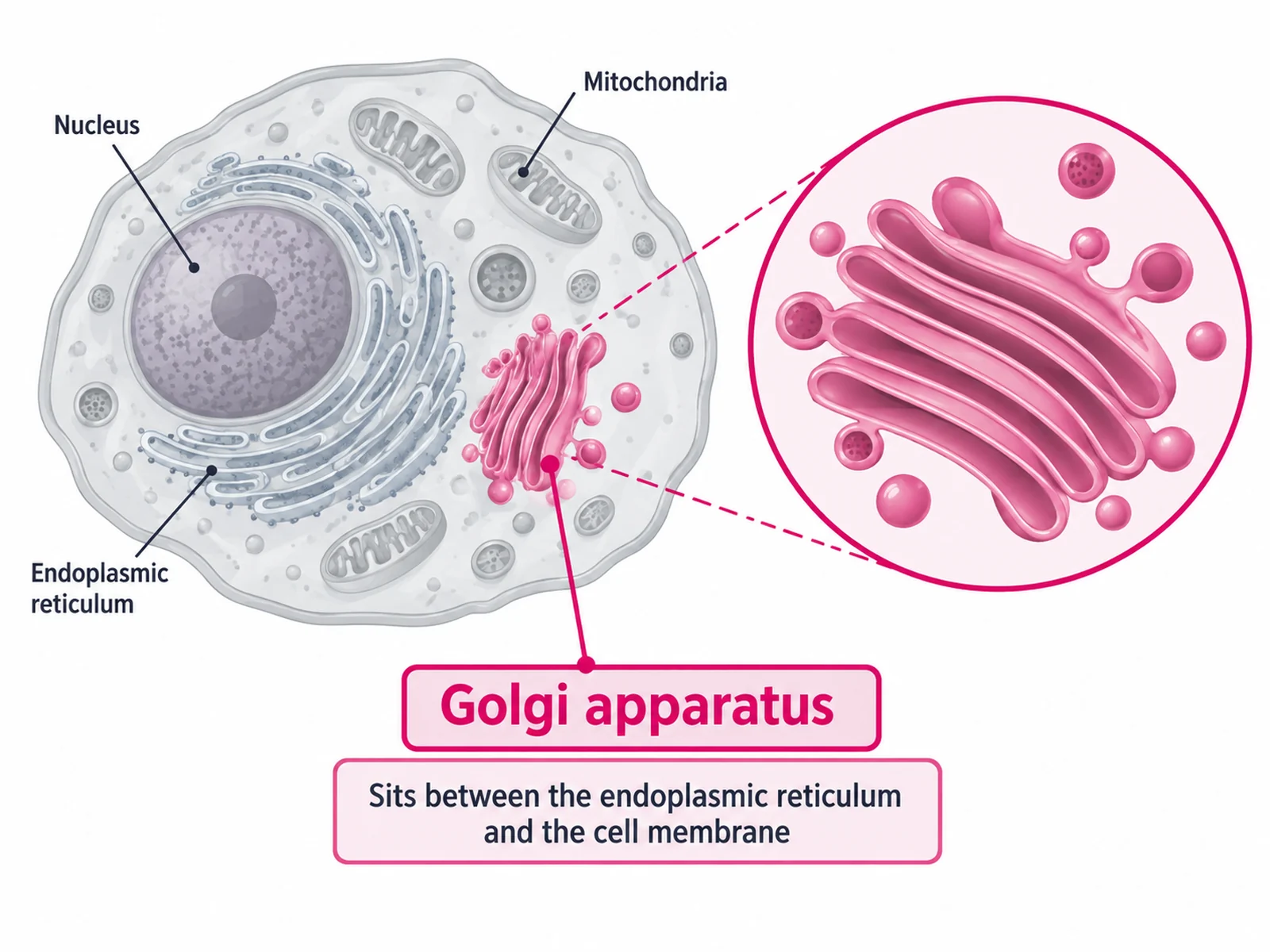
Animal cell diagram with the Golgi apparatus highlighted in position between the nucleus and the cell membrane

Prompt

Create a labeled animal cell diagram with the Golgi apparatus highlighted. Draw the whole rounded cell in muted greys and pale tones with the nucleus, mitochondria, and endoplasmic reticulum lightly labeled, then render the Golgi apparatus in full saturated pink with a bright outline and a magnified callout circle beside the cell showing its stacked cisternae and vesicles in detail. Label "Golgi apparatus" prominently and add a short caption reading "Sits between the endoplasmic reticulum and the cell membrane".

Golgi Inside the Cell

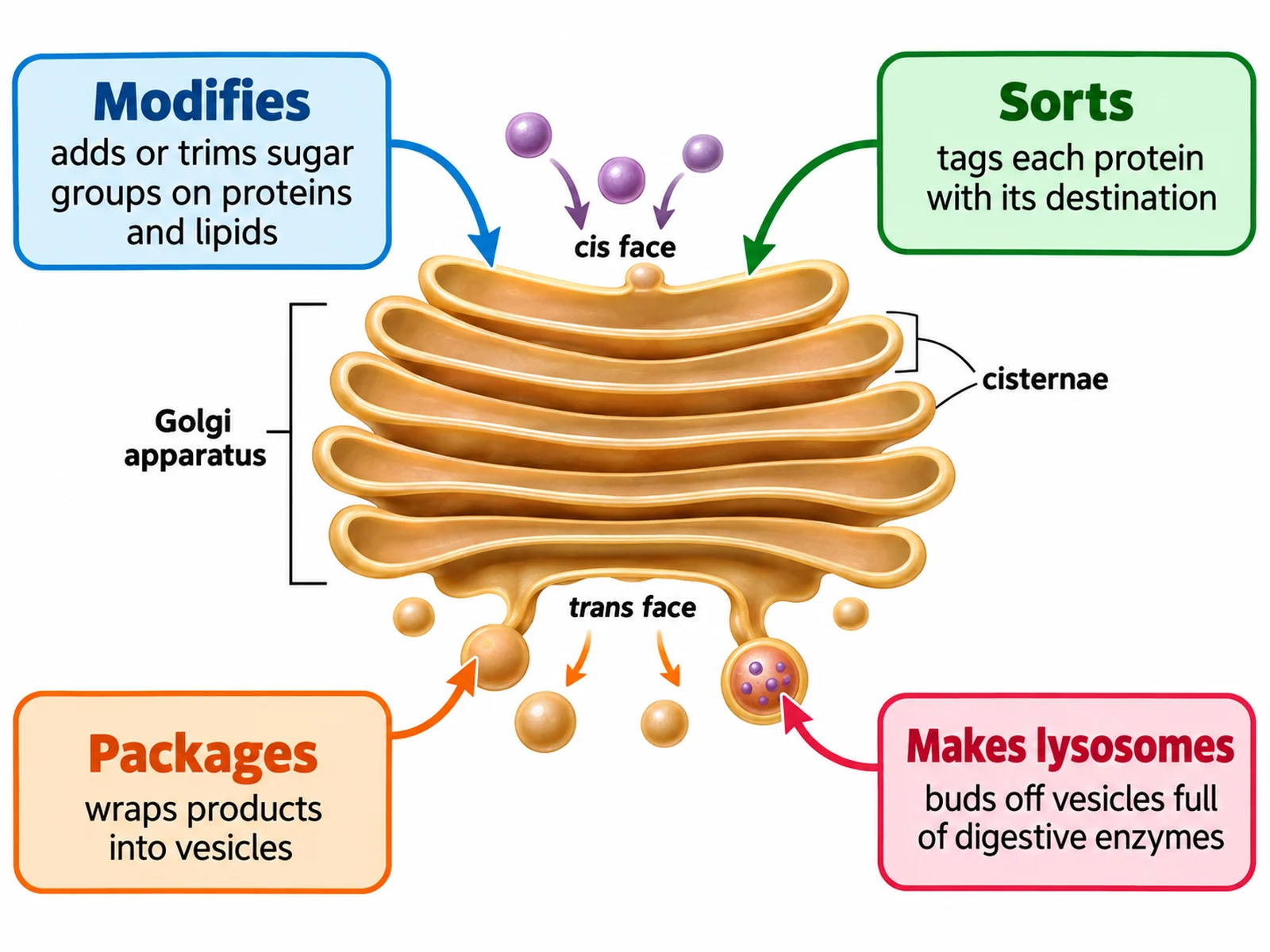
Golgi apparatus diagram with callout cards describing modification, sorting, packaging, and lysosome formation

Prompt

Create a Golgi apparatus function diagram. Draw the labeled Golgi stack in the centre and surround it with four rounded callout cards connected by thin leader lines, each with a bold heading and a one-line description: "Modifies" — "adds or trims sugar groups on proteins and lipids"; "Sorts" — "tags each protein with its destination"; "Packages" — "wraps products into vesicles"; "Makes lysosomes" — "buds off vesicles full of digestive enzymes". Point the first three cards at the cisternae and the last at a vesicle leaving the trans face.

Golgi Functions

How to draw a labeled Golgi apparatus

1

Choose the detail level

A simple stack of sacs for younger classes, a full labeled organelle with cis and trans faces, or the whole secretory pathway from ribosome to cell membrane.

2

Generate the diagram

The generator stacks the cisternae correctly, keeps the cis face toward the ER, and buds vesicles from the trans face.

3

Check the orientation, then export

Confirm the cis face points at the endoplasmic reticulum and the trans face toward the membrane, then download the PNG.

What is the Golgi apparatus?

The Golgi apparatus is a stack of flattened membrane sacs, called cisternae, that finishes and dispatches the proteins and lipids made by the endoplasmic reticulum. It is the sorting office of the cell: material enters at one face, is chemically modified as it moves through the stack, and leaves at the other face in vesicles addressed to a specific destination. Cells that secrete heavily, such as pancreatic and goblet cells, have especially large Golgi stacks.

Parts of the Golgi apparatus

  • Cis face: the convex receiving side facing the endoplasmic reticulum, where transport vesicles fuse.
  • Cisternae: the flattened membrane sacs themselves, usually four to eight in a stack.
  • Medial cisternae: the middle sacs where most sugar modification takes place.
  • Trans face: the concave shipping side facing the cell membrane, where vesicles bud off.
  • Transport vesicles: small spheres carrying material from the ER to the cis face.
  • Secretory vesicles: larger spheres leaving the trans face for the membrane, a lysosome, or export.

What the Golgi does, step by step

  • Receives: transport vesicles from the rough ER fuse with the cis face and empty their contents.
  • Modifies: enzymes in each cisterna add, trim, or alter sugar chains as the protein moves through.
  • Sorts: molecular tags mark each product with its destination.
  • Packages: the trans face buds off vesicles containing the finished product.
  • Dispatches: vesicles travel to the cell membrane for secretion, become lysosomes, or deliver to other organelles.

Golgi Apparatus Diagram FAQ

The cis face (receiving side, nearest the endoplasmic reticulum), the medial cisternae in the middle, the trans face (shipping side), the flattened membrane sacs called cisternae, transport vesicles arriving from the ER, and secretory vesicles budding off the trans face. A fuller diagram also labels the cis-Golgi network and trans-Golgi network.

Need other biology figures?

Generate organelle close-ups, cell diagrams, pathway figures, and worksheet-ready illustrations with the same SciFigure AI workflow.