Meiosis Diagram AI

Meiosis Diagram Generator

Draw a labeled meiosis diagram covering meiosis I and meiosis II, crossing over, and every phase. Ask for a blank worksheet and students can label the stages themselves.

Meiosis I and IICrossing over shownLabeled or blank

AI Meiosis Diagram Generator

Describe which meiosis stages to show and the style you need.

Costs 5 credits368 / 2000 characters

Generate a publication-ready PNG from your description.

Preview

Meiosis Diagram Example Previews

Target aspect ratio: 16:9

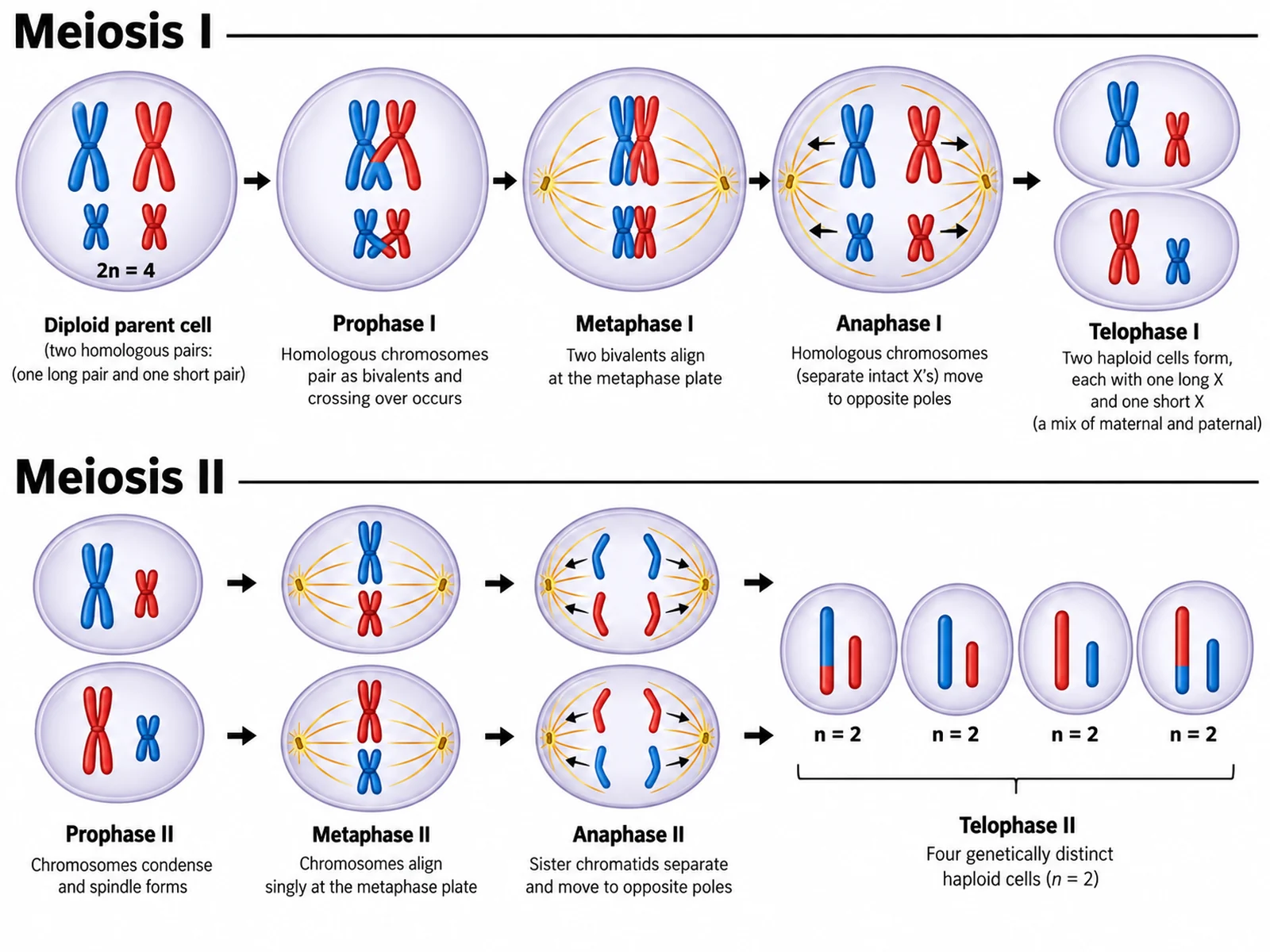
Labeled meiosis diagram showing the full sequence from a diploid parent cell through meiosis I and meiosis II to four haploid daughter cells
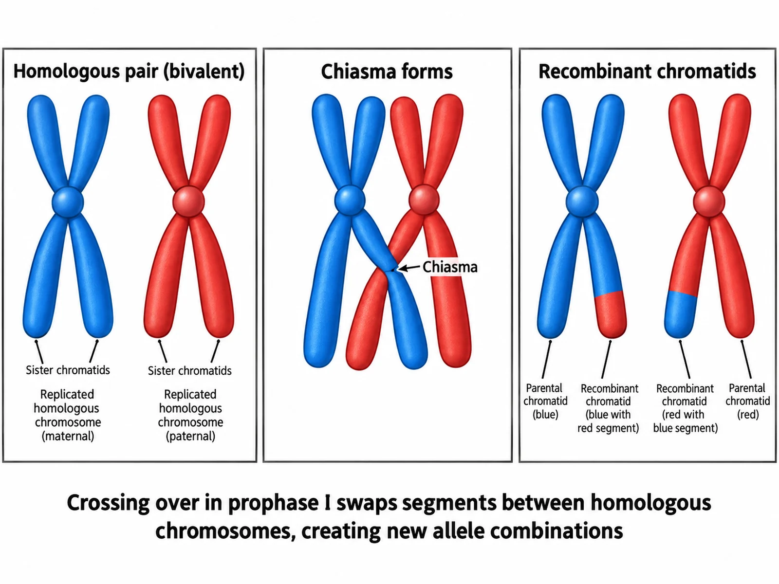
Magnified diagram of crossing over in prophase I showing a bivalent, chiasma, and recombinant chromatids
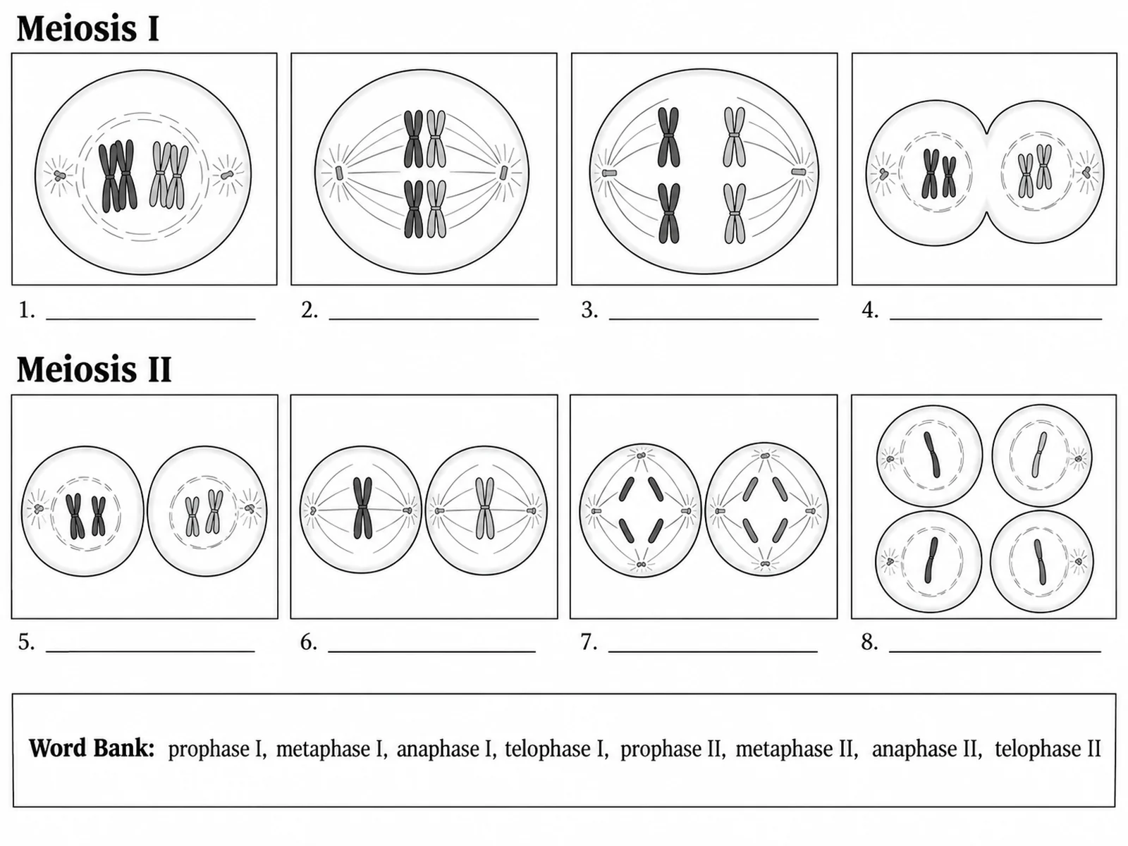
Blank meiosis worksheet with eight empty phase panels and ruled label lines for students to complete

Sample previews. Describe your diagram to generate your own meiosis figure.

Meiosis Diagram Examples

Use these as visual directions for the full sequence, crossing over close-ups, stage-by-stage breakdowns, mitosis comparisons, and blank worksheets.

Labeled meiosis diagram showing the full sequence from a diploid parent cell through meiosis I and meiosis II to four haploid daughter cells

Prompt

Create a fully labeled meiosis overview diagram on a white background. Draw the complete sequence left to right in two rows. Row one, headed "Meiosis I", shows a diploid parent cell with 2n = 4 (two blue chromosomes from one parent, two red from the other), then prophase I with paired bivalents and visible crossing over, metaphase I with pairs aligned on the plate, anaphase I with whole homologues moving apart each keeping two chromatids, and telophase I with two cells. Row two, headed "Meiosis II", shows prophase II, metaphase II with chromosomes aligned singly, anaphase II with sister chromatids separating, and telophase II ending in four small haploid cells with mixed colours. Label every phase beneath its cell and add "2n = 4" at the start and "n = 2" at the end.
Meiosis Overview

Magnified diagram of crossing over in prophase I showing a bivalent, chiasma, and recombinant chromatids

Prompt

Create a magnified labeled diagram of crossing over on a white background. Show three panels left to right. Panel 1: two homologous chromosomes, one solid blue and one solid red, each with two sister chromatids, pairing up, labeled "Homologous pair (bivalent)". Panel 2: the same pair with two non-sister chromatids overlapping at an X-shaped junction, labeled "Chiasma forms". Panel 3: the chromosomes separated, with two chromatids now part blue and part red, labeled "Recombinant chromatids". Add a caption beneath reading "Crossing over in prophase I swaps segments between homologous chromosomes, creating new allele combinations".

Crossing Over

Blank meiosis worksheet with eight empty phase panels and ruled label lines for students to complete

Prompt

Create a blank meiosis worksheet. Draw eight panels in two rows of four, each containing a cell outline with chromosomes drawn in clean line art and no text. Beneath each panel place a short horizontal ruled blank line, numbered 1 to 8. Head the first row "Meiosis I" and the second row "Meiosis II" but leave no phase names. Add a word bank box below reading "prophase I, metaphase I, anaphase I, telophase I, prophase II, metaphase II, anaphase II, telophase II". Generous white space, no answer key.

Blank Meiosis Worksheet

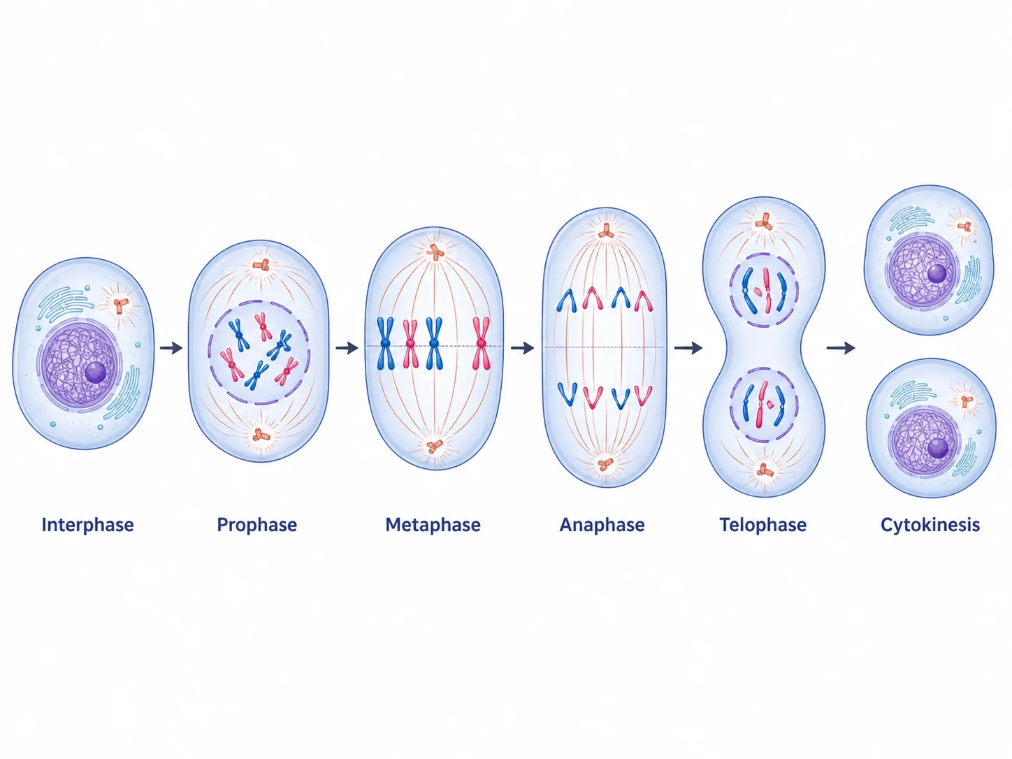
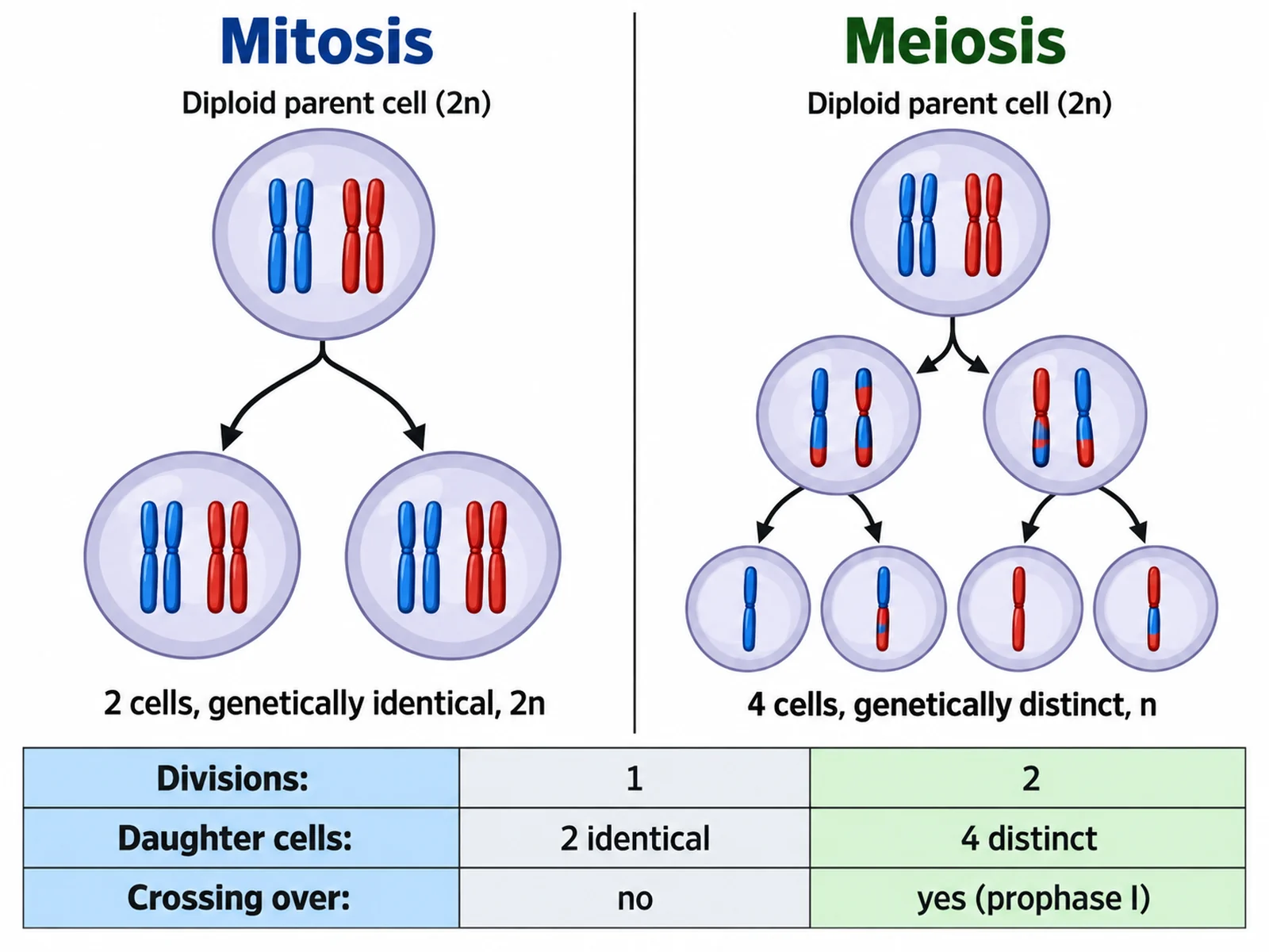
Side-by-side comparison of mitosis producing two identical diploid cells and meiosis producing four distinct haploid cells

Prompt

Create a side-by-side comparison of mitosis and meiosis on a white background. On the left, headed "Mitosis", show one diploid parent cell dividing once into two identical diploid daughter cells, with a caption "2 cells, genetically identical, 2n". On the right, headed "Meiosis", show one diploid parent cell dividing twice into four haploid cells with mixed chromosome colours, captioned "4 cells, genetically distinct, n". Below, add a three-row comparison strip reading "Divisions: 1 vs 2", "Daughter cells: 2 identical vs 4 distinct", and "Crossing over: no vs yes (prophase I)".

Meiosis vs Mitosis

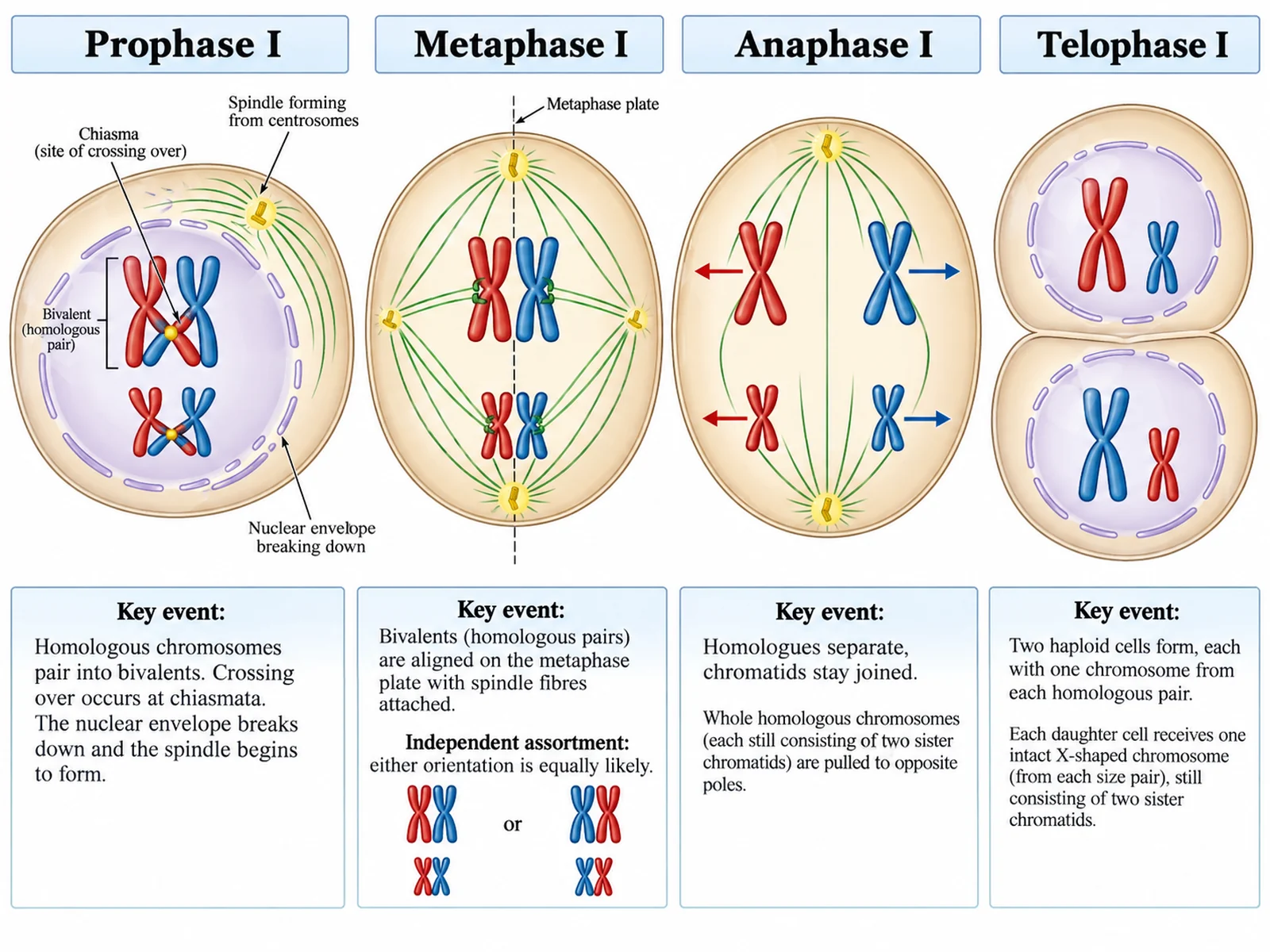
Detailed labeled diagram of prophase I, metaphase I, anaphase I, and telophase I with chromosome behaviour annotated

Prompt

Create a detailed labeled diagram of meiosis I on a white background, four large panels left to right. "Prophase I": homologous chromosomes pair into bivalents, crossing over at chiasmata, nuclear envelope breaking down, spindle forming. "Metaphase I": bivalents aligned on the metaphase plate with spindle fibres attached, annotated "independent assortment: either orientation is equally likely". "Anaphase I": whole homologous chromosomes pulled to opposite poles, each still holding two sister chromatids, annotated "homologues separate, chromatids stay joined". "Telophase I": two cells forming, each with one chromosome from each pair. Label each panel and annotate the key event beneath it.

Meiosis I Stages

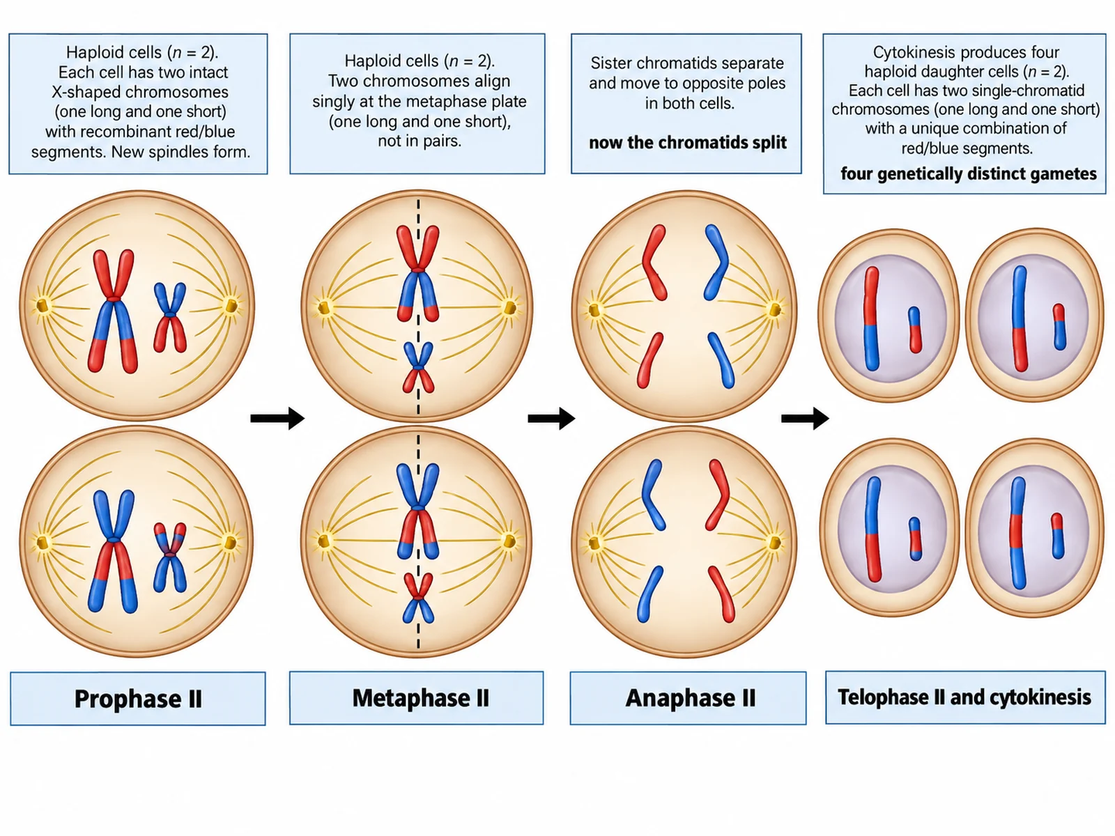
Detailed labeled diagram of prophase II, metaphase II, anaphase II, and telophase II ending in four haploid gametes

Prompt

Create a detailed labeled diagram of meiosis II on a white background, four large panels left to right. "Prophase II": chromosomes recondense in each of the two haploid cells, new spindles form. "Metaphase II": chromosomes align singly on the plate, not in pairs. "Anaphase II": sister chromatids separate and move to opposite poles, annotated "now the chromatids split". "Telophase II and cytokinesis": four haploid daughter cells form, each with a different combination of coloured chromosome segments, annotated "four genetically distinct gametes". Label each panel beneath its cells.

Meiosis II Stages

How to draw a labeled meiosis diagram

1

Choose the scope

The full eight-phase sequence, meiosis I or II alone, a crossing over close-up, or a mitosis comparison. Say whether you want labels or blank lines.

2

Generate the diagram

The generator keeps homologues separating in anaphase I and chromatids separating in anaphase II, and shows crossing over in prophase I.

3

Check the chromosomes, then export

Confirm anaphase I separates whole homologues, not chromatids, and that the result is four cells rather than two, then download the PNG.

What is a meiosis diagram?

A meiosis diagram traces one diploid cell through two rounds of division into four haploid cells. It matters because it explains two separate things at once: how chromosome number is halved so fertilisation restores it, and how genetic variation is generated through crossing over and independent assortment. Most diagrams use 2n = 4 and two colours so both ideas stay visible.

The phases of meiosis

  • Prophase I: chromosomes condense, homologues pair into bivalents, and crossing over exchanges segments at chiasmata.
  • Metaphase I: bivalents line up on the metaphase plate; their random orientation is independent assortment.
  • Anaphase I: homologous chromosomes are pulled apart; sister chromatids stay joined at the centromere.
  • Telophase I and cytokinesis: two haploid cells form, each with one chromosome from every pair.
  • Prophase II: chromosomes recondense and new spindles form in both cells.
  • Metaphase II: chromosomes align singly on the plate, not as pairs.
  • Anaphase II: sister chromatids finally separate and move to opposite poles.
  • Telophase II and cytokinesis: four genetically distinct haploid cells form.

Where genetic variation comes from

  • Crossing over in prophase I: homologues swap segments, producing recombinant chromatids.
  • Independent assortment in metaphase I: each bivalent orients randomly, so maternal and paternal chromosomes mix.
  • Random fertilisation: any gamete can fuse with any other, multiplying the combinations again.

Meiosis Diagram Generator FAQ

Meiosis I: prophase I (homologous chromosomes pair and cross over), metaphase I (pairs line up on the plate), anaphase I (homologues separate), telophase I and cytokinesis. Meiosis II: prophase II, metaphase II, anaphase II (sister chromatids separate), telophase II and cytokinesis, producing four genetically distinct haploid cells.

Need other biology figures?

Generate cell division sequences, cell diagrams, genetics figures, and worksheet-ready illustrations with the same SciFigure AI workflow.