Heart Diagram AI

Heart Diagram Generator

Generate a labeled human heart with its four chambers, four valves, and the great vessels named, or a blank heart diagram with numbered lines for students to label. Describe the detail level you need, then download a clean figure for worksheets, revision notes, and slides.

Labeled or blankChambers and valvesBlood flow marked

AI Heart Diagram Generator

Describe the detail level and whether you want labels or blank lines.

Costs 5 credits466 / 2000 characters

Generate a publication-ready PNG from your description.

Preview

Heart Diagram Example Previews

Target aspect ratio: 4:3

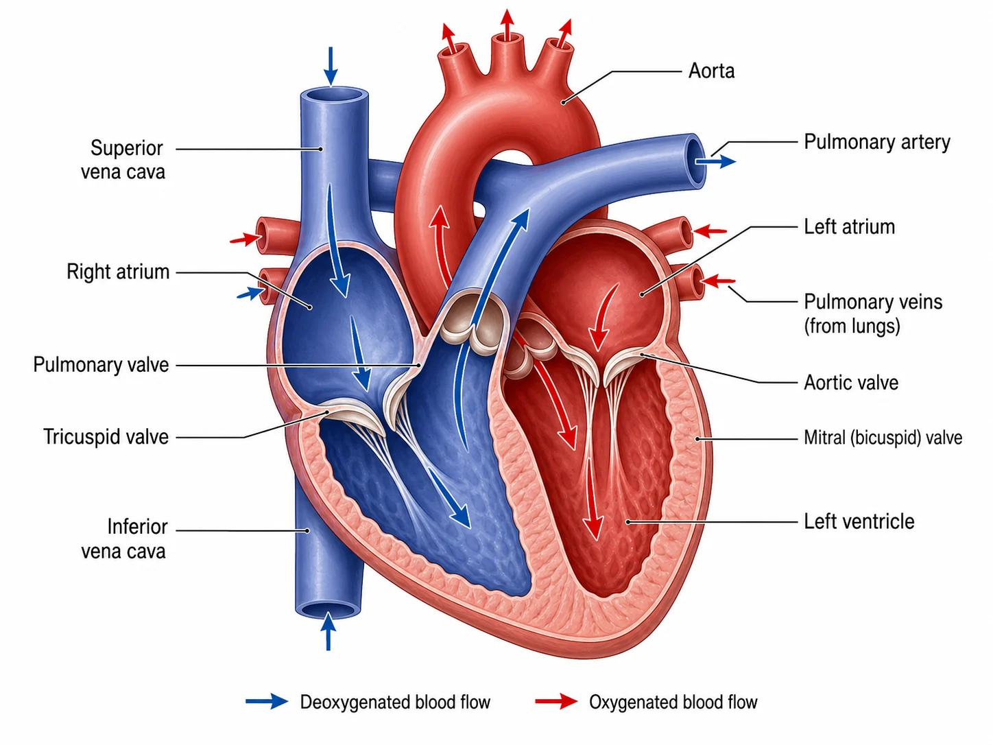
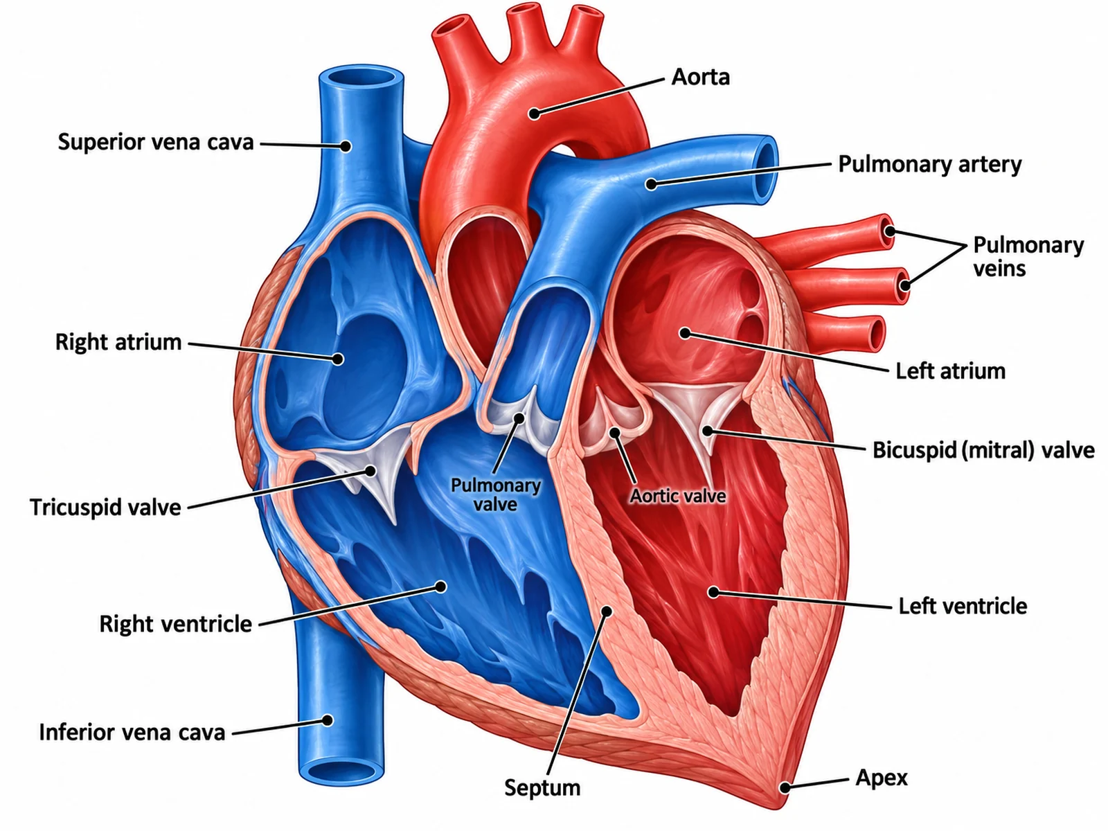
Labeled human heart diagram naming the four chambers, the tricuspid, pulmonary, mitral, and aortic valves, the vena cavae, pulmonary artery, pulmonary veins, aorta, septum, and apex
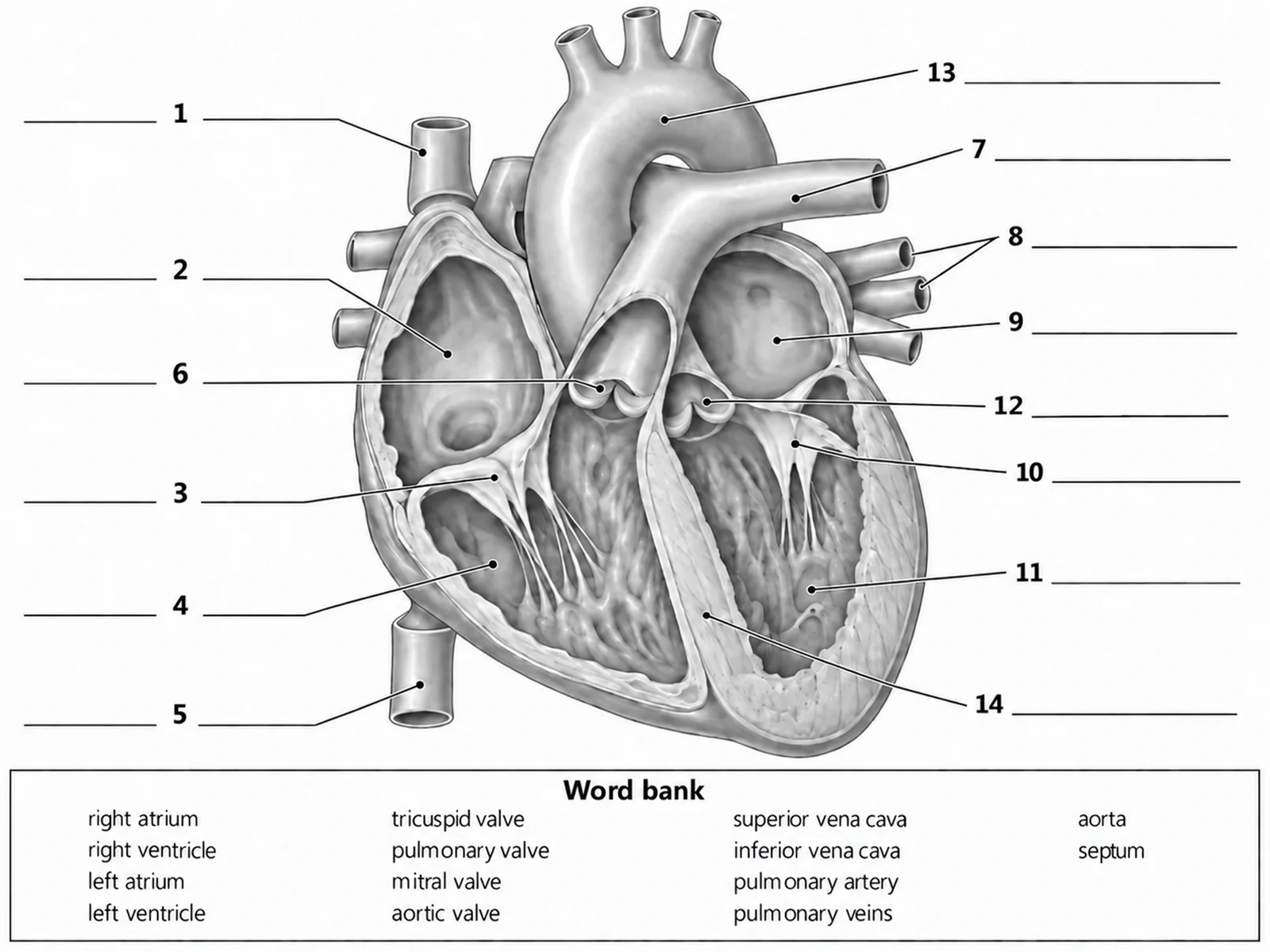
Blank unlabeled heart diagram with numbered empty label lines and a word bank for students to complete
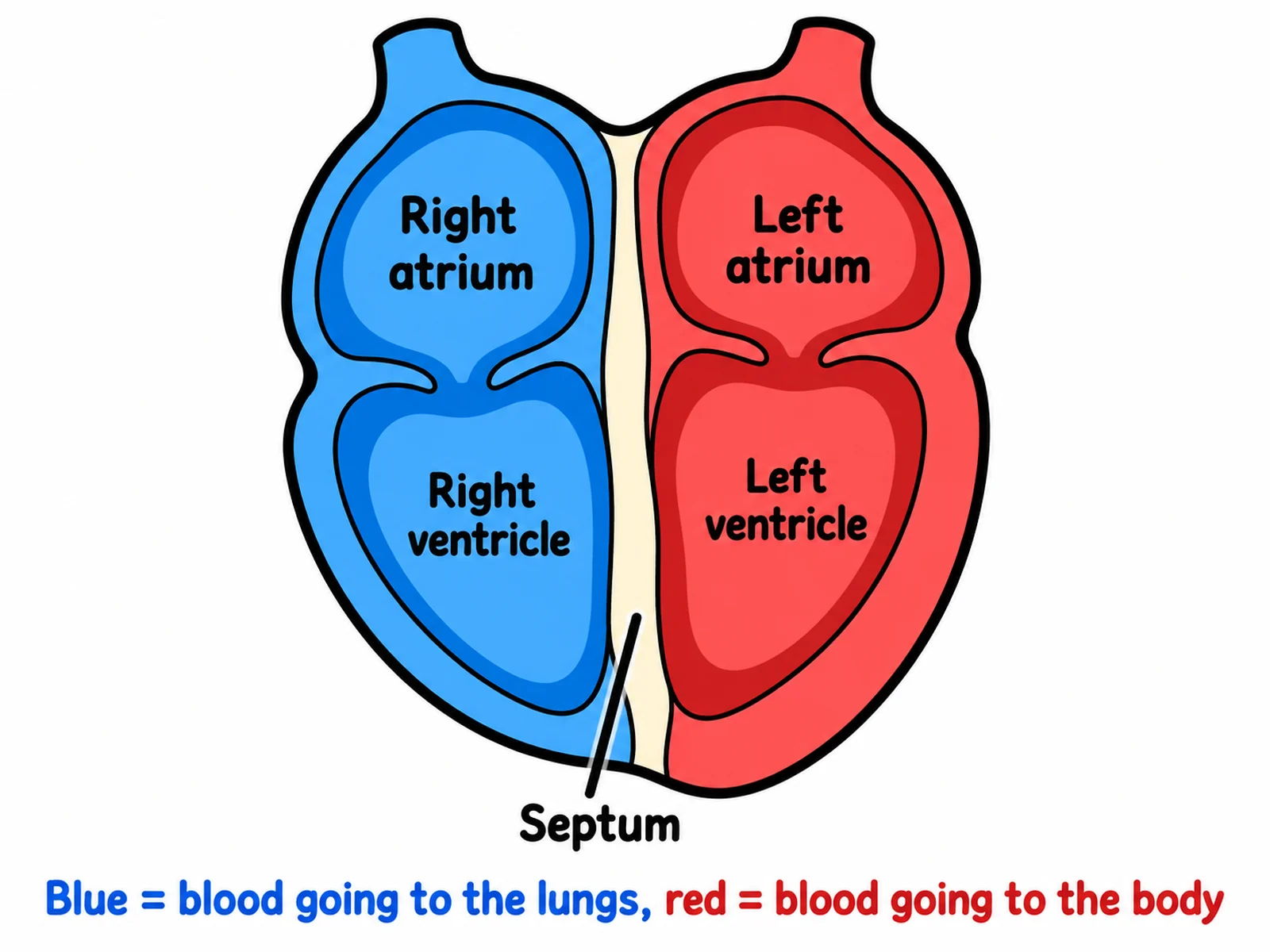
Simple easy-to-draw heart diagram for younger students labeling only the four chambers and the septum

Sample previews. Describe your diagram to generate your own heart figure.

Labeled and Blank Heart Diagram Examples

Use these as visual directions for labeled anatomy, blank worksheets with numbered lines, simple versions for younger classes, blood flow figures, valve close-ups, and double circulation.

Labeled human heart diagram naming the four chambers, the tricuspid, pulmonary, mitral, and aortic valves, the vena cavae, pulmonary artery, pulmonary veins, aorta, septum, and apex

Prompt

Create a fully labeled human heart diagram on a white background, drawn as an anterior view cut away so all four chambers are open. Because the view is from the front of the patient, place the right atrium and right ventricle on the VIEWER’S LEFT in blue, and the left atrium and left ventricle on the viewer’s right in red, with the left ventricle wall clearly thicker. Label right atrium, right ventricle, left atrium, left ventricle, tricuspid valve, pulmonary valve, bicuspid (mitral) valve, aortic valve, superior vena cava, inferior vena cava, pulmonary artery, pulmonary veins, aorta, septum, and apex. Colour the pulmonary artery blue and the pulmonary veins red. Use thin dark leader lines and readable sans-serif text.
Labeled Human Heart

Blank unlabeled heart diagram with numbered empty label lines and a word bank for students to complete

Prompt

Create a blank heart diagram worksheet. Draw the same anterior cut-away heart in clean line art with pale uniform grey fills and no colour coding, so nothing reveals which side is oxygenated, and keep the anatomy exact: the patient’s right chambers on the VIEWER’S LEFT, the left ventricle wall thickest. Include no text anywhere on the figure. Draw thin leader lines from each part out to a short horizontal ruled blank line, numbered 1 to 14. Add a word bank box below reading "right atrium, right ventricle, left atrium, left ventricle, tricuspid valve, pulmonary valve, mitral valve, aortic valve, superior vena cava, inferior vena cava, pulmonary artery, pulmonary veins, aorta, septum". Generous white space, no answer key.

Blank Heart Diagram to Label

Simple easy-to-draw heart diagram for younger students labeling only the four chambers and the septum

Prompt

Create a simple, easy-to-draw heart diagram for younger students. Draw the heart as a rounded cut-away shape in two bold flat colours with thick dark outlines: the two right chambers blue on the VIEWER’S LEFT, the two left chambers red on the viewer’s right, separated by a clear central septum. Label only five things in large readable text: "Right atrium", "Right ventricle", "Left atrium", "Left ventricle", and "Septum", plus one short caption reading "Blue = blood going to the lungs, red = blood going to the body". Omit the valves and the vessel names, and leave plenty of white space.

Simple Heart Drawing

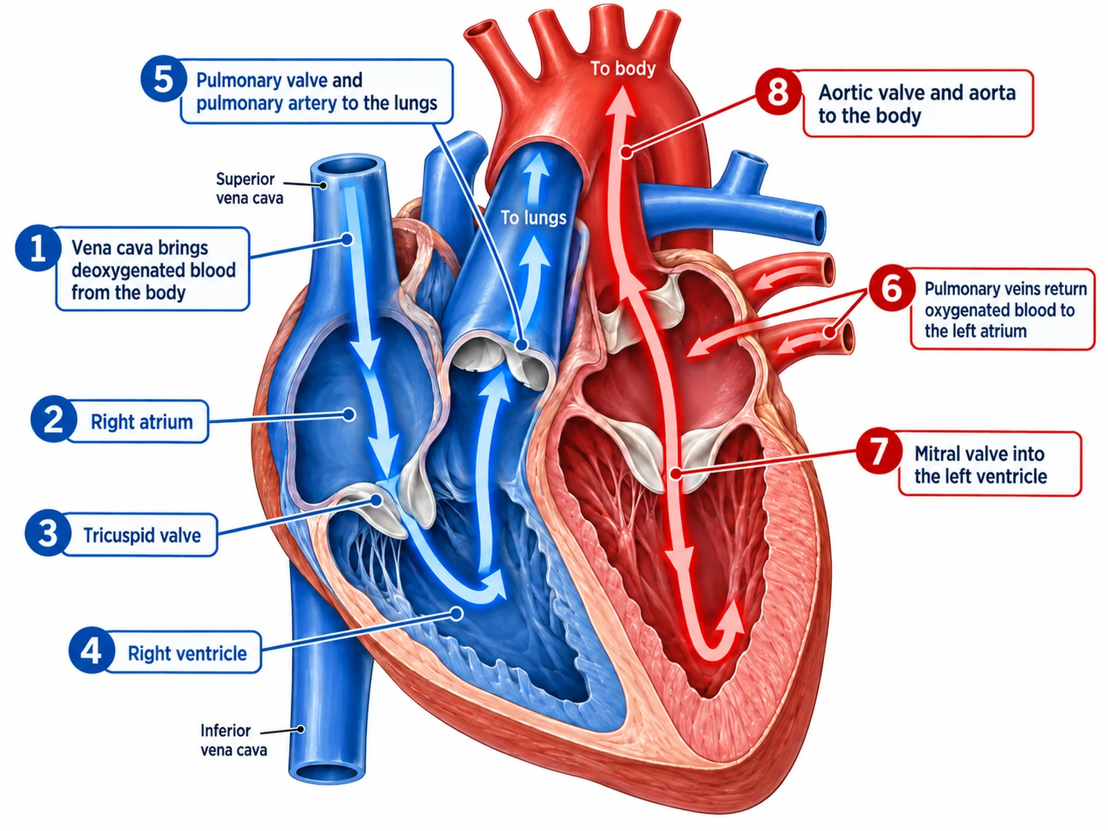
Heart diagram tracing the numbered path of blood from the vena cava through both sides of the heart to the aorta

Prompt

Create a labeled blood flow diagram of the human heart on a white background, using thick numbered arrows drawn through an anterior cut-away heart. Number eight stages in order with short captions: "1 Vena cava brings deoxygenated blood from the body", "2 Right atrium", "3 Tricuspid valve", "4 Right ventricle", "5 Pulmonary valve and pulmonary artery to the lungs", "6 Pulmonary veins return oxygenated blood to the left atrium", "7 Mitral valve into the left ventricle", "8 Aortic valve and aorta to the body". Draw stages 1 to 5 in blue on the VIEWER’S LEFT, stages 6 to 8 in red on the viewer’s right, and colour the pulmonary artery blue and the pulmonary veins red.

Blood Flow Through the Heart

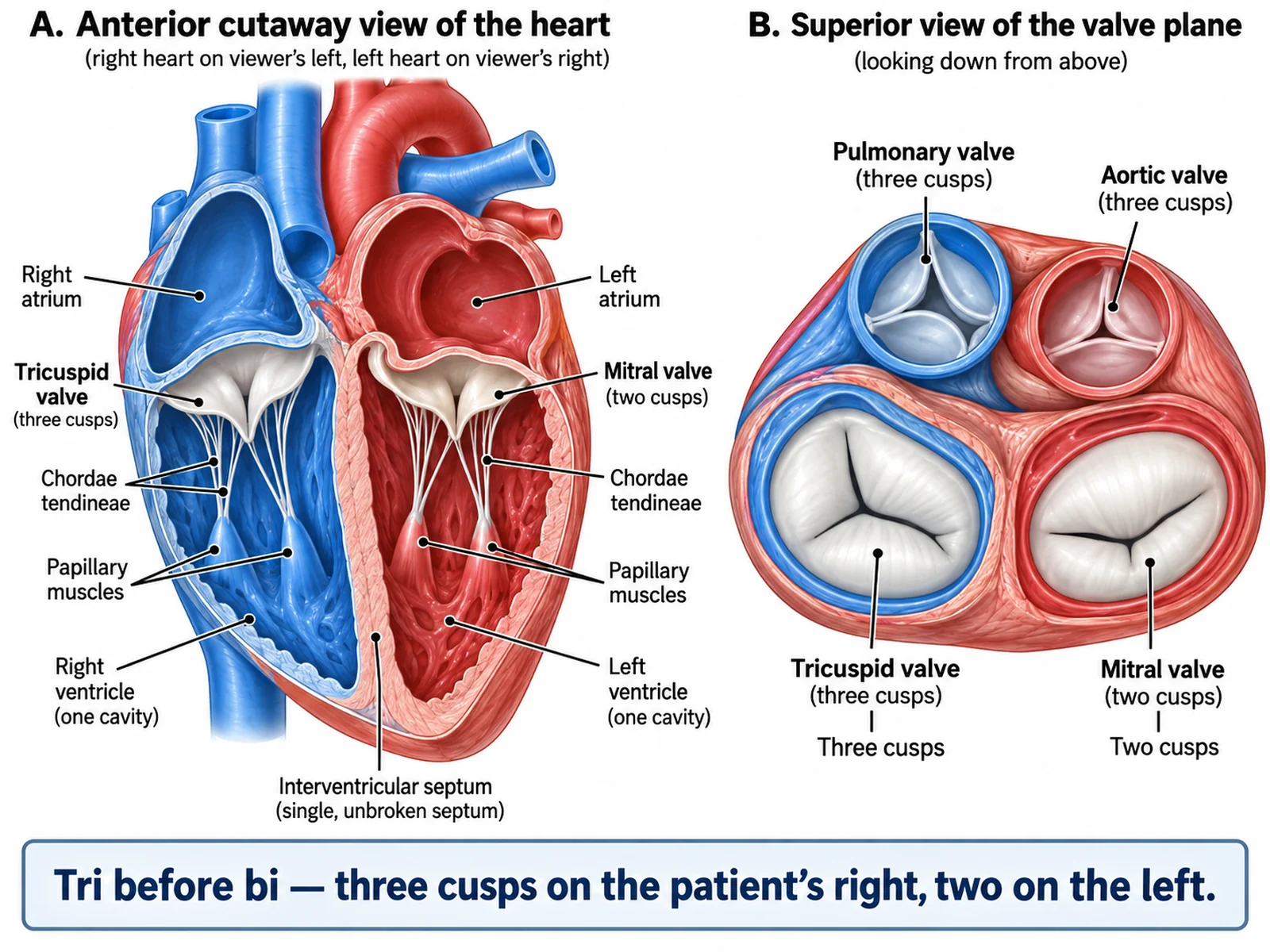
Detailed heart valve diagram showing the tricuspid, mitral, pulmonary, and aortic valves with chordae tendineae and papillary muscles

Prompt

Create a detailed heart valve diagram with two linked panels on a white background. On the left, an anterior cut-away heart showing the tricuspid valve between the patient’s right atrium and right ventricle (drawn on the VIEWER’S LEFT) and the bicuspid or mitral valve between the patient’s left atrium and left ventricle, each with chordae tendineae running down to labeled papillary muscles. On the right, a superior view looking down on the valve plane with all four valves labeled: tricuspid with three cusps, mitral with two cusps, and the pulmonary and aortic semilunar valves with three cusps each. Add a caption reading "Tri before bi — three cusps on the patient’s right, two on the left".

Heart Valves in Detail

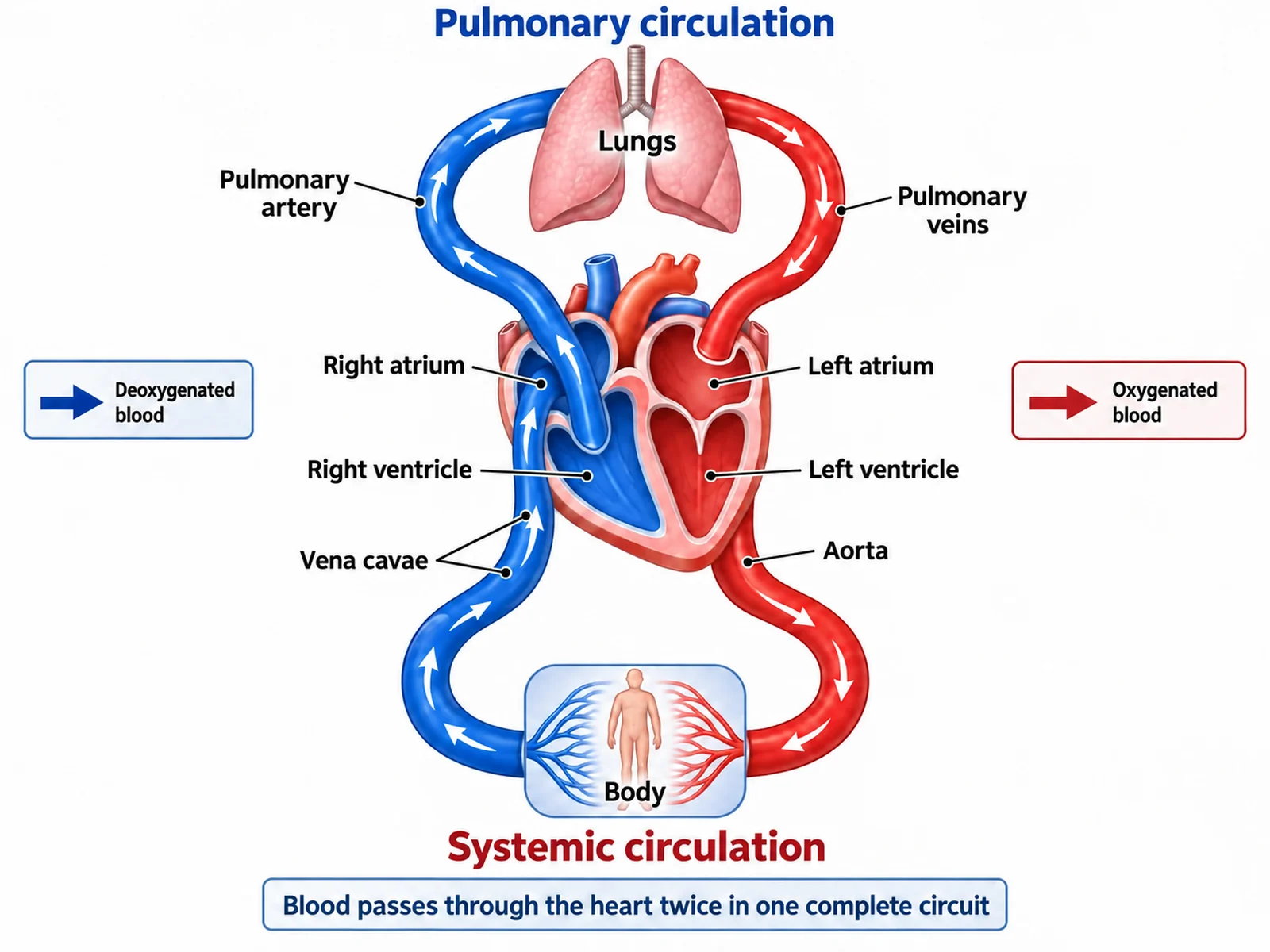
Double circulation diagram showing the pulmonary and systemic circuits passing through the heart twice in one complete circuit

Prompt

Create a labeled double circulation diagram on a white background, drawn as a figure-of-eight loop with the heart in the centre. Draw the pulmonary circuit above: right ventricle out through a blue pulmonary artery to a pair of labeled lungs, returning through red pulmonary veins to the left atrium. Draw the systemic circuit below: left ventricle out through a red aorta to a labeled body block, returning through blue vena cavae to the right atrium. Label "Pulmonary circulation", "Systemic circulation", "Deoxygenated blood" in blue, "Oxygenated blood" in red, and add a caption reading "Blood passes through the heart twice in one complete circuit".

Double Circulation Diagram

How to make a labeled or blank heart diagram

1

Choose labeled or blank

Say whether you want every part named, or a blank heart diagram with numbered empty lines for students to fill in. Ask for both from the same description and the labeled one becomes your answer key.

2

Generate the diagram

The generator draws the anterior cut-away view, keeps the patient’s right chambers on the viewer’s left, thickens the left ventricle wall, and colours the pulmonary artery blue and the pulmonary veins red.

3

Check the anatomy, then export

Confirm the sides, the valve names, the ventricle wall thickness, and the pulmonary vessel colours, then download the PNG for a worksheet, slide, or revision sheet.

What is a heart diagram?

A heart diagram is a labeled cross-section of the human heart, drawn as an anterior view cut away so the four chambers, the four valves, and the great vessels are all visible at once. It is the standard figure for teaching double circulation, and it comes in two forms that classrooms use together: a fully labeled version that names every structure, and a blank heart diagram with numbered empty lines that students complete themselves. Generating both from one description gives you the worksheet and the answer key in a single pass.

Parts of the heart to label

  • Right atrium and right ventricle: receive deoxygenated blood from the body and pump it to the lungs. In a front view they appear on the viewer’s left.
  • Left atrium and left ventricle: receive oxygenated blood from the lungs and pump it to the body. The left ventricle has the thickest wall of the four chambers.
  • Tricuspid valve: three cusps, between the patient’s right atrium and right ventricle.
  • Bicuspid or mitral valve: two cusps, between the patient’s left atrium and left ventricle.
  • Pulmonary and aortic valves: semilunar valves guarding the exits from the right and left ventricles.
  • Great vessels: superior and inferior vena cava in, pulmonary artery out to the lungs, pulmonary veins back in, aorta out to the body.
  • Septum and apex: the muscular wall separating left from right, and the pointed tip at the bottom of the heart.

How blood flows through the heart

  • Deoxygenated blood arrives from the body through the superior and inferior vena cava into the right atrium.
  • It passes the tricuspid valve into the right ventricle.
  • It leaves through the pulmonary valve and pulmonary artery, which carries deoxygenated blood to the lungs.
  • Oxygenated blood returns through the pulmonary veins into the left atrium.
  • It passes the bicuspid (mitral) valve into the left ventricle.
  • It leaves through the aortic valve and the aorta to the rest of the body, completing a circuit that crosses the heart twice.

Using a blank heart diagram in class

  • Numbered lines, no text: ask for the same anatomy with every label replaced by a ruled blank, numbered so answers can be marked quickly.
  • Pale uniform fills: keep the blank version uncoloured so red and blue do not reveal which side carries oxygenated blood.
  • Word bank optional: include one for a scaffolded lesson, leave it out for an assessment.
  • Matching answer key: generate the labeled version from the same description so the numbering lines up exactly.
  • Difficulty levels: four chambers only for a starter, chambers plus valves for a mid-unit check, chambers, valves, and all five great vessels for revision.

Common errors to check before you print

  • Sides mirrored. In a front view the patient’s right chambers appear on the viewer’s left; a diagram that puts the right ventricle on the right of the page is wrong.
  • Equal ventricle walls. The left ventricle wall is the thickest because it pumps to the whole body, not just the lungs.
  • Valves swapped. Tricuspid on the patient’s right, bicuspid (mitral) on the patient’s left — the "tri before bi" mnemonic only works once the sides are right.
  • Pulmonary vessels miscoloured. The pulmonary artery carries deoxygenated blood and should be blue; the pulmonary veins carry oxygenated blood and should be red.
  • Blood crossing the septum. In a healthy heart the septum is unbroken and nothing passes between the left and right sides.
  • Colour taken literally. Blue and red are a teaching convention for deoxygenated and oxygenated blood; real blood is never blue.

Heart Diagram Generator FAQ

The four chambers (right atrium, right ventricle, left atrium, left ventricle), the four valves (tricuspid, pulmonary, bicuspid or mitral, aortic), the great vessels (superior vena cava, inferior vena cava, pulmonary artery, pulmonary veins, aorta), the septum dividing left from right, and the apex at the bottom. Fuller diagrams also label the chordae tendineae and papillary muscles that anchor the tricuspid and mitral valves.

Need other anatomy figures?

Generate body system diagrams, organ close-ups, blank labeling worksheets, and revision-ready biology illustrations with the same SciFigure AI workflow.