Neuron AI

Neuron Diagram Generator

Draw a labeled neuron with its dendrites, cell body, axon, myelin sheath, nodes of Ranvier, and axon terminals. Ask for a simple easy-to-draw version, a synapse close-up, or a blank one for students to label.

Dendrites to axon terminalsNodes of Ranvier shownLabeled or blank

AI Neuron Diagram Generator

Describe the level of detail and which parts to label.

Costs 5 credits562 / 2000 characters

Generate a publication-ready PNG from your description.

Preview

Neuron Diagram Example Previews

Target aspect ratio: 16:9

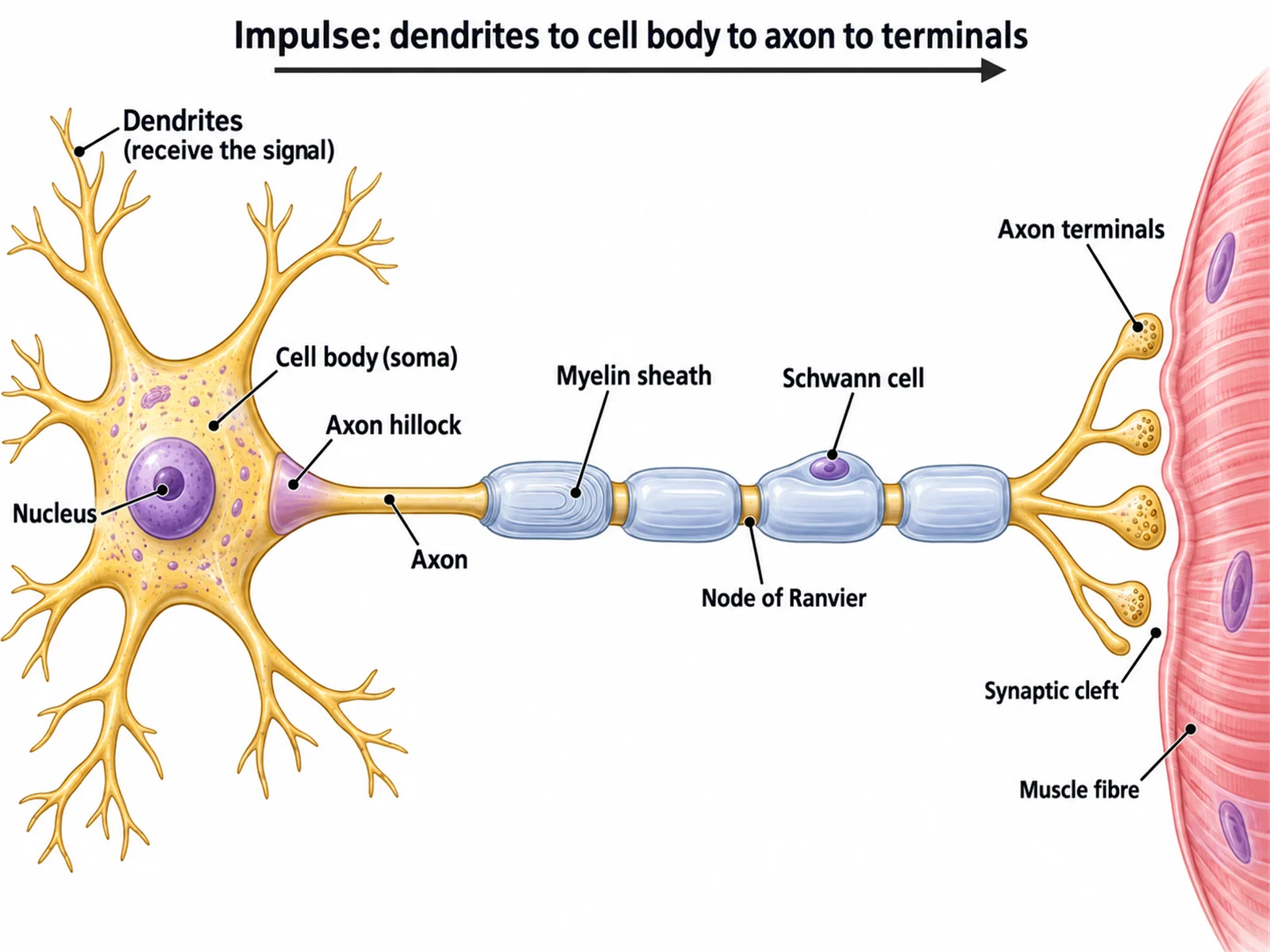
Labeled motor neuron diagram naming the dendrites, cell body, nucleus, axon hillock, axon, myelin sheath, Schwann cell, nodes of Ranvier, and axon terminals
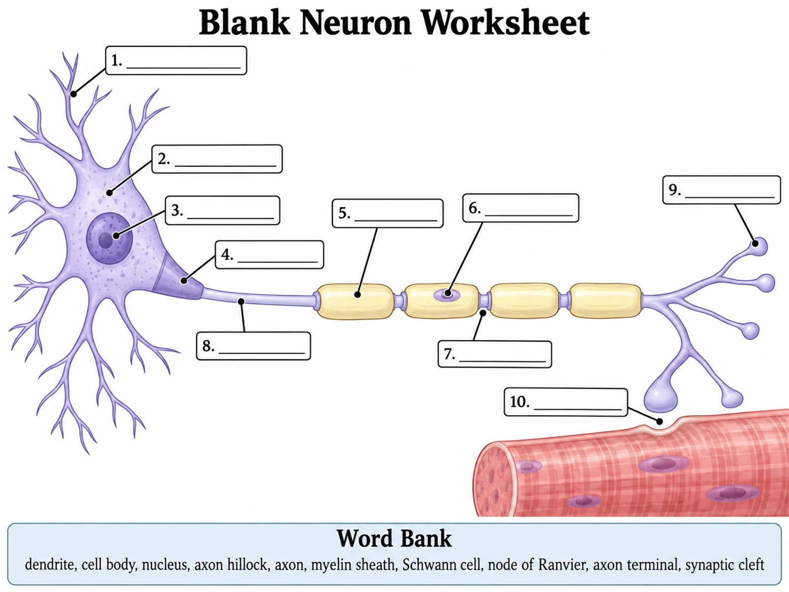
Blank unlabeled neuron diagram with numbered empty label lines and a word bank for students to complete
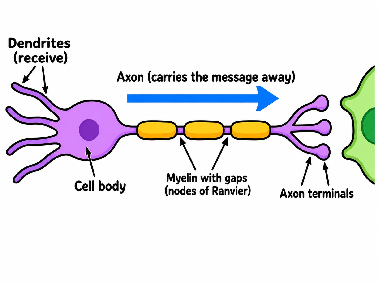
Simple easy-to-draw neuron with a cell body, axon, three myelin blocks, and a few basic labels

Sample previews. Describe your diagram to generate your own neuron figure.

Neuron Diagram Examples

Use these as visual directions for labeled motor neurons, blank worksheets, simple drawings, synapse close-ups, neuron type comparisons, and impulse pathway figures.

Labeled motor neuron diagram naming the dendrites, cell body, nucleus, axon hillock, axon, myelin sheath, Schwann cell, nodes of Ranvier, and axon terminals

Prompt

Create a fully labeled motor neuron on a white background, drawn left to right in clean biology textbook style. On the left draw a star-shaped cell body with a round nucleus clearly inside it and five branching processes; label "Dendrites (receive the signal)", "Cell body (soma)" and "Nucleus". Label the cone where the axon leaves "Axon hillock". Run one long axon right, wrapped in four separate myelin segments with bare gaps between them; label "Myelin sheath", "Schwann cell (wraps the axon)" and "Node of Ranvier" at one gap. End in branching "Axon terminals" stopping short of a muscle fibre, leaving a visible gap labeled "Synaptic cleft". Add an arrow above reading "Impulse: dendrites to cell body to axon to terminals".
Labeled Motor Neuron

Blank unlabeled neuron diagram with numbered empty label lines and a word bank for students to complete

Prompt

Create a blank neuron worksheet. Draw the same motor neuron in clean line art with pale uniform fills and no text on the figure: dendrites and a nucleated cell body on the left, the axon hillock, a long axon wrapped in four separate myelin segments with clearly visible bare gaps between them, and branching terminals on the right stopping short of a muscle fibre so a synaptic cleft stays open. Draw thin leader lines from each part to a short horizontal ruled blank line, numbered 1 to 10. Add a word bank box below reading "dendrite, cell body, nucleus, axon hillock, axon, myelin sheath, Schwann cell, node of Ranvier, axon terminal, synaptic cleft". Generous white space, no answer key.

Blank Neuron Worksheet

Simple easy-to-draw neuron with a cell body, axon, three myelin blocks, and a few basic labels

Prompt

Create a simple, easy-to-draw neuron for younger students. Draw a rounded cell body in one flat colour with a dot inside for the nucleus and four thick branching dendrites on the left, one long axon running right with three separate sausage-shaped myelin blocks and obvious bare gaps between them, and three short branches at the far end. Label only five things in large readable text: "Dendrites (receive)", "Cell body", "Axon (carries the message away)", "Myelin with gaps (nodes of Ranvier)", and "Axon terminals". Add one big arrow from the cell body to the terminals, and leave an open synaptic cleft between the terminals and the next cell so they never touch. Bold flat colours, thick outlines, plenty of white space.

Simple Neuron Drawing

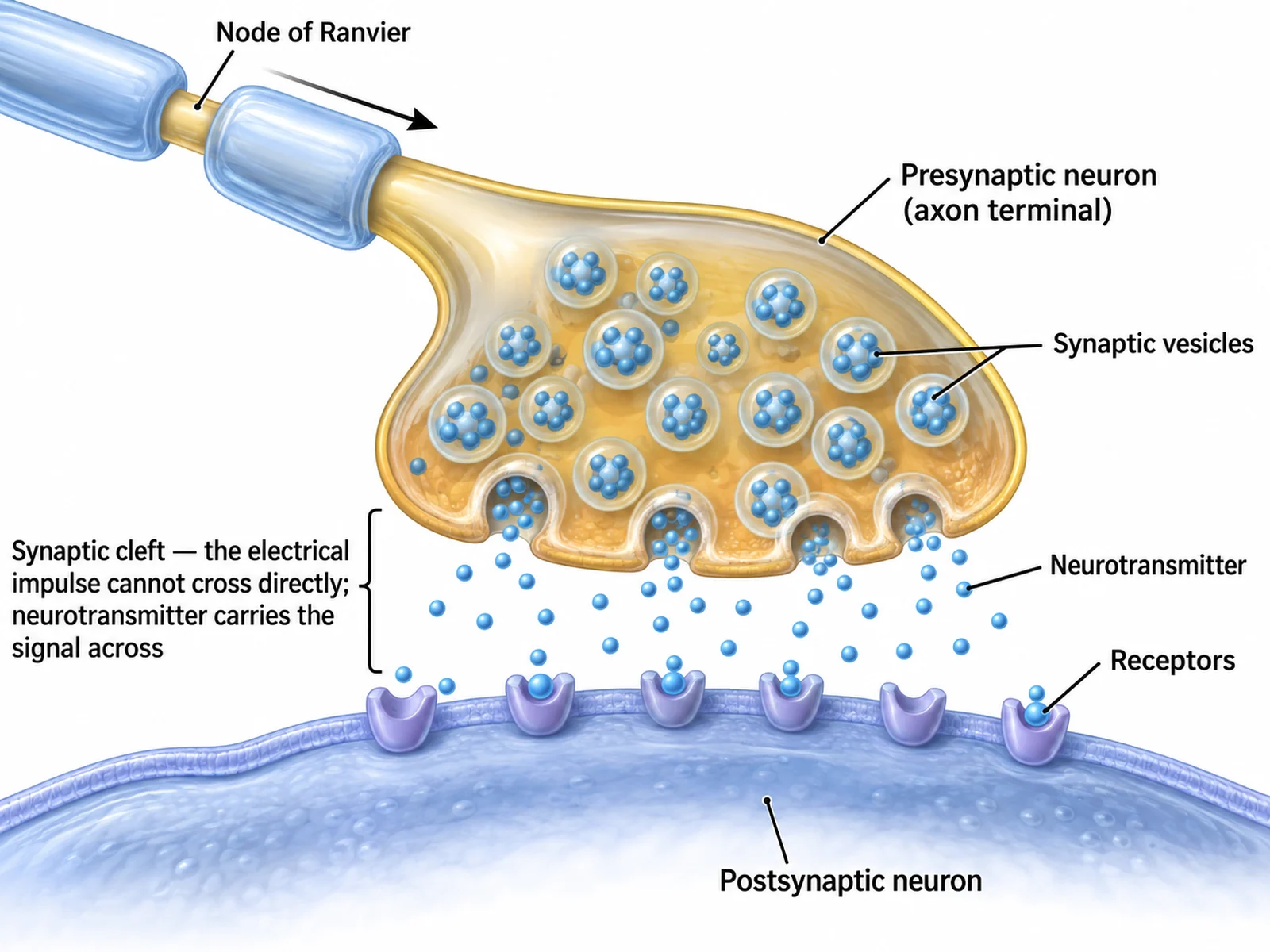
Close-up synapse diagram showing synaptic vesicles, neurotransmitter crossing the synaptic cleft, and receptors on the postsynaptic membrane

Prompt

Create a labeled synapse close-up on a white background. At the upper left show the incoming myelinated axon with two separate myelin segments and a visible bare gap between them labeled "Node of Ranvier", with an arrow running toward the swollen ending labeled "Presynaptic neuron (axon terminal)". Inside it draw many small circles labeled "Synaptic vesicles" and show several fusing with the membrane and spilling dots labeled "Neurotransmitter". Draw a clear open channel between the two membranes labeled "Synaptic cleft — the electrical impulse cannot cross directly; neurotransmitter carries the signal across" with neurotransmitter dots diffusing across it and locking into cup-shaped "Receptors" on the "Postsynaptic neuron" membrane below. The two cells must never touch.

Synapse in Detail

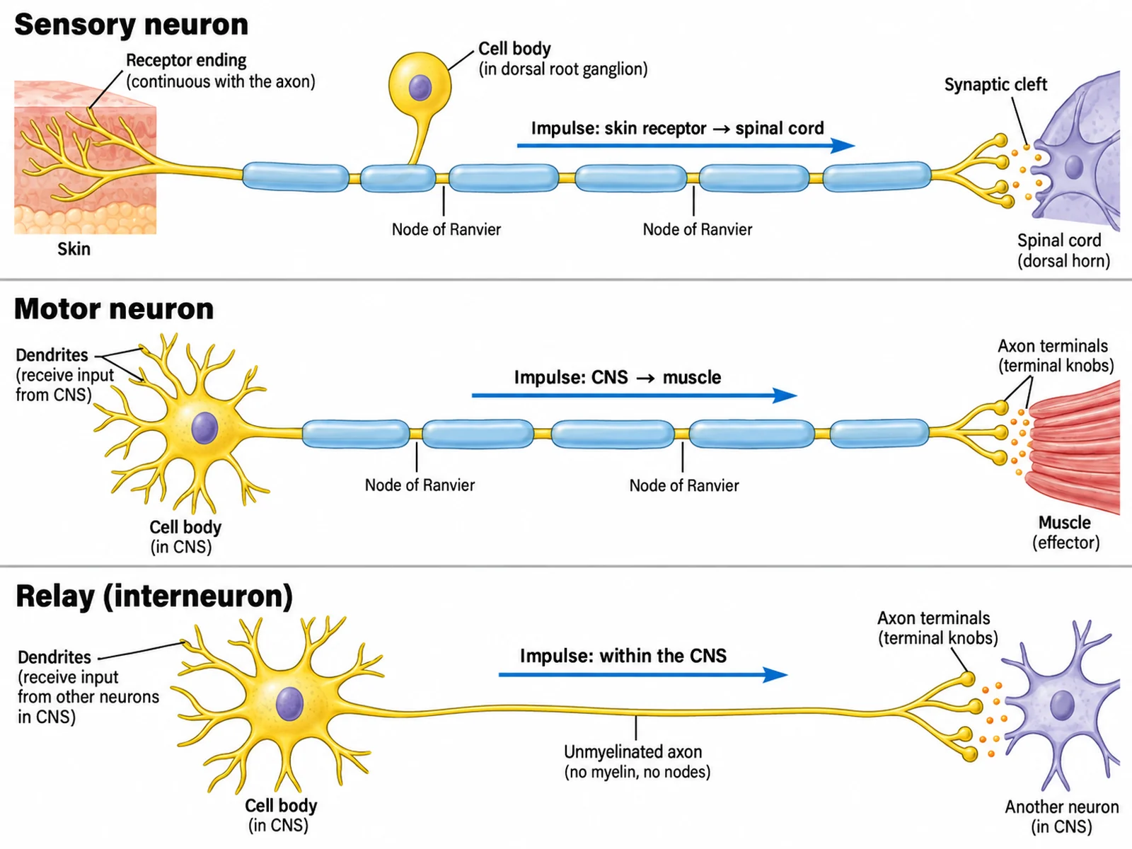
Comparison diagram of sensory, motor, and relay neurons showing cell body position, myelination, and direction of the impulse

Prompt

Create a comparison of three neuron types as labeled rows on white. Row 1 "Sensory neuron": the cell body sits on a short side branch partway along the cell, an arrow runs from a skin receptor to the spinal cord, and the myelin is drawn as separate segments with visible bare gaps labeled "Node of Ranvier". Row 2 "Motor neuron": a cell body with many dendrites at one end, a long axon in separate myelin segments with the same visible gaps, and an arrow running from the CNS to a muscle. Row 3 "Relay (interneuron)": short, inside the CNS, with a bare unmyelinated axon and no nodes. Where neurons meet, leave a visible synaptic cleft — the terminals never touch.

Sensory, Motor and Relay Neurons

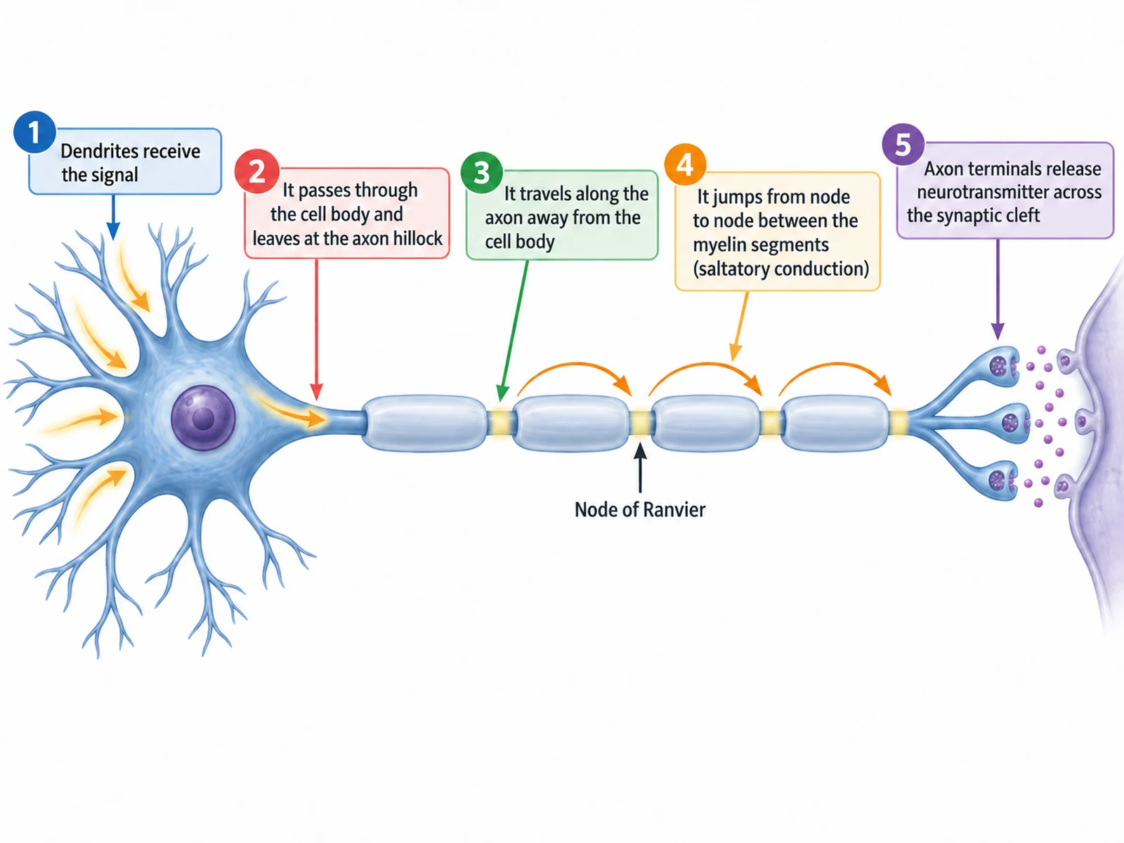
Diagram tracing a nerve impulse from the dendrites along a myelinated axon, jumping between nodes of Ranvier, to release at the synapse

Prompt

Create a labeled nerve impulse diagram running left to right with five numbered stages and captions: "1 Dendrites receive the signal", "2 It passes through the cell body and leaves at the axon hillock", "3 It travels along the axon away from the cell body", "4 It jumps from node to node between the myelin segments (saltatory conduction)", "5 Axon terminals release neurotransmitter across the synaptic cleft". Draw the myelin as four separate segments with bright bare gaps labeled "Node of Ranvier" and curved arrows arcing from gap to gap. At the far right show the terminal separated from the next cell by a visible synaptic cleft with neurotransmitter dots crossing it. Every arrow points away from the cell body.

How a Nerve Impulse Travels

How to draw a labeled neuron

1

Choose the detail level

A simple cell body and axon for younger classes, a full labeled motor neuron with nodes of Ranvier, a synapse close-up, or a comparison of sensory, motor, and relay neurons.

2

Generate the diagram

The generator keeps the dendrites on the cell body, the nucleus inside the soma, the myelin in separate segments with visible nodes, and the impulse arrow pointing away from the cell body.

3

Check the direction, then export

Confirm the arrow runs dendrites to axon terminals, the myelin is broken by nodes of Ranvier, and the synapse shows a cleft rather than contact, then download the PNG.

What is a neuron?

A neuron is a nerve cell built to carry an electrical impulse in one direction. Signals arrive on the branching dendrites, pass into the cell body where the nucleus sits, leave at the axon hillock, and travel down a single long axon to the axon terminals at the far end. Most axons outside the brain are insulated by a myelin sheath made of Schwann cells, broken at intervals by bare gaps called nodes of Ranvier. Because the impulse jumps from node to node rather than creeping along the whole membrane, a myelinated axon conducts a signal many times faster than a bare one. That direction — in at the dendrites, out at the terminals — is the cell’s defining feature, and it is the first thing to check on any neuron drawing.

Parts of a neuron

  • Dendrites: short branching processes on the cell body that receive signals from other neurons.
  • Cell body (soma): the rounded region holding the nucleus and most of the cell’s organelles.
  • Axon hillock: the cone-shaped point where the axon leaves the cell body and the impulse begins.
  • Axon: the single long fibre that carries the impulse away from the cell body.
  • Myelin sheath and Schwann cells: each Schwann cell wraps a short stretch of axon, and its wound membrane is the myelin sheath.
  • Nodes of Ranvier: the bare gaps between neighbouring myelin segments, where the impulse jumps.
  • Axon terminals: the branched endings, also called synaptic knobs, that release neurotransmitter.

How a nerve impulse travels, step by step

  • Receives: neurotransmitter from another neuron binds to the dendrites and starts a change in charge.
  • Sums: the signal spreads across the cell body to the axon hillock, where the impulse either fires or does not.
  • Conducts: the impulse runs along the axon away from the cell body, never back toward the dendrites.
  • Jumps: on a myelinated axon it leaps from one node of Ranvier to the next, which is why myelin speeds it up.
  • Releases: at the axon terminal, synaptic vesicles fuse with the membrane and empty neurotransmitter into the synaptic cleft.
  • Crosses: the neurotransmitter diffuses across the gap and binds to receptors on the postsynaptic membrane, starting the signal again in the next cell.

Neuron Diagram FAQ

The dendrites, the cell body (soma) with the nucleus inside it, the axon hillock where the axon leaves the cell body, the axon, the myelin sheath, the Schwann cells that make it, the nodes of Ranvier between the myelin segments, and the axon terminals at the far end. A fuller diagram also labels the synaptic cleft between one neuron and the next.

Need other biology figures?

Generate nerve cell close-ups, organ diagrams, cell figures, and worksheet-ready illustrations with the same SciFigure AI workflow.