Brain AI

Brain Diagram Generator

Draw a labeled brain with the frontal, parietal, temporal, and occipital lobes, the cerebellum, and the brain stem named. Ask for a blank one for students to label, a simple drawing, a sagittal section, or a plain silhouette outline to use as a graphic.

Four lobes in the right orderLabeled or blankSilhouette and outline shapes

AI Brain Diagram Generator

Describe the view you need and which structures to label.

Costs 5 credits538 / 2000 characters

Generate a publication-ready PNG from your description.

Preview

Brain Diagram Example Previews

Target aspect ratio: 16:9

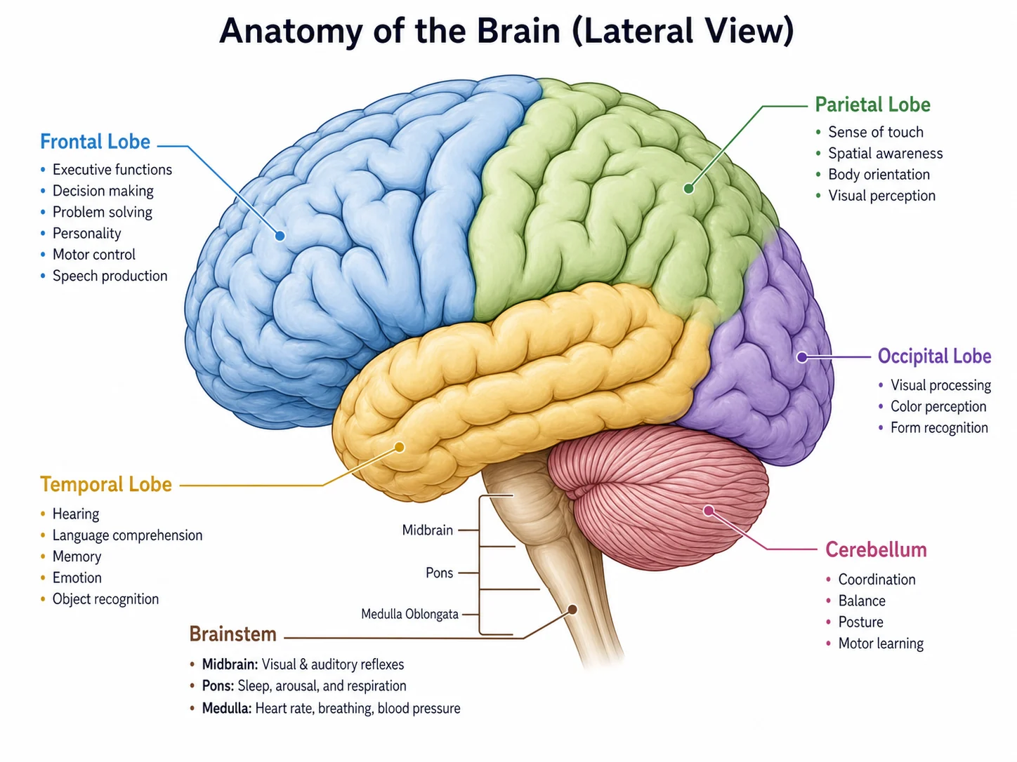
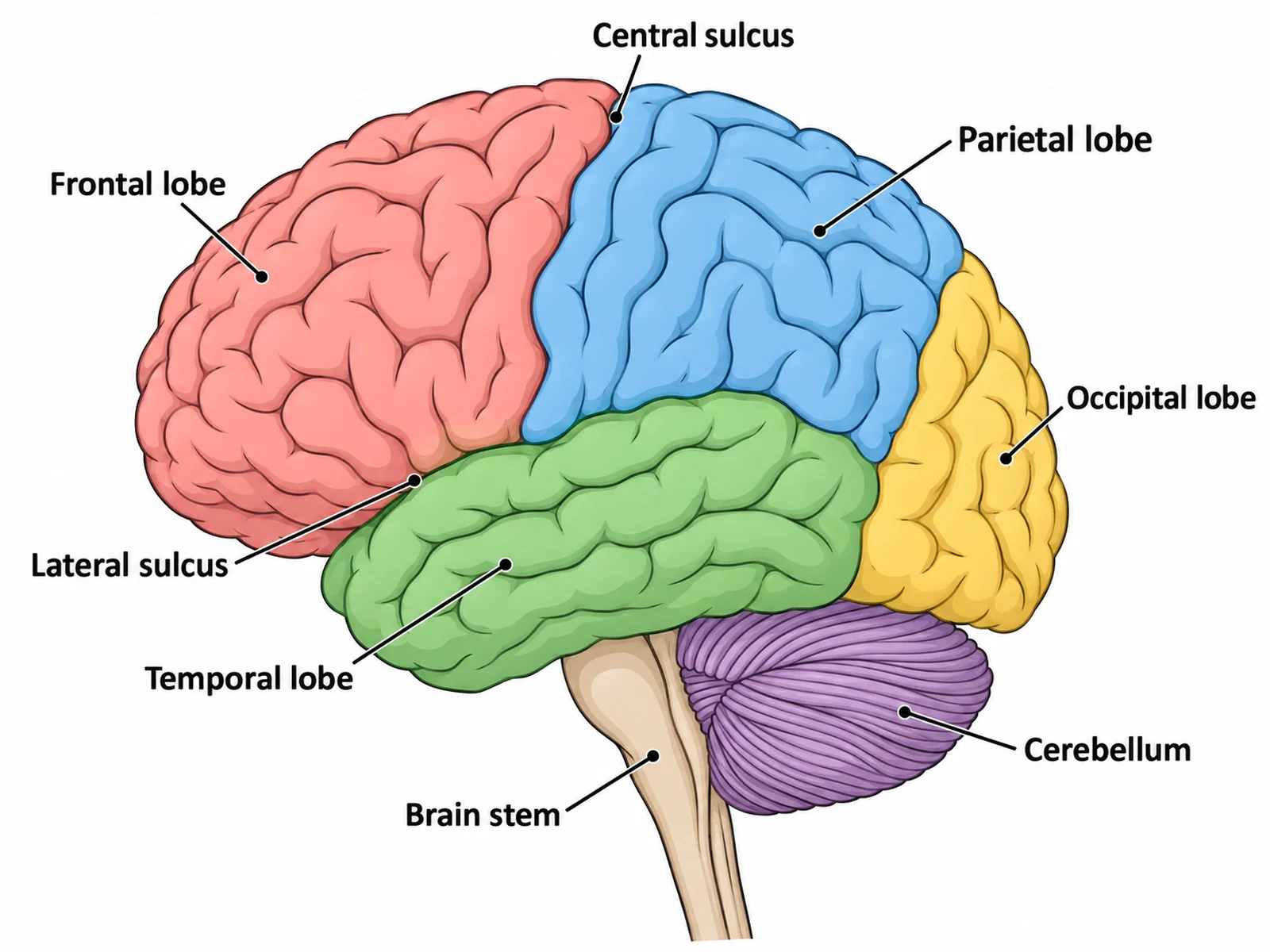
Labeled brain diagram in left lateral view naming the frontal, parietal, temporal, and occipital lobes, the cerebellum, and the brain stem
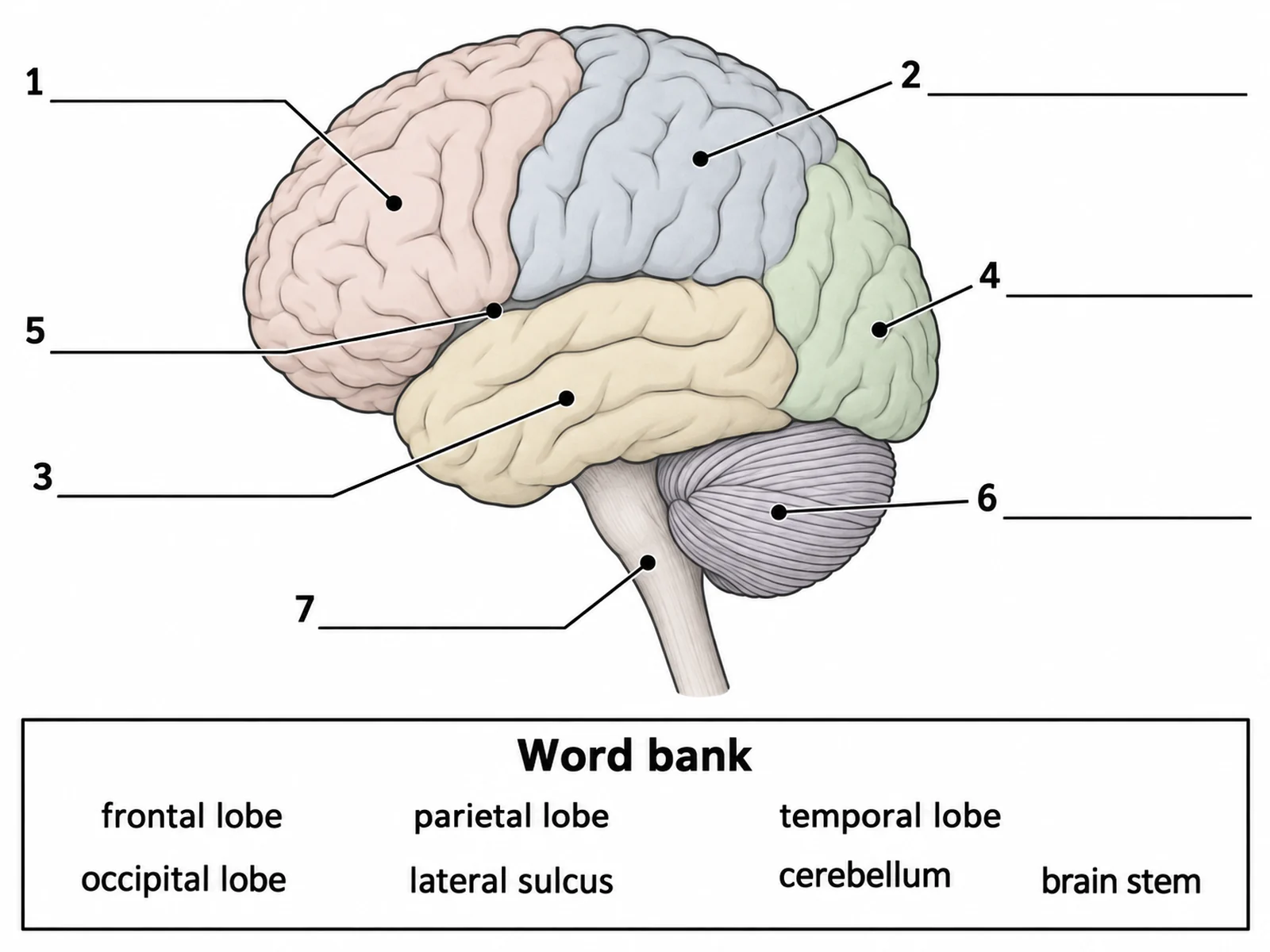
Blank unlabeled brain diagram with numbered empty label lines and a word bank for students to complete
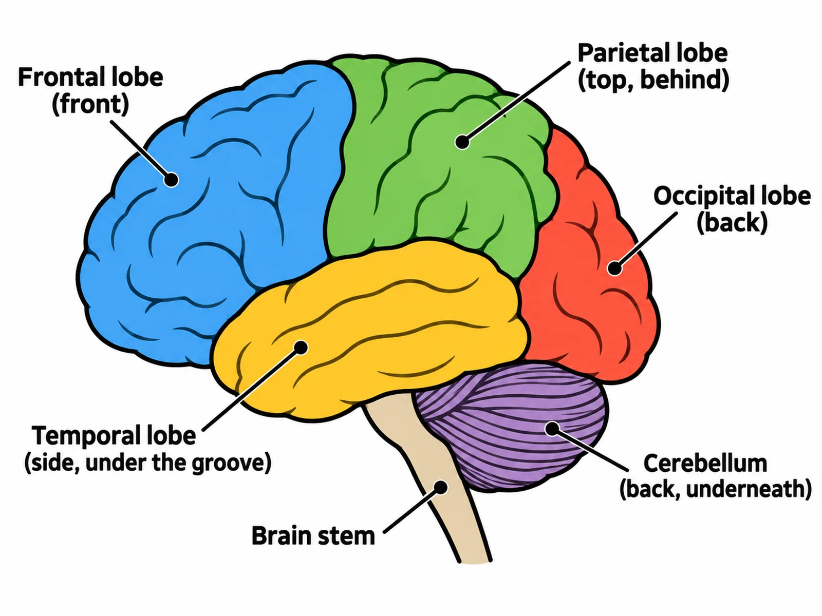
Simple easy-to-draw brain with four coloured lobes, the cerebellum at the back, and the brain stem below

Sample previews. Describe your diagram to generate your own brain figure.

Brain Diagram Examples

Use these as visual directions for labeled lateral views, blank worksheets, simple drawings, four-lobe reference figures, plain silhouette and outline shapes for design work, and midsagittal sections.

Labeled brain diagram in left lateral view naming the frontal, parietal, temporal, and occipital lobes, the cerebellum, and the brain stem

Prompt

Create a fully labeled human brain on a white background in clean biology textbook style, drawn as a left lateral view with the brain facing left. Shade the four lobes of the cerebrum in four soft flat colours and label "Frontal lobe" at the front, "Parietal lobe" behind and above it, "Occipital lobe" rearmost at the back, and "Temporal lobe" low at the side, below a labeled "Lateral sulcus". Label "Central sulcus" on the groove between frontal and parietal. Below the occipital lobe draw the "Cerebellum" with much finer tightly packed folds, and label the "Brain stem" descending in front of it. No other labels.
Labeled Brain Diagram

Blank unlabeled brain diagram with numbered empty label lines and a word bank for students to complete

Prompt

Create a blank brain worksheet. Draw the same left lateral view in clean line art with pale uniform fills and no text on the figure: the four cerebral lobes separated by visible sulci, the temporal lobe below the lateral sulcus, the cerebellum below the occipital lobe at the back drawn with finer folds, and the brain stem descending in front of the cerebellum. Draw thin leader lines from each region to a short horizontal ruled blank line, numbered 1 to 7. Add a word bank box below reading "frontal lobe, parietal lobe, temporal lobe, occipital lobe, lateral sulcus, cerebellum, brain stem". Generous white space, no answer key.

Blank Brain Diagram to Label

Simple easy-to-draw brain with four coloured lobes, the cerebellum at the back, and the brain stem below

Prompt

Create a simple, easy-to-draw brain for younger students. Draw a left lateral view facing left with thick outlines and four bold flat colours for the lobes, with only a few wide folds so it is easy to copy. Label just five things in large readable text: "Frontal lobe (front)", "Parietal lobe (top, behind)", "Occipital lobe (back)", "Temporal lobe (side, under the groove)", and "Cerebellum (back, underneath)". Add "Brain stem" on the stalk running down in front of the cerebellum. Draw the cerebellum with closer, finer lines than the cerebrum so the two read as different structures. Plenty of white space.

Simple Brain Drawing

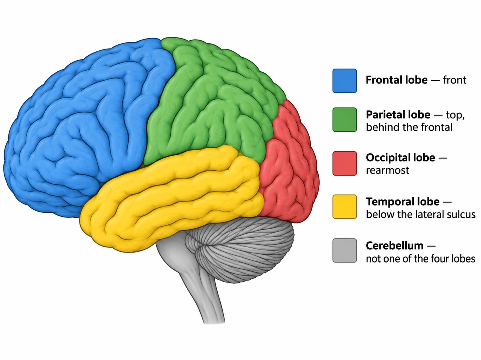
Brain lobes diagram showing the frontal, parietal, temporal, and occipital lobes in separate colours with the cerebellum shown as a separate structure

Prompt

Create a lobes-only reference figure of the brain on white, left lateral view facing left. Fill each of the four cerebral lobes with a distinct flat colour and a matching key at the side: "Frontal lobe — front", "Parietal lobe — top, behind the frontal", "Occipital lobe — rearmost", "Temporal lobe — below the lateral sulcus". Draw the boundaries on the real sulci: the central sulcus between frontal and parietal, and the lateral sulcus above the temporal lobe. Show the cerebellum below the occipital lobe in plain grey with much finer folds, captioned "Cerebellum — not one of the four lobes". No Broca or Wernicke areas.

The Four Lobes

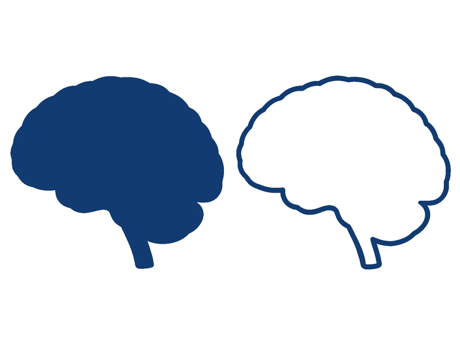
Plain brain silhouette as a flat single-colour shape beside a matching unfilled brain outline, both with no internal detail or labels

Prompt

Create two clean brain shapes side by side on a plain white background, both in left lateral profile facing left, as graphic design assets rather than teaching figures. On the left, a solid silhouette: the brain profile filled in one flat dark colour, with the cerebellum and a short brain stem included in the same shape, and absolutely no gyri, sulci, internal divisions, gradients, shading, or text. On the right, the identical profile drawn as a single smooth unfilled outline of even stroke weight with a transparent white interior, ready to colour in or place text inside. Crisp edges, no drop shadow, no background elements, no labels anywhere.

Brain Silhouette and Outline

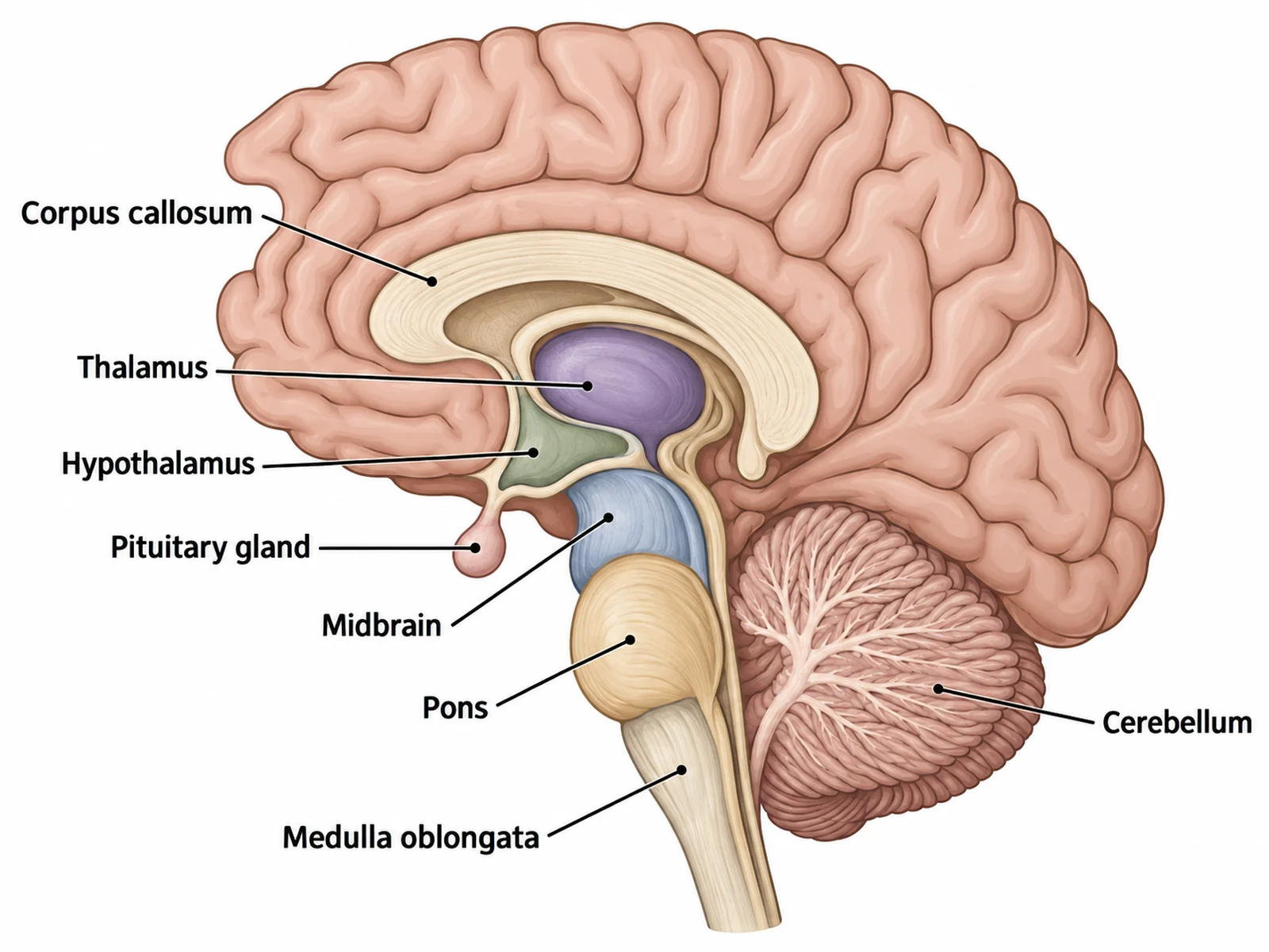
Midsagittal section of the brain labeling the corpus callosum, thalamus, hypothalamus, pituitary gland, midbrain, pons, medulla oblongata, and cerebellum

Prompt

Create a labeled midsagittal section of the human brain on white, cut down the midline and facing left, so the internal structures are exposed. Label the "Corpus callosum" as the thick arched band above the centre, the "Thalamus" directly below it, the "Hypothalamus" below that, and the "Pituitary gland" hanging beneath the hypothalamus on a short stalk. Running downward label the "Midbrain", then the "Pons", then the "Medulla oblongata" as the brain stem, with the "Cerebellum" behind the pons drawn with fine tightly packed folia. Show the cut cerebral cortex around the outside with its broader gyri for contrast.

Brain in Sagittal Section

How to draw a labeled, blank, or outline brain diagram

1

Choose the version you need

A labeled lateral view for reference, a blank one with numbered lines for students, a simple drawing for younger classes, a sagittal section for the midline structures, or a plain silhouette or outline to use as a graphic element.

2

Generate the figure

The generator keeps the lobes in order front to back, the temporal lobe below the lateral sulcus, the cerebellum below the occipital lobe with its own finer folds, and the brain stem descending in front of the cerebellum.

3

Check the layout, then export

Confirm the parietal and occipital lobes are not swapped, the cerebellum is at the back and not counted as a lobe, and no midline structure is labeled on a surface view, then download the PNG.

What is a brain diagram?

A brain diagram is a labeled map of the brain, usually drawn as a left lateral view with the brain facing left. That single view shows the four lobes of the cerebrum — frontal at the front, parietal behind and above it, occipital rearmost, and temporal below the lateral sulcus — together with the cerebellum below the occipital lobe and the brain stem descending in front of the cerebellum. A midsagittal section is the second standard view: cutting down the midline exposes the corpus callosum, thalamus, hypothalamus, and pituitary gland, none of which can honestly be labeled on a surface drawing. The same outline also gets used a third way, with no anatomy in it at all: as a flat brain silhouette or a clean outline shape for posters, icons, and slide graphics.

Parts of the brain to label

  • Frontal lobe: the frontmost lobe of the cerebrum, ahead of the central sulcus.
  • Parietal lobe: behind and above the frontal lobe, on top of the brain toward the back — not the rearmost lobe.
  • Occipital lobe: the rearmost lobe, at the very back of the cerebrum.
  • Temporal lobe: low at the side, below the lateral sulcus that separates it from the frontal and parietal lobes.
  • Cerebellum: a separate structure below the occipital lobe at the back, with much finer and more tightly packed folds than the cerebrum.
  • Brain stem: the midbrain, pons, and medulla oblongata running downward in front of the cerebellum.
  • Midline structures: the corpus callosum, thalamus, hypothalamus, and pituitary gland — label these only on a sagittal section.

Choosing between a labeled diagram, a blank, and a silhouette

  • Labeled lateral view: use it when the figure has to name structures — a reference slide, a revision sheet, or an answer key.
  • Blank version: the same anatomy with every label replaced by a numbered ruled blank, pale uniform fills so the colours give nothing away, and an optional word bank.
  • Silhouette: use it when the brain is a graphic element rather than the subject — a flat single-colour shape with no internal detail and no labels, for an icon, a logo mark, a poster section marker, or a container to place text or data inside. It is a design asset, so never ask it to teach anatomy.
  • Outline: the same profile as a clean unfilled line of even stroke weight, for colouring in, tracing, cutting as a stencil, or overlaying on a photograph.
  • Sagittal section: the only view that can legitimately show the corpus callosum, thalamus, hypothalamus, and pituitary gland.

Common errors to check before you print

  • The cerebellum at the front, or counted as a lobe. It belongs below the occipital lobe at the back, and the four lobes are all cerebrum — this is the single most common mistake on this figure.
  • The parietal and occipital lobes swapped. Parietal is on top toward the back; occipital is the rearmost lobe.
  • The temporal lobe drawn above the lateral sulcus. It sits below it, low at the side.
  • The brain stem behind the cerebellum. It descends in front of it, not out of its back.
  • The cerebellum given the cerebrum’s folds. Its folia are much finer and more tightly packed, and that difference is how the two structures are told apart at a glance.
  • The corpus callosum labeled on a lateral surface view. It is a midline band, visible only once the brain is cut sagittally.
  • A silhouette returned with gyri, shading, or a label on it. A silhouette is one flat shape and nothing else.

Brain Diagram FAQ

The four lobes of the cerebrum — frontal, parietal, temporal, and occipital — plus the cerebellum below the occipital lobe at the back and the brain stem running down in front of the cerebellum. A fuller lateral view also names the lateral sulcus that separates the temporal lobe from the frontal and parietal lobes, and the central sulcus between the frontal and parietal lobes. Midline structures such as the corpus callosum, thalamus, and pituitary gland cannot be labeled on a surface view at all — they only appear in a sagittal section.

Need other anatomy figures?

Generate nerve cell close-ups, organ diagrams, cross-sections, and worksheet-ready illustrations with the same SciFigure AI workflow.