Protein Synthesis AI

Transcription and Translation Diagram Generator

Draw the central dogma the way your class needs to see it: DNA to mRNA in the nucleus, mRNA to polypeptide at the ribosome. Describe the step, the labels, and the worksheet style, then generate a clean protein synthesis figure.

Transcription in the nucleusTranslation at the ribosomeCodon and anticodon labels

AI Transcription and Translation Diagram Generator

Describe the stage, the structures you want labeled, and the teaching style you need.

Costs 5 credits485 / 2000 characters

Generate a publication-ready PNG from your description.

Preview

Transcription and Translation Diagram Example Previews

Target aspect ratio: 16:9

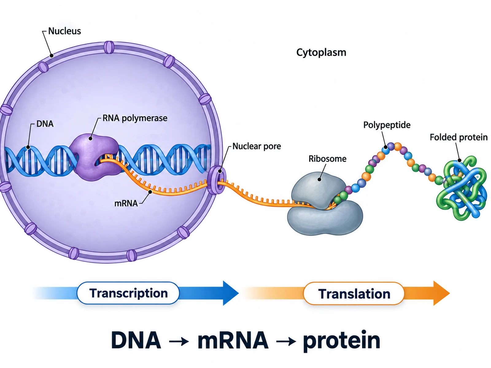
Labeled central dogma diagram showing DNA transcribed to mRNA in the nucleus and mRNA translated to protein at a ribosome in the cytoplasm
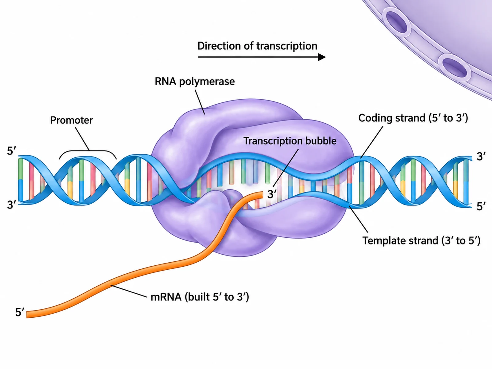
Transcription diagram showing RNA polymerase reading the DNA template strand and synthesizing an mRNA strand inside the nucleus
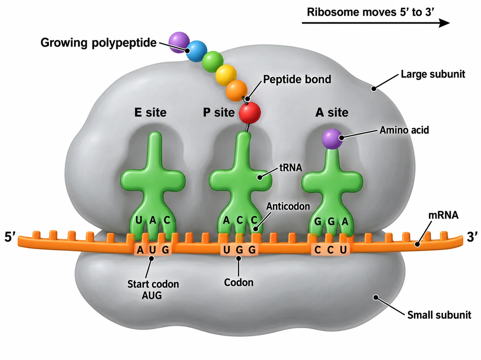
Translation diagram showing mRNA threaded through a ribosome with tRNA delivering amino acids and a polypeptide chain growing

Sample previews. Describe your diagram to generate your own protein synthesis figure.

Labeled Transcription and Translation Diagram Examples

Use these examples as visual directions for central dogma figures, single-stage close-ups, codon charts, and blank protein synthesis worksheets.

Labeled central dogma diagram showing DNA transcribed to mRNA in the nucleus and mRNA translated to protein at a ribosome in the cytoplasm

Prompt

Create a labeled central dogma diagram running left to right: DNA, then mRNA, then protein. Draw the DNA double helix and RNA polymerase inside a nucleus, the finished mRNA strand leaving through a nuclear pore, and a ribosome in the cytoplasm building a polypeptide chain. Label transcription over the DNA-to-mRNA arrow and translation over the mRNA-to-protein arrow, mark the nucleus and cytoplasm, and keep a clean white biology textbook style.
Central Dogma Overview

Transcription diagram showing RNA polymerase reading the DNA template strand and synthesizing an mRNA strand inside the nucleus

Prompt

Create a labeled transcription diagram set inside the nucleus. Show RNA polymerase bound at the promoter, the DNA double helix unwound into a transcription bubble, and the enzyme moving right while reading the lower template strand from 3' at left to 5' at right. Show the complementary mRNA growing 5' to 3', with its 5' end trailing left and its 3' end nearest the enzyme; the upper coding strand runs 5' at left to 3' at right. Label the promoter, RNA polymerase, template strand, coding strand, and growing mRNA. Keep the labels short and the background white.

Transcription in the Nucleus

Translation diagram showing mRNA threaded through a ribosome with tRNA delivering amino acids and a polypeptide chain growing

Prompt

Create a labeled translation diagram centered on one ribosome in the cytoplasm. Show the mRNA strand threaded between the large and small ribosomal subunits, read 5' to 3', with the AUG start codon marked. Draw one tRNA in the A site carrying an amino acid, one tRNA in the P site attached to the growing polypeptide, and one tRNA leaving the E site. Label the ribosomal subunits, mRNA codons, tRNA anticodons, amino acids, the peptide bond, and the polypeptide chain.

Translation at the Ribosome

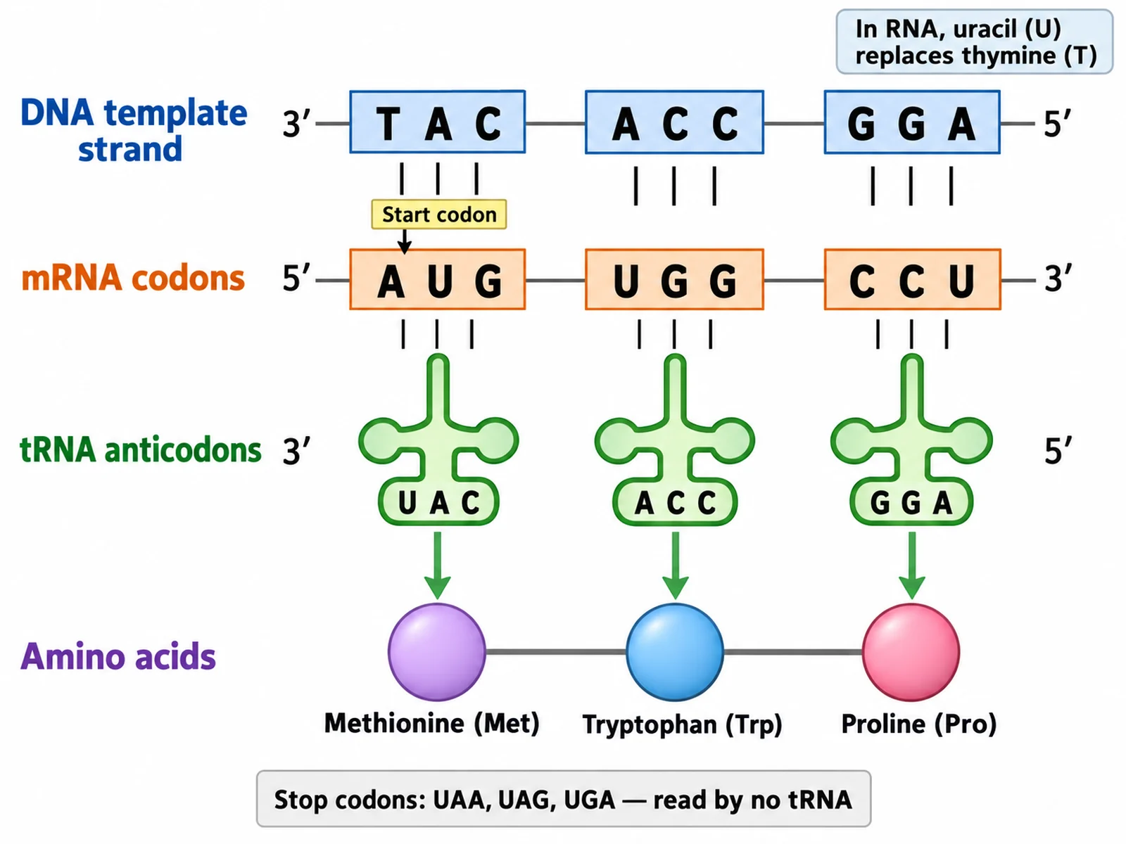
Close-up diagram of mRNA codons pairing with tRNA anticodons and the matching amino acids in the polypeptide chain

Prompt

Create a close-up diagram of codon and anticodon pairing. Show one DNA template strand above an mRNA strand above three tRNA molecules. Use a worked example: DNA template TAC ACC GGA, mRNA AUG UGG CCU, anticodons UAC ACC GGA, amino acids methionine, tryptophan, proline. Label the start codon, each codon, each anticodon, the amino acid each tRNA carries, and note that uracil replaces thymine in RNA. Add a short caption line listing the three stop codons UAA, UAG, and UGA.

Codon and Anticodon Pairing

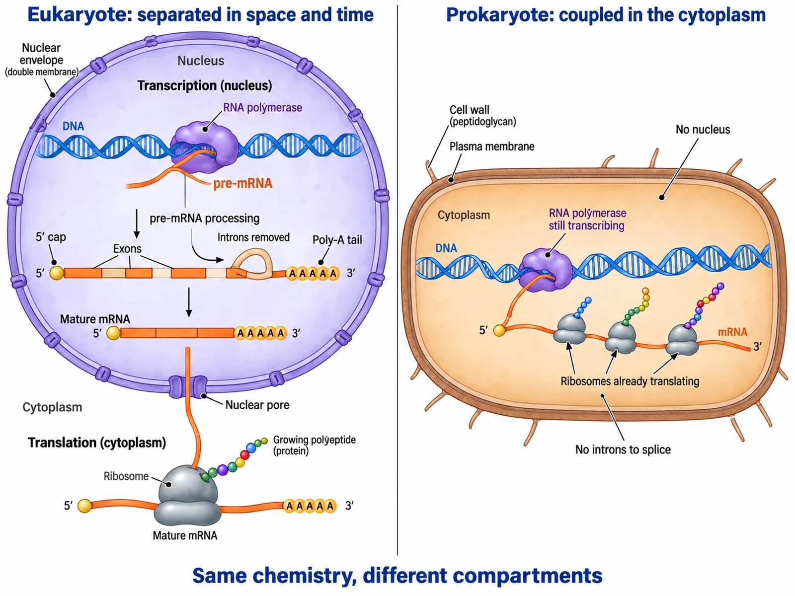
Side-by-side comparison of coupled prokaryotic transcription and translation and the separated eukaryotic process split by the nuclear envelope

Prompt

Create a two-panel comparison figure of protein synthesis. In the eukaryotic panel, show transcription inside a nucleus, pre-mRNA processing with a 5' cap, poly-A tail and intron splicing, the mature mRNA leaving through a nuclear pore, and translation at a separate cytoplasmic ribosome. In the prokaryotic panel, show no nucleus and several ribosomes already translating an mRNA strand while RNA polymerase is still transcribing it. Head the panels Eukaryote: separated in space and time and Prokaryote: coupled in the cytoplasm.

Prokaryotic and Eukaryotic Compared

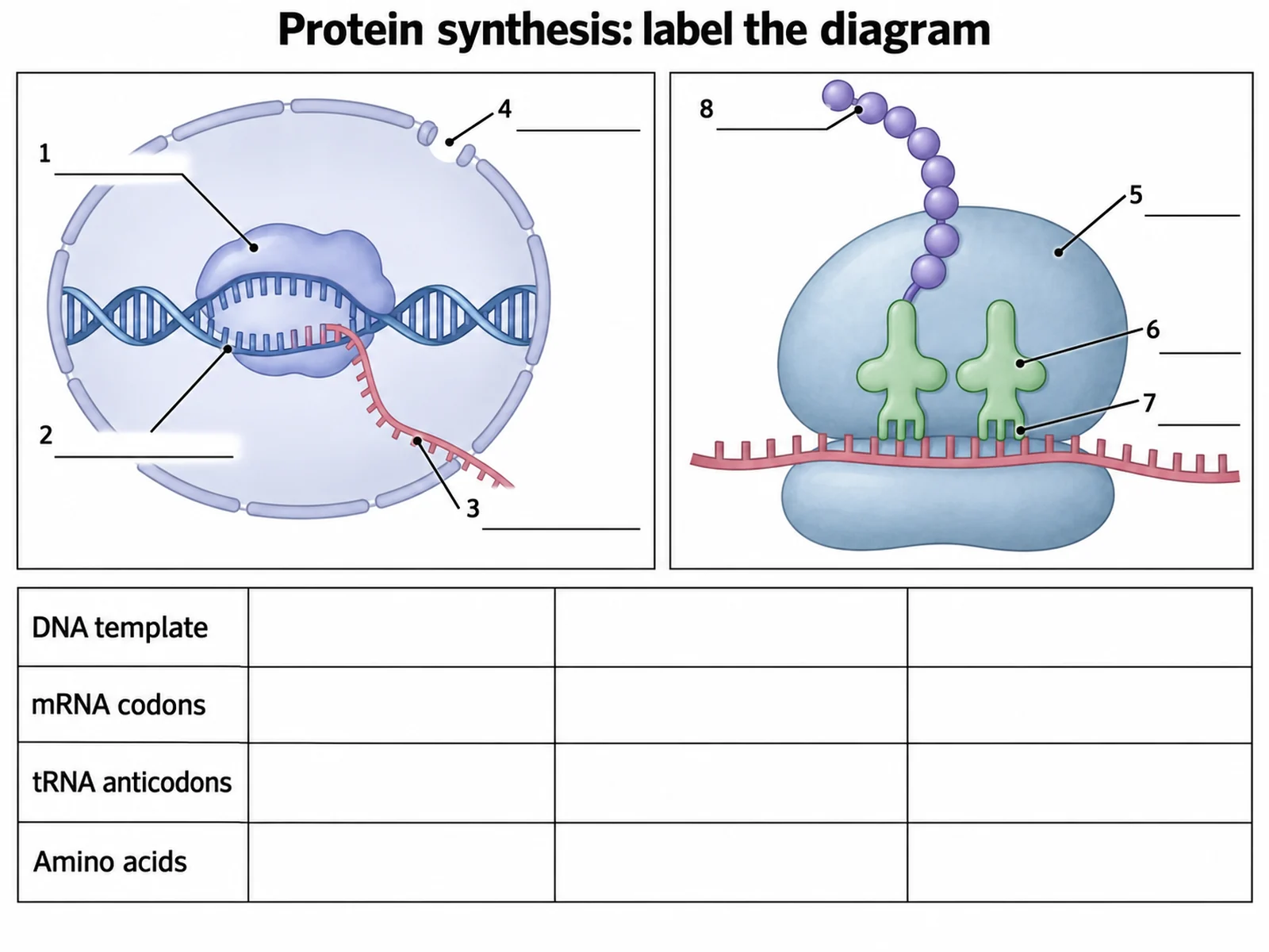
Blank protein synthesis worksheet with unlabeled transcription and translation panels, empty label lines, and blank codon boxes

Prompt

Create a blank protein synthesis worksheet. Draw an unlabeled transcription panel inside a nucleus outline and an unlabeled translation panel with a ribosome in the cytoplasm, with numbered leader lines pointing at RNA polymerase, the template strand, the mRNA, the nuclear pore, the ribosomal subunits, the tRNA, the anticodon, and the polypeptide, and nothing written on them. Below the panels add a short empty table with one row for a DNA template sequence, one for the mRNA codons, one for the tRNA anticodons, and one for the amino acids. Leave generous white space and no answer key.

Blank Protein Synthesis Worksheet

How to make a transcription and translation diagram

1

Pick the stage

Say whether you want transcription only, translation only, or the full DNA to RNA to protein sequence, and name the structures you want labeled.

2

Generate the diagram

Create a clean biology figure with the nucleus and cytoplasm separated, RNA polymerase and the ribosome in the right places, and readable leader lines.

3

Check and export

Confirm the strand directions, codon sequences, and label placement, then download the result for slides, worksheets, lab reports, or study guides.

What is a transcription and translation diagram?

A transcription and translation diagram shows how a gene becomes a protein. It is the picture of the central dogma: DNA is transcribed into messenger RNA, and that mRNA is translated into a polypeptide. A clear version keeps the two steps in different places, because in a eukaryotic cell they happen in different compartments. Transcription takes place in the nucleus, where RNA polymerase copies one DNA strand into mRNA. Translation takes place at a ribosome in the cytoplasm, where transfer RNA reads the mRNA three bases at a time and adds the matching amino acid to a growing chain.

Transcription: DNA to mRNA

  • RNA polymerase binds the promoter and unwinds a short stretch of the double helix into a transcription bubble.
  • The enzyme reads the template strand in the 3' to 5' direction and builds the mRNA in the 5' to 3' direction.
  • RNA uses uracil where DNA uses thymine, so a template adenine is copied as uracil rather than thymine.
  • In eukaryotes the new transcript is processed before it leaves: a 5' cap and a poly-A tail are added and introns are spliced out.
  • The mature mRNA passes through a nuclear pore into the cytoplasm, where translation can begin.

Translation: mRNA to protein

  • A ribosome assembles on the mRNA and scans in the 5' to 3' direction to the AUG start codon, which codes for methionine.
  • Each codon is three mRNA bases; a tRNA whose anticodon is complementary to that codon delivers the matching amino acid.
  • A peptide bond joins the incoming amino acid to the chain, the ribosome moves one codon along, and the empty tRNA is released.
  • The 64 codons specify 20 amino acids, so most amino acids answer to more than one codon.
  • Translation ends at a stop codon, UAA, UAG, or UGA, which no tRNA reads, and the finished polypeptide is released and folded.

Why use a protein synthesis diagram generator

  • Draw the nucleus and cytoplasm split that students most often get wrong, instead of sketching it on a board each lesson.
  • Keep RNA polymerase, the ribosome, codons, and anticodons labeled the same way across a whole unit of lessons.
  • Produce a close-up of one step, such as codon and anticodon pairing, without redrawing the whole pathway.
  • Turn any figure into a blank worksheet with numbered leader lines and an empty codon table.

Transcription and translation compared

  • Location: transcription is in the nucleus of a eukaryotic cell; translation is at a ribosome in the cytoplasm.
  • Template: transcription reads a DNA strand; translation reads the mRNA that transcription produced.
  • Machinery: transcription needs RNA polymerase; translation needs a ribosome, tRNA, and amino acids.
  • Product: transcription makes an RNA strand; translation makes a chain of amino acids.
  • Prokaryotes are the exception that proves the rule: with no nucleus, ribosomes can start translating an mRNA while RNA polymerase is still transcribing it, so the two steps are coupled rather than separated.

Transcription and Translation Diagram FAQ

You can create a central dogma overview, a transcription close-up inside the nucleus, a translation close-up at the ribosome, a codon and anticodon chart, a prokaryote and eukaryote comparison, or a blank protein synthesis worksheet.

Need other biology figures?

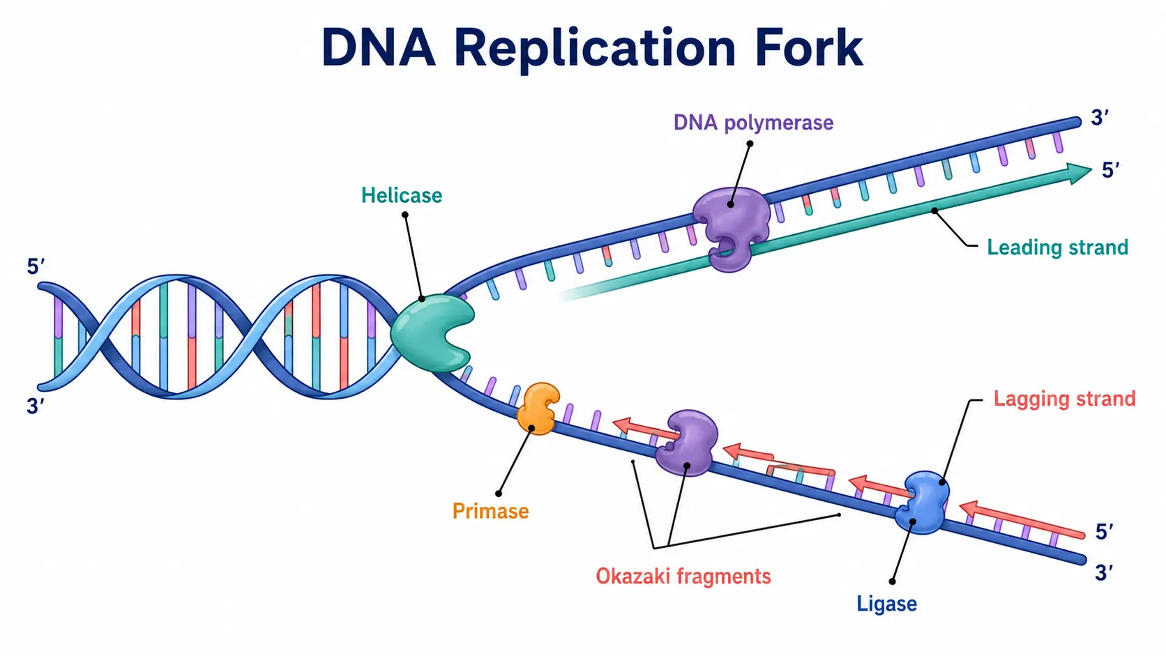
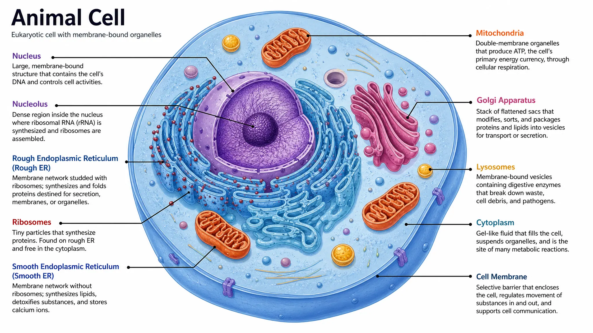
Generate DNA diagrams, cell diagrams, mitosis figures, photosynthesis visuals, and worksheet-ready biology illustrations with the same SciFigure AI workflow.