Generate a skin diagram without labels for students to fill in, or a labeled cross section of the epidermis, dermis, and hypodermis with the hair follicle, glands, vessels, and receptors named, plus a matching answer key. Zoom into the epidermal layers or the hair follicle when you need detail.
Without labels or labeledThree layers, five strataMatching answer key
Checked against OpenStax A&P
Worksheet + answer key pairs
Print-ready PNG
AI Skin Diagram Generator
Describe the section and the parts to label, or pick an unlabeled or blank version from the Labels menu.
534 / 2000 characters
Generate a publication-ready PNG from your description.
Preview
Skin Diagram Example Previews
Target aspect ratio: 4:3
Sample previews. Describe your diagram to generate your own skin figure.
Unlabeled and Labeled Skin Diagram Examples
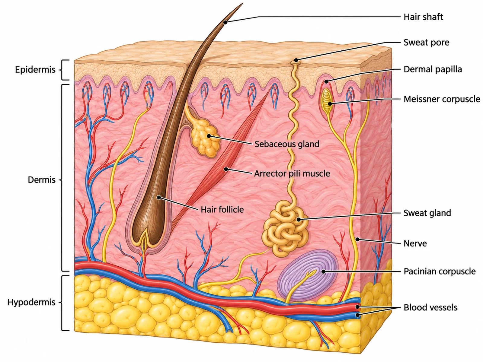
A labeled cross section of the skin layers, a skin diagram without labels with its answer key, the five layers of the epidermis, a hair follicle close-up, and an easy version for younger classes.
Prompt
Create a labeled skin diagram on a white background: a cut-away block of thin, hairy human skin showing a thin pale epidermis with a wavy lower border, a much thicker pink dermis, and a yellowish hypodermis of rounded fat cells. Show a slanting hair in its follicle with a bulb at the base, a sebaceous gland opening into the follicle, and an arrector pili muscle from the follicle up to the top of the dermis; a coiled sweat gland whose duct spirals up to its own pore; dermal papillae with capillary loops; a Meissner corpuscle in a papilla; a Pacinian corpuscle deep in the dermis; a nerve; and a red artery and blue vein in the deep dermis, with no vessels in the epidermis. Mark the layers with brackets labeled "Epidermis", "Dermis", "Hypodermis", and label "Hair shaft", "Hair follicle", "Sebaceous gland", "Arrector pili muscle", "Sweat gland", "Sweat pore", "Dermal papilla", "Meissner corpuscle", "Pacinian corpuscle", "Nerve", "Blood vessels".
Use this example
Prompt
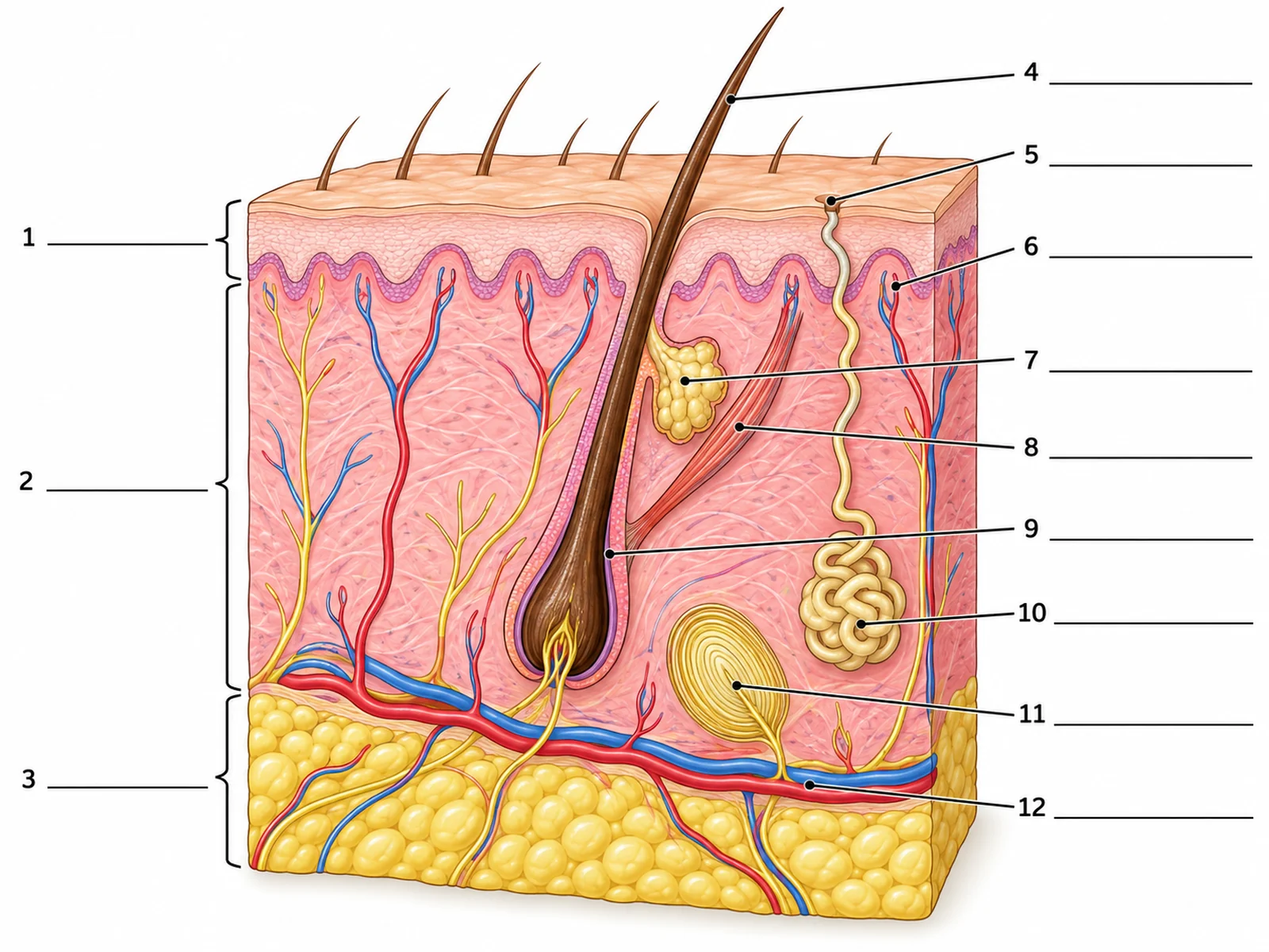
Create a blank skin diagram worksheet without labels, on a white background. Draw a rectangular block of thin, hairy human skin cut away to show its three layers from the surface down: a thin pale epidermis with a wavy lower border, a much thicker pink dermis below it, and a yellowish hypodermis of rounded fat cells at the bottom. Show one hair growing at a slant from a hair follicle that reaches down into the deep dermis, with a hair bulb at its base, a small sebaceous gland opening into the follicle, and a thin arrector pili muscle running from the follicle up to the top of the dermis on the side the hair slopes toward. Show one coiled sweat gland deep in the dermis with a duct spiralling up through the epidermis to a pore on the surface, finger-like dermal papillae pushing up into the epidermis, a Pacinian corpuscle drawn as a layered oval deep in the dermis, and a red artery and blue vein running across the bottom of the dermis with small branches into the papillae. No blood vessels inside the epidermis. Put three brackets on the viewer’s left marking the three layers, and nine thin leader lines on the right, each bracket and line ending at a short horizontal ruled answer line with a number beside it and no text, in this order: 1 = Epidermis (bracket on the left), 2 = Dermis (bracket on the left), 3 = Hypodermis (bracket on the left), 4 = Hair shaft, 5 = Sweat pore, 6 = Dermal papilla, 7 = Sebaceous gland, 8 = Arrector pili muscle, 9 = Hair follicle, 10 = Sweat gland, 11 = Pacinian corpuscle, 12 = Blood vessel. Put no names, no key and no word bank anywhere on the image.
Blank worksheet
Use this example
Prompt
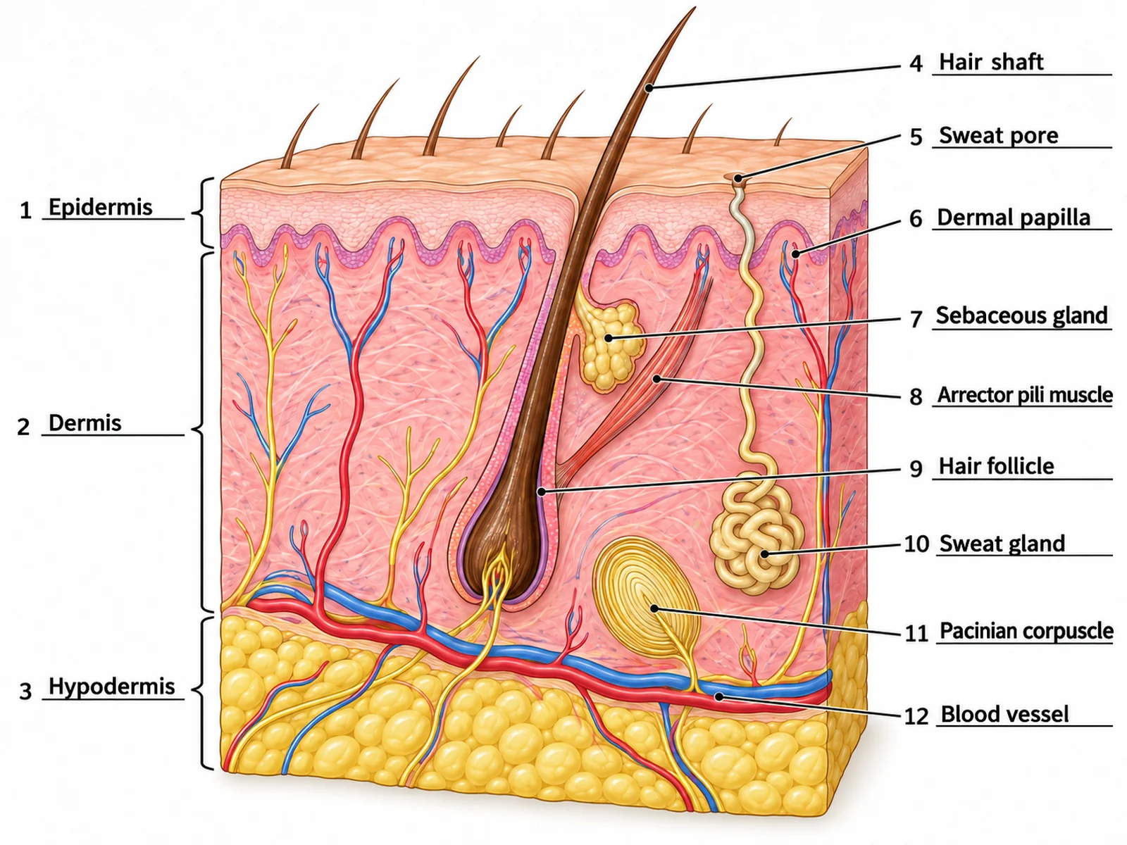
Create the answer key for a blank skin diagram worksheet, on a white background. Draw a rectangular block of thin, hairy human skin cut away to show its three layers from the surface down: a thin pale epidermis with a wavy lower border, a much thicker pink dermis below it, and a yellowish hypodermis of rounded fat cells at the bottom. Show one hair growing at a slant from a hair follicle that reaches down into the deep dermis, with a hair bulb at its base, a small sebaceous gland opening into the follicle, and a thin arrector pili muscle running from the follicle up to the top of the dermis on the side the hair slopes toward. Show one coiled sweat gland deep in the dermis with a duct spiralling up through the epidermis to a pore on the surface, finger-like dermal papillae pushing up into the epidermis, a Pacinian corpuscle drawn as a layered oval deep in the dermis, and a red artery and blue vein running across the bottom of the dermis with small branches into the papillae. No blood vessels inside the epidermis. Put three brackets on the viewer’s left marking the three layers, and nine thin leader lines on the right, each ending at a short ruled line with its number, and write the answer on each line in dark sans-serif text: 1 = Epidermis (bracket on the left), 2 = Dermis (bracket on the left), 3 = Hypodermis (bracket on the left), 4 = Hair shaft, 5 = Sweat pore, 6 = Dermal papilla, 7 = Sebaceous gland, 8 = Arrector pili muscle, 9 = Hair follicle, 10 = Sweat gland, 11 = Pacinian corpuscle, 12 = Blood vessel. Keep the numbering in this order so it matches the worksheet line for line.
Use this example
Prompt
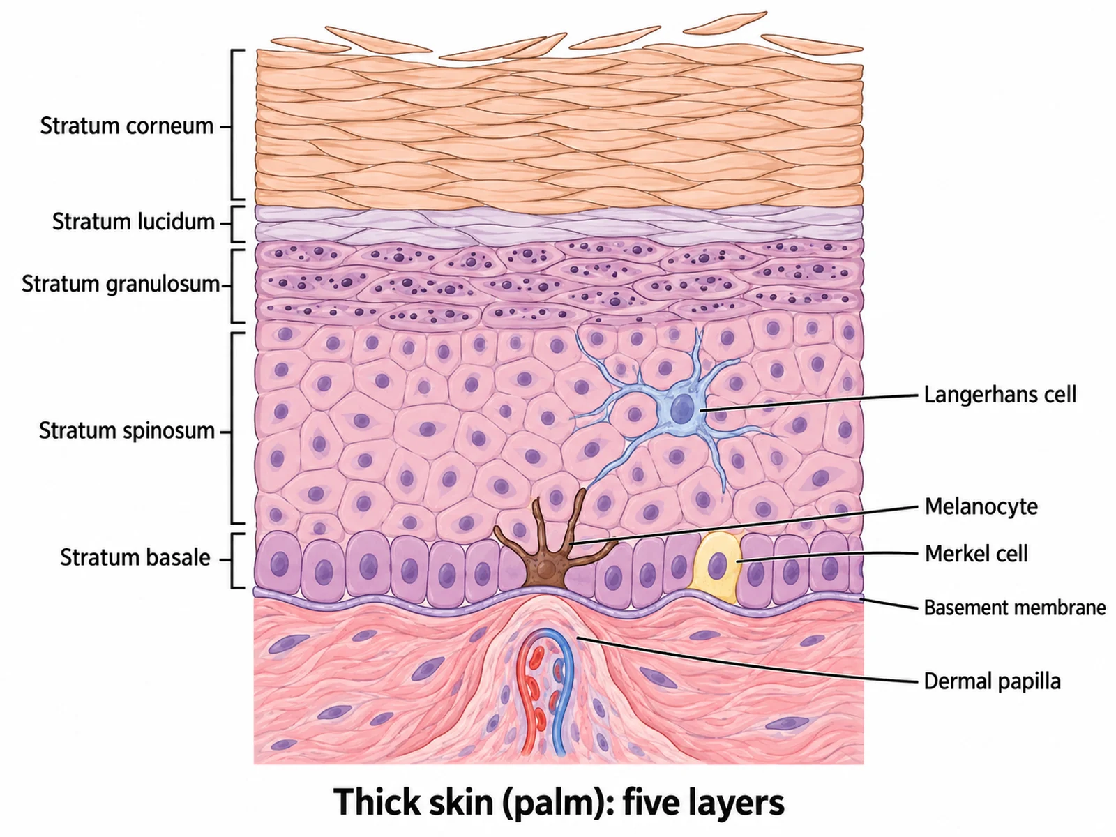
Create a labeled close-up of the epidermis of THICK skin from the palm on a white background, drawn as a magnified column of cells. From the bottom up: the dermis with a dermal papilla and a capillary loop; the STRATUM BASALE, one row of upright cuboidal cells with a dark melanocyte and a Merkel cell among them; the STRATUM SPINOSUM, several layers of polygonal cells with a branched Langerhans cell; the STRATUM GRANULOSUM, a few layers of flatter cells with dark granules; the STRATUM LUCIDUM, a thin clear band; and the STRATUM CORNEUM, many layers of flat dead cells flaking at the top. Mark each stratum with a bracket and label "Stratum basale", "Stratum spinosum", "Stratum granulosum", "Stratum lucidum", "Stratum corneum", "Melanocyte", "Merkel cell", "Langerhans cell", "Dermal papilla". No hair and no blood vessels inside the epidermis.
Epidermis layers
Use this example
Prompt
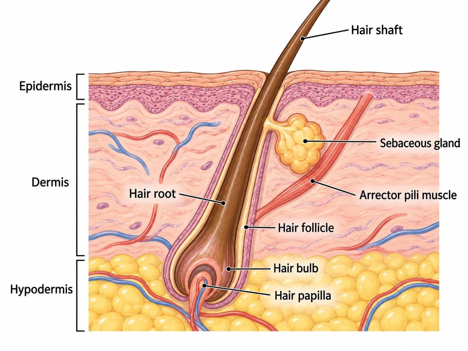
Create a labeled close-up of a single hair follicle on a white background, cut lengthwise. The follicle slants down through the epidermis and dermis into the hypodermis. Show the hair shaft above the skin surface, the hair root inside the follicle, the onion-shaped hair bulb at the base wrapped around the hair papilla, which holds a small capillary. Beside the follicle draw a lobed sebaceous gland whose short duct opens into the follicle, and a thin arrector pili muscle running from the follicle, below the sebaceous gland, up to the top of the dermis on the side the hair slopes toward. Label "Hair shaft", "Hair root", "Hair follicle", "Hair bulb", "Hair papilla", "Sebaceous gland", "Arrector pili muscle", "Epidermis", "Dermis", "Hypodermis".
Hair follicle and glands
Use this example
Prompt
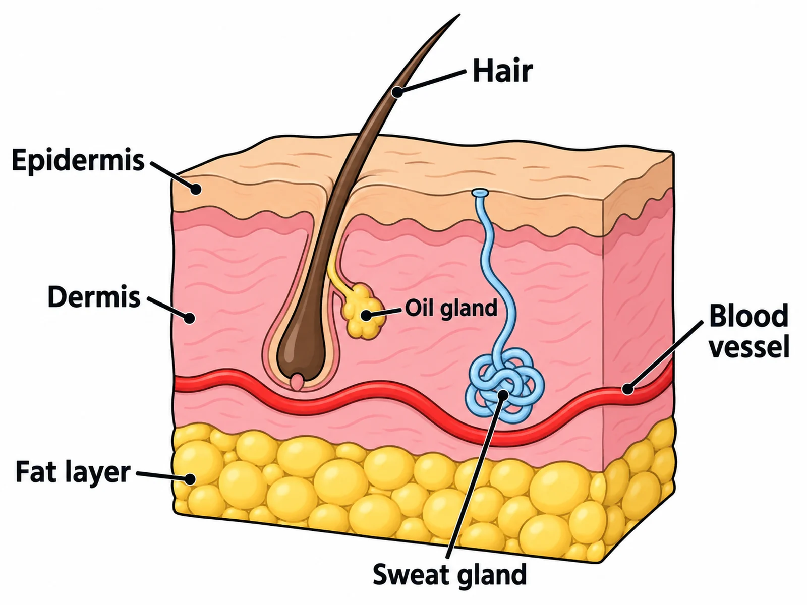
Create an easy, simple skin diagram for younger students on a white background: a clean cut-away block of skin with thick outlines, a thin top layer, a thicker middle layer and a bottom layer of round fat cells. Show one slanting hair with a small oil gland opening into its follicle, one coiled sweat gland with a tube reaching up to a pore on the surface, and a red blood vessel in the middle layer only. Label only seven things in large readable text: "Epidermis", "Dermis", "Fat layer", "Hair", "Sweat gland", "Oil gland", "Blood vessel". Plenty of white space.
Middle school
Use this example
How to make a skin diagram with or without labels
1
Pick the view, level, and labels
A full cross section, the epidermal layers, the hair follicle, the receptors, or thick vs thin skin. Then choose labeled, numbered, a blank worksheet, a word bank, or a fully unlabeled drawing.
2
Generate the diagram
The generator keeps the epidermis thin and free of blood vessels, opens the sebaceous gland into the follicle and the sweat duct onto its own pore, and puts each receptor at its real depth.
3
Make the answer key, check, and export
For a worksheet, make the answer key from the same result so the numbers match. Check the layer order and the gland openings, then download the PNG.
Skin key facts
Layers
Epidermis, dermis, hypodermis
Epidermal strata (thick skin)
5: basale, spinosum, granulosum, lucidum, corneum
Epidermal strata (thin skin)
4: no stratum lucidum
Dermis layers
Papillary (loose) and reticular (dense)
Blood vessels in the epidermis
None
Thick skin
Palms, soles, fingertips; no hair
Pigment cells
Melanocytes in the stratum basale
Surface layer renewed
About every four weeks
What is a skin diagram?
A skin diagram is a cross section through the skin, usually drawn as a cut-away block so the layers and the structures inside them show at once. The standard version shows thin, hairy skin with its three layers: the epidermis on top, the dermis below, and the fatty hypodermis at the bottom. Hair follicles, glands, blood vessels, nerves, and sensory receptors are set into them.
Most people searching for one want a skin diagram without labels. That means either a blank worksheet with numbered lines for students to fill in, or a clean unlabeled drawing for a slide or a quiz. This page makes both, along with the labeled version that serves as the answer key.
Parts of the skin to label
Epidermis: the outer layer of stacked epithelial cells. It is waterproofed by keratin and has no blood vessels.
Dermis: the thick connective-tissue layer beneath, with an upper papillary layer and a deeper reticular layer.
Hypodermis (subcutaneous layer): mostly fat. It cushions and insulates and anchors the skin to the muscle below.
Hair shaft, hair follicle, and hair bulb: the visible hair, the tube it grows in, and the growing base around the hair papilla.
Sebaceous gland: an oil gland beside each follicle that empties sebum into it.
Arrector pili muscle: a small smooth muscle that stands the hair up.
Sweat gland, duct, and pore: a coiled gland deep in the dermis with a duct spiralling up to a pore on the surface.
Dermal papillae: finger-like ridges of dermis pushing up into the epidermis, carrying capillary loops and touch receptors. On the fingertips they form fingerprints.
Blood vessels and nerves: networks in the dermis and hypodermis, with fine branches up into the papillae.
The epidermis is built from the bottom up. New cells are made in the deepest layer and pushed toward the surface, changing as they go, until they die and flake off. OpenStax gives about four weeks for the surface layer to be replaced.
Stratum basale: a single row of dividing stem cells on the basement membrane. Melanocytes sit among them and pass melanin up to the cells above; Merkel cells here sense touch.
Stratum spinosum: several layers of keratinocytes joined by strong connections that look spiny under the microscope. It also holds the Langerhans cells of the immune system.
Stratum granulosum: a few layers of flattening cells filling with keratin and waterproofing lipids. The cells here begin to die.
Stratum lucidum: a thin, clear layer of dead cells found only in thick skin.
Stratum corneum: many layers of flat, dead, keratin-filled cells. It is the actual barrier, and it is shed constantly.
The dermis and hypodermis
The dermis is the skin’s working layer. Its upper papillary layer is loose connective tissue. It forms the dermal papillae, which interlock with the epidermis like the teeth of a zip and hold its capillary loops and Meissner corpuscles. The deeper reticular layer is dense irregular connective tissue, a mesh of collagen for strength and elastic fibres for recoil. It houses the hair follicles, the sebaceous and sweat glands, larger blood vessels, and nerves. The loss of collagen and elastin here with age and sun exposure is what causes wrinkles.
The hypodermis below is not strictly part of the skin, but every skin diagram includes it. It is mostly adipose tissue, which stores energy and insulates, with the larger blood vessels and nerves that supply the dermis. Pacinian corpuscles and the bulbs of the deepest hair follicles often sit at the junction of dermis and hypodermis. Its thickness varies more than any other layer, both between body regions and between people.
Hair follicles, glands, and nails
Hair follicle: a sleeve of epidermis that dips down into the dermis. The hair grows from the matrix cells in the bulb, fed by the capillary in the hair papilla. The hair emerges at a slant, not straight up.
Sebaceous gland: produces sebum, an oily mix that softens the hair and skin. It almost always opens into a follicle, which is why thick skin, with no hair, has no sebaceous glands.
Arrector pili muscle: runs from the follicle to the upper dermis. Its contraction makes goose bumps, and in furry animals it fluffs the coat for insulation.
Eccrine sweat glands: the most numerous sweat glands, especially dense on the palms, soles, and forehead. Their watery sweat cools the body as it evaporates.
Apocrine sweat glands: in the armpits and groin. They open into hair follicles and become active at puberty; their secretion is what skin bacteria turn into body odour.
Nails: plates of hard keratin made by the nail matrix under the cuticle. A nail diagram is a separate close-up.
Sensory receptors in the skin
The skin is a sense organ as well as a covering, and receptor diagrams test whether each receptor sits at the right depth.
Free nerve endings: bare branches of sensory neurons that reach into the epidermis. They sense pain, heat, cold, and itch.
Merkel cells: in the stratum basale, paired with nerve endings. They sense light, steady touch, such as texture and shape.
Meissner (tactile) corpuscles: small oval capsules in the dermal papillae, most common in hairless skin such as the fingertips and lips. They sense light touch.
Pacinian (lamellated) corpuscles: large onion-like capsules deep in the dermis and hypodermis. They sense deep pressure and vibration.
Ruffini endings: in the dermis. They sense stretching of the skin.
Hair follicle receptors: nerve endings wrapped around the follicle. They sense the hair being moved.
Thick skin and thin skin
The words refer to the epidermis. Thick skin covers the palms, the soles, and the fingertips. It has five epidermal layers including the stratum lucidum, a very thick stratum corneum, many sweat glands, and friction ridges, but no hair follicles and no sebaceous glands. Thin skin covers everywhere else. It has four epidermal layers, a thinner stratum corneum, and hair follicles with their sebaceous glands and arrector pili muscles. That gives two easy checks for any diagram. If it shows hair, it should not show a stratum lucidum. If it is labeled as palm skin, it should show no hair.
What the skin does
Protection: the stratum corneum and its lipids block microbes and stop the body drying out. Melanin absorbs ultraviolet light before it can damage the dividing cells of the stratum basale.
Temperature control: sweating cools the body by evaporation, and the dermal blood vessels widen to shed heat or narrow to keep it.
Sensation: receptors for touch, pressure, vibration, stretch, temperature, and pain make the skin the body’s largest sense organ.
Vitamin D: ultraviolet light converts a cholesterol-based molecule in the skin into a precursor of vitamin D. The liver and kidneys then turn it into the active hormone.
Immune defense: Langerhans cells in the epidermis catch invading microbes and present them to the immune system.
Reading a skin cross section: scale and proportion
A skin diagram is drawn hugely magnified, and the proportions are where generated figures most often go wrong. The epidermis ranges from about 0.05 mm on the eyelids to about 1.5 mm on the soles. The dermis below is usually several times thicker, from about 0.3 mm to around 3 mm. The hypodermis varies most of all, from almost nothing to several centimetres of fat. A cross section of thin skin should therefore show a narrow epidermis over a broad dermis, never two bands of equal height.
Structures follow the same logic. Hair follicles and sweat glands are rooted deep in the dermis or at the top of the hypodermis, so they are drawn reaching well down the section, while their ducts and shafts pass up through the narrow epidermis. Capillaries reach only as far as the dermal papillae. Blocks of skin are usually drawn with the surface at the top, the cut faces at the front and side, and the hypodermis at the bottom resting on a hint of muscle.
Skin colour and melanin
Skin colour comes mainly from melanin, made by melanocytes in the stratum basale and passed in small packets to the keratinocytes above, where it forms a cap over each nucleus that shields its DNA from ultraviolet light. People of all skin colours have roughly the same number of melanocytes. The difference is how much melanin they make and which kind: dark brown to black eumelanin, or reddish-yellow pheomelanin. Carotene from the diet adds a yellow-orange tint, and blood in the dermal capillaries shows through as pink, which is why skin flushes with heat and pales with cold. Sun exposure makes melanocytes increase their output, producing a tan. A diagram that shows melanocytes should put them in the deepest layer, with their branches reaching between the cells above.
How skin heals
A scrape that stays within the epidermis heals completely, because the stem cells of the stratum basale divide and cover it from below and from the edges. A cut that reaches the dermis heals in stages. Bleeding vessels form a clot, which dries into a scab. White blood cells clear debris and bacteria. Fibroblasts in the dermis lay down new collagen, and new capillaries grow in as granulation tissue, while the epidermis creeps across from the wound edges under the scab. Over the following months the collagen is remodelled. Because the new dermis is mostly collagen, it lacks the original hair follicles and glands, and that patch is a scar. A deep wound that destroys the epidermis across a wide area cannot be covered from its edges in time and may need a skin graft.
Burns and skin depth
Health courses often use the skin diagram to explain burn depth, because the classification follows the layers. A superficial (first-degree) burn involves only the epidermis; it is red and painful but heals without scarring. A partial-thickness (second-degree) burn extends into the dermis and blisters. A full-thickness (third-degree) burn destroys the epidermis and the whole dermis, including the nerve endings, so it may be less painful at the centre. It cannot regrow from its own edges and often needs a graft. Ask for a burn-depth panel if you need one; the generator will not add it unasked.
Skin diagram labels by level
Middle school: about seven parts with everyday names, such as epidermis, dermis, fat layer, hair, sweat gland, oil gland, and blood vessel.
High school: the three layers plus the hair shaft and follicle, sebaceous gland, arrector pili muscle, sweat gland and pore, blood vessels, a nerve, and one sensory receptor.
College: the high-school set plus the papillary and reticular dermis, the dermal papillae, the hair bulb and papilla, and the Meissner and Pacinian corpuscles.
Medical: the five epidermal strata and their cell types, the receptor types at their depths, and the vascular plexuses of the dermis.
The skin as an organ
The skin is usually called the body’s largest organ. In an adult it covers roughly 1.5 to 2 square metres and makes up a large share of body weight. With the hair, nails, and glands it forms the integumentary system. It counts as an organ, not just a covering, because it is built from all four tissue types working together: epithelium in the epidermis, connective tissue in the dermis and hypodermis, smooth muscle in the arrector pili and vessel walls, and nervous tissue in its receptors and nerves. A good skin diagram shows all four.
Using a skin diagram without labels in class
Blank first, key second: hand out the skin diagram without labels, then show the answer key made from the same result so the numbers line up.
Layers as brackets, structures as lines: twelve blanks work well, three layer brackets and nine structures.
Use the unlabeled drawing for discussion: project it and have students name parts aloud before the worksheet.
Add the epidermis close-up for older students, who are expected to name the five strata in order.
Use thick vs thin skin as a check: the two sections side by side show why palms have no hair and no oil glands.
Most common mistakes on a skin diagram
These are the errors image models make most often on skin cross sections. Check each one before a diagram goes into graded work.
MistakeCorrect
MistakeBlood vessels running into the epidermis
CorrectThe epidermis is avascular; capillary loops stop in the dermal papillae
MistakeAn epidermis as thick as the dermis
CorrectIn thin skin the epidermis is a thin band over a much thicker dermis
MistakeA sweat duct opening into a hair follicle
CorrectAn eccrine sweat duct reaches its own pore on the surface
MistakeA sebaceous gland floating free or opening on the surface
CorrectIt sits beside the follicle and empties into it
MistakeArrector pili attached to the skin surface or drawn as a nerve
CorrectSmooth muscle from the follicle to the top of the dermis
MistakeStratum lucidum in hairy skin
CorrectOnly thick skin (palms, soles) has it, and thick skin has no hair
Capillary loops in the papillae, larger vessels deeper
Large vessels
Main cells
Keratinocytes, melanocytes, Merkel and Langerhans cells
Fibroblasts, with collagen and elastic fibres
Fat cells (adipocytes)
Structures inside
Sweat ducts and hair shafts pass through it
Hair follicles, glands, nerves, receptors
Deep follicle bulbs, Pacinian corpuscles
Main job
Barrier and UV protection
Strength, elasticity, blood supply, sensation
Insulation, energy store, anchoring
Skin Diagram FAQ
The three layers, epidermis, dermis, and hypodermis (subcutaneous layer), and the structures inside them. Those are a hair with its shaft, follicle, and bulb, the sebaceous gland, the arrector pili muscle, a sweat gland with its duct and pore, the dermal papillae, blood vessels, nerves, and sensory receptors such as Meissner and Pacinian corpuscles. Higher-level diagrams also name the five layers of the epidermis.
Generate body system diagrams, tissue cross sections, blank labeling worksheets with matching answer keys, and revision-ready biology figures with the same SciFigure AI workflow.