Generate a lymphatic system diagram with labels for the lymph nodes, vessels, ducts, spleen, thymus, and tonsils, or a blank lymphatic system diagram to label, with a matching answer key. Choose a full-body view, a lymph node close-up, the drainage map, or a lymph flow chart.
With labels or to labelNodes, ducts and organsMatching answer key
Checked against OpenStax A&P
Worksheet + answer key pairs
Print-ready PNG
AI Lymphatic System Diagram Generator
Describe the view and the parts to label, or pick a blank worksheet from the Labels menu.
526 / 2000 characters
Generate a publication-ready PNG from your description.
Preview
Lymphatic System Diagram Example Previews
Target aspect ratio: 3:4
Sample previews. Describe your diagram to generate your own lymphatic system figure.
Lymphatic System Diagram Examples
A lymphatic system diagram with labels, a blank diagram to label with its answer key, a lymph node cross section, the drainage map of the two ducts, and a simple drawing for younger classes.
Prompt
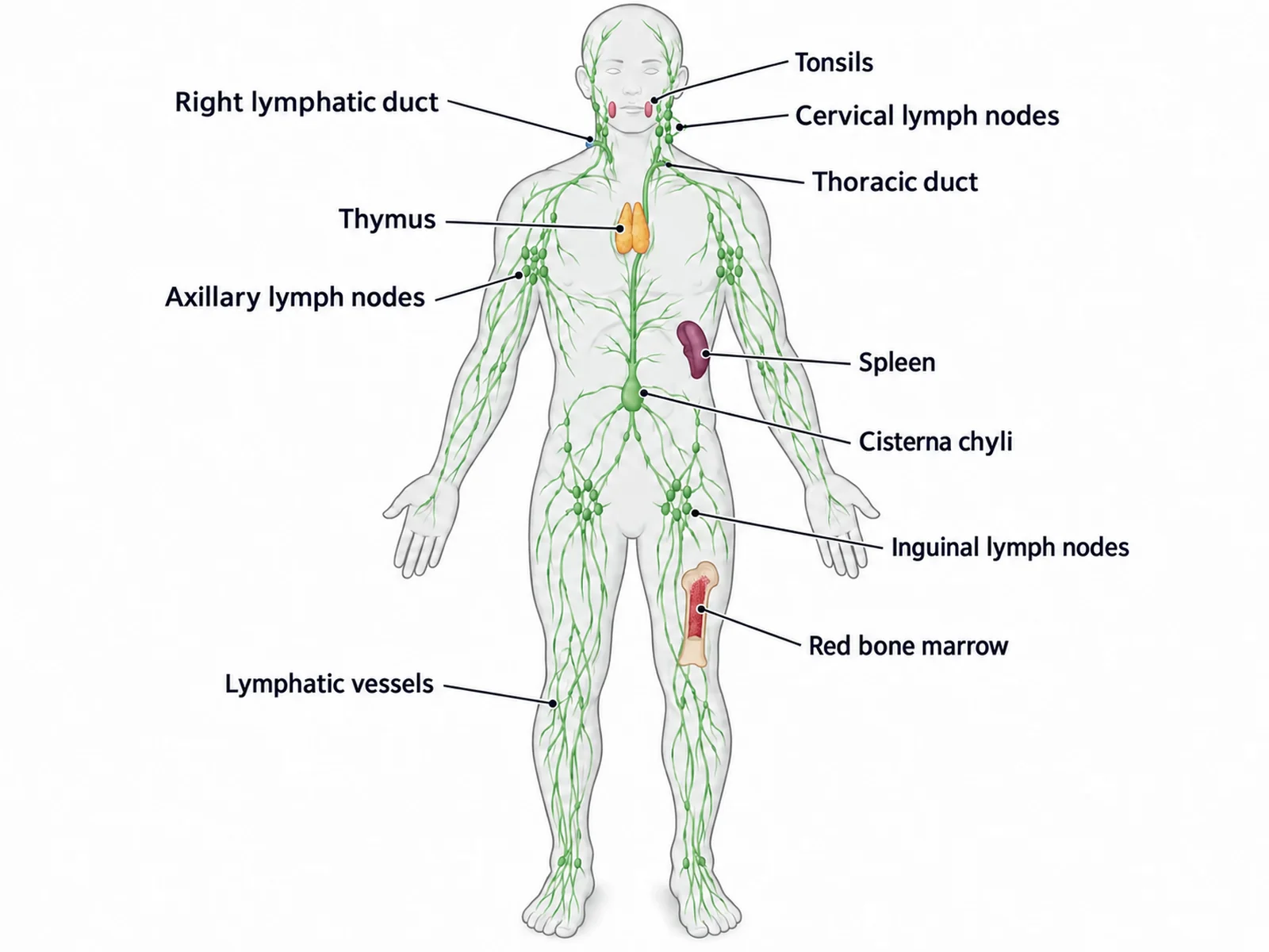
Create a lymphatic system diagram with labels on a white background: a front-facing human body outline in pale grey, the person’s right side on the viewer’s left, with the lymphatic system drawn in green. Show clusters of small bean-shaped lymph nodes in the neck, both armpits and both groins; branching lymphatic vessels up the arms and legs; the tonsils; the thymus behind the breastbone above the heart; the spleen in the upper left abdomen, on the viewer’s right; the cisterna chyli in front of the upper lumbar spine; the thoracic duct rising from it to the base of the neck on the person’s LEFT; and the short right lymphatic duct on the person’s right. Label with thin leader lines: "Tonsils", "Cervical lymph nodes", "Right lymphatic duct", "Thoracic duct", "Thymus", "Axillary lymph nodes", "Spleen", "Cisterna chyli", "Inguinal lymph nodes", "Lymphatic vessels", "Red bone marrow".
Use this example
Prompt
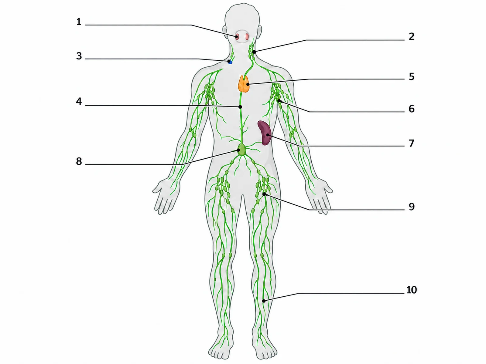
Create a blank lymphatic system diagram to label, on a white background. Draw a front-facing human body outline in pale grey, the person’s right side on the viewer’s left, with the lymphatic system drawn in green on top: clusters of small bean-shaped lymph nodes in the neck, both armpits and both groins; branching lymphatic vessels running up both arms and both legs; the tonsils at the back of the throat; the thymus in the upper chest behind the breastbone, above the heart; the spleen in the upper left abdomen under the lower ribs, which in this front view is on the viewer’s right; the cisterna chyli as a small sac in front of the upper lumbar spine; the long thoracic duct rising from it up the front of the spine to empty into the veins at the base of the neck on the person’s LEFT (viewer’s right); and the short right lymphatic duct at the base of the neck on the person’s right (viewer’s left). Draw exactly 10 thin leader lines, each ending at a short horizontal ruled answer line with a number beside it and no text, pointing at these targets in this order: 1 = Tonsils, 2 = Cervical lymph nodes, 3 = Right lymphatic duct, 4 = Thoracic duct, 5 = Thymus, 6 = Axillary lymph nodes, 7 = Spleen, 8 = Cisterna chyli, 9 = Inguinal lymph nodes, 10 = Lymphatic vessel. Put no structure names, no key and no word bank anywhere on the image. Leave generous white space.
Blank worksheet
Use this example
Prompt
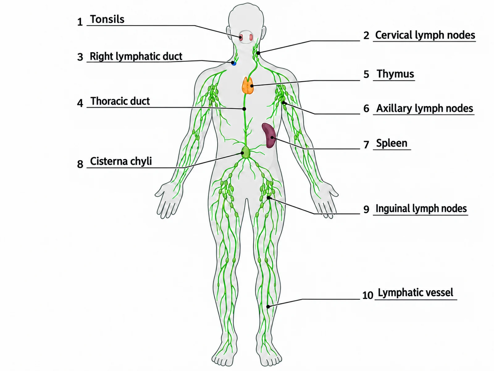
Create the answer key for a blank lymphatic system worksheet, on a white background. Draw a front-facing human body outline in pale grey, the person’s right side on the viewer’s left, with the lymphatic system drawn in green on top: clusters of small bean-shaped lymph nodes in the neck, both armpits and both groins; branching lymphatic vessels running up both arms and both legs; the tonsils at the back of the throat; the thymus in the upper chest behind the breastbone, above the heart; the spleen in the upper left abdomen under the lower ribs, which in this front view is on the viewer’s right; the cisterna chyli as a small sac in front of the upper lumbar spine; the long thoracic duct rising from it up the front of the spine to empty into the veins at the base of the neck on the person’s LEFT (viewer’s right); and the short right lymphatic duct at the base of the neck on the person’s right (viewer’s left). Draw exactly 10 thin leader lines, each ending at a short horizontal ruled line with its number, and write the answer on each line in dark sans-serif text: 1 = Tonsils, 2 = Cervical lymph nodes, 3 = Right lymphatic duct, 4 = Thoracic duct, 5 = Thymus, 6 = Axillary lymph nodes, 7 = Spleen, 8 = Cisterna chyli, 9 = Inguinal lymph nodes, 10 = Lymphatic vessel. Keep the numbering in this order so it matches the worksheet line for line.
Use this example
Prompt
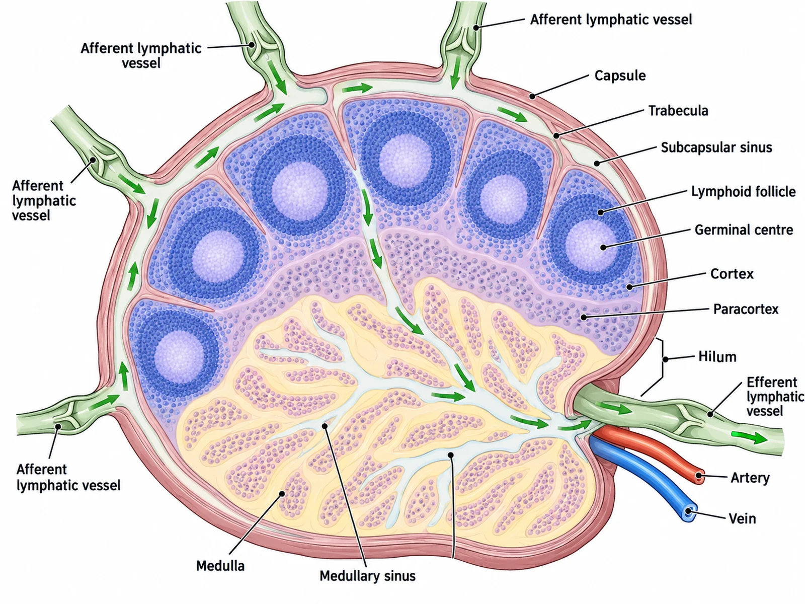
Create a labeled cross section of a single lymph node on a white background, drawn large and bean-shaped. Show a thin capsule with trabeculae reaching inward, the subcapsular sinus just under the capsule, the outer cortex with round lymphoid follicles each containing a pale germinal centre, the paracortex beneath them, and the inner medulla with medullary cords and medullary sinuses. Draw four afferent lymphatic vessels with small valves entering around the curved outer side, and one efferent lymphatic vessel leaving at the hilum, the dent on the concave side, beside a small artery and vein. Add arrows showing lymph flowing in through the afferent vessels and out through the efferent vessel. Label each part with a thin leader line.
Lymph node close-up
Use this example
Prompt
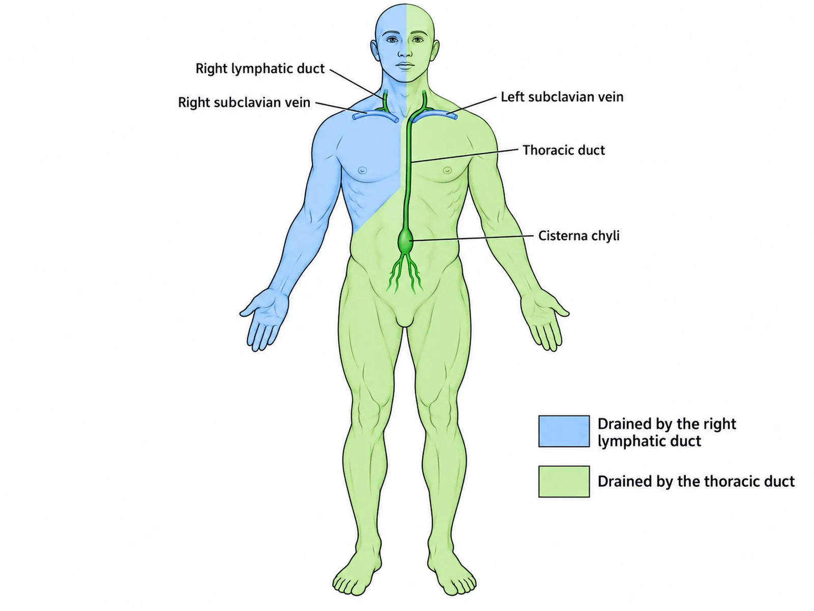
Create a lymphatic drainage map on a white background: a front-facing body outline, the person’s right side on the viewer’s left. Shade the person’s right arm and the right side of the head, neck and chest in light blue, labeled "Drained by the right lymphatic duct". Shade all the rest of the body, both legs, the abdomen, the left arm and the left side of the head, neck and chest, in light green, labeled "Drained by the thoracic duct". Draw the short right lymphatic duct ending at the base of the neck on the person’s right, and the long thoracic duct rising from the cisterna chyli in front of the upper lumbar spine to end at the base of the neck on the person’s left, where the left subclavian and internal jugular veins meet. Label "Cisterna chyli", "Thoracic duct", "Right lymphatic duct", "Left subclavian vein", "Right subclavian vein".
Drainage map
Use this example
Prompt
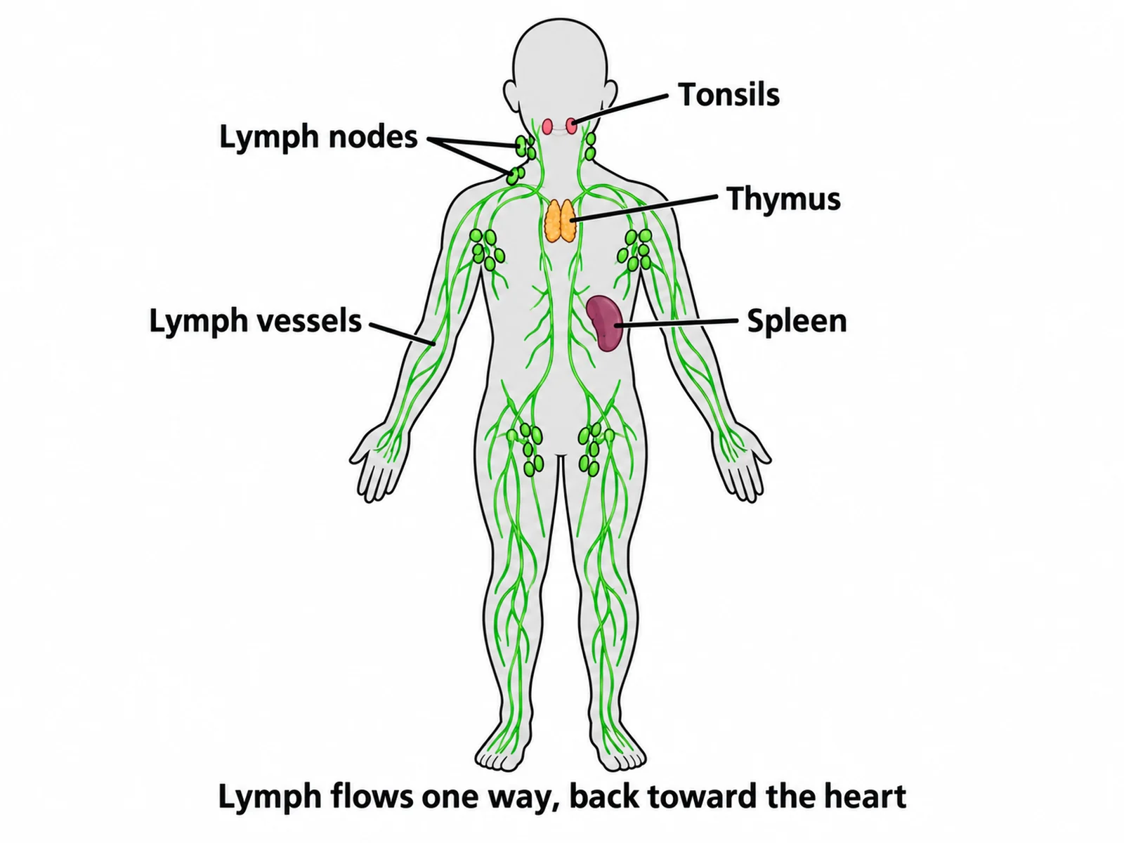
Create a simple, easy lymphatic system drawing for younger students on a white background. Draw a plain front-facing body outline with thick clean outlines, the lymph vessels as green lines in the arms, legs and trunk, and green bean-shaped lymph nodes grouped in the neck, armpits and groin. Show the tonsils in the throat, the thymus in the upper chest and the spleen in the upper left abdomen, on the viewer’s right. Label only five things in large readable text: "Lymph nodes", "Lymph vessels", "Tonsils", "Thymus", "Spleen". Add a short caption reading "Lymph flows one way, back toward the heart".
Middle school
Use this example
How to make a lymphatic system diagram with labels or to label
1
Pick the view, level, and labels
Full body, a lymph node close-up, the drainage map, the head and neck, or a lymph flow chart. Then choose labeled, numbered with a key, a blank worksheet to label, or a worksheet with a word bank.
2
Generate the diagram
The generator draws the system front-facing, puts the spleen and the end of the thoracic duct on the person’s left, keeps the two ducts unequal, and clusters the nodes where they really gather.
3
Make the answer key, check, and export
For a worksheet, make the answer key from the same result so the numbers match. Check the duct endings, the spleen, and the flow direction, then download the PNG.
Lymphatic system key facts
Lymph returned to the blood
About 3 liters a day
Lymph nodes
Several hundred, roughly 500 to 700
Primary lymphoid organs
Red bone marrow and thymus
Largest lymphoid organ
Spleen (filters blood)
Thoracic duct drains
Everything except the right arm and the right side of the head, neck and chest
Ducts empty into
The subclavian and internal jugular veins at the base of the neck
Lymph capillaries in the gut
Lacteals, which absorb fats as chyle
Pump
None: muscles, breathing, and valves keep lymph moving
What is a lymphatic system diagram?
A lymphatic system diagram is a labeled drawing of the vessels, nodes, and organs that drain fluid from the body’s tissues and return it to the blood. The usual version shows a front-facing body outline with the lymphatic system drawn in green on top. It shows clusters of lymph nodes in the neck, armpits, and groin, vessels running up the limbs, the thoracic duct climbing beside the spine, and the lymphoid organs: tonsils, thymus, spleen, and bone marrow.
Teachers use it in two forms. A lymphatic system diagram with labels names every part and works as a reference or answer key. A lymphatic system diagram to label keeps the drawing and the leader lines but leaves numbered blanks for students to fill in. This page generates both from one description, plus a numbered version with a key and a worksheet with a word bank.
Parts of the lymphatic system to label
Lymphatic capillaries: blind-ended tubes among the tissue cells. Their overlapping wall cells act as one-way flaps, letting tissue fluid in but not out.
Lymphatic vessels: larger collecting vessels with valves, running alongside veins. They carry lymph toward the trunk.
Lymph nodes: small bean-shaped filters in clusters along the vessels. They trap pathogens and debris and house lymphocytes.
Lymphatic trunks: the jugular, subclavian, bronchomediastinal, intestinal, and lumbar trunks. They gather lymph from large regions.
Cisterna chyli: a small sac in front of the upper lumbar spine where the lymph from the legs and gut collects.
Thoracic duct: the main channel. It rises from the cisterna chyli through the chest and empties into the veins at the base of the neck on the person’s left.
Right lymphatic duct: a short duct that drains the right arm and the right side of the head, neck, and chest into the veins on the person’s right.
Lymphoid organs: red bone marrow and the thymus, where lymphocytes are made and mature, plus the spleen, the tonsils, and patches of lymphoid tissue in the gut wall.
How lymph flows: from tissue fluid back to the blood
Most of the plasma that leaks out of the blood capillaries is reabsorbed. OpenStax gives about 20 liters leaving the blood each day and about 17 liters taken back, which leaves roughly 3 liters in the tissues for the lymphatic system to return. Without it the tissues would swell, which is what happens in lymphedema.
Fluid leaves the blood capillaries and bathes the cells as tissue fluid.
Tissue fluid enters the lymphatic capillaries and from then on is called lymph.
Lymph moves through collecting vessels, whose valves stop it from flowing back.
It passes through one or more lymph nodes, where it is filtered and checked by lymphocytes.
Efferent vessels from the nodes join lymphatic trunks, which feed either the right lymphatic duct or the thoracic duct.
Both ducts empty into the large veins at the base of the neck, where the subclavian and internal jugular veins meet. The lymph rejoins the blood on its way back to the heart.
Nothing pumps lymph. It is squeezed along by contracting skeletal muscles, by pressure changes in the chest during breathing, and by smooth muscle in the larger vessel walls. The valves make every squeeze push it the same way.
The right lymphatic duct and the thoracic duct
The most tested fact on a lymphatic system diagram is that the drainage is lopsided. The right lymphatic duct is only a centimeter or two long. It collects lymph from the right arm and the right side of the head, neck, and chest, roughly the upper right quarter of the body. The thoracic duct handles everything else: both legs, the pelvis and abdomen, the left arm, and the left side of the head, neck, and chest.
The thoracic duct begins at the cisterna chyli in the upper abdomen, passes up through the diaphragm and the back of the chest beside the vertebral column, and crosses to the left. It empties at the junction of the left subclavian and left internal jugular veins. On a front-view diagram that ending is on the viewer’s right, because the person’s left is on your right. A diagram that draws two matching ducts, or ends the thoracic duct on the person’s right, is wrong.
A lymph node body chart: where the nodes cluster
Lymph nodes are not spread evenly through the body. They gather where vessels from a region converge, often near a joint or the root of a limb. A lymph node body chart marks those groups on an outline of the body.
Cervical nodes: along the jaw and down the sides of the neck. They drain the head and neck, and swell with throat infections.
Supraclavicular nodes: just above the collarbone, close to where the ducts empty.
Axillary nodes: in the armpit. They drain the arm and much of the chest wall and breast, which is why they matter in breast cancer staging.
Mediastinal nodes: in the chest around the airways and great vessels.
Abdominal nodes: the mesenteric nodes along the gut, and the para-aortic nodes beside the aorta.
Inguinal nodes: in the groin. They drain the leg, the lower abdominal wall, and the skin of the genital area.
Popliteal and cubital nodes: smaller groups behind the knee and near the elbow.
Inside a lymph node
A lymph node close-up is the second most common lymphatic figure. The node is bean-shaped, wrapped in a fibrous capsule that sends partitions called trabeculae inward. Lymph arrives through several afferent vessels around the curved outer surface and spreads through the subcapsular sinus under the capsule. It filters through the outer cortex, where B cells gather in round lymphoid follicles with pale germinal centres, then the paracortex, where T cells cluster, then the medulla, with its medullary cords and sinuses. It leaves through the efferent vessel at the hilum, the indented side where an artery and a vein also enter and leave. Because more vessels bring lymph in than take it out, flow through the node slows, which gives its immune cells time to find anything foreign.
Lymphoid organs: marrow, thymus, spleen, tonsils, and gut tissue
Red bone marrow: a primary lymphoid organ. It makes all blood cells, including lymphocytes; B cells also mature here.
Thymus: the other primary lymphoid organ, where T cells mature. It sits in the upper chest behind the breastbone. It is largest relative to body size in childhood and is gradually replaced by fat after puberty.
Spleen: the largest secondary lymphoid organ, in the upper left abdomen under ribs 9 to 11. It filters blood, not lymph, removing worn-out red cells and responding to blood-borne infections.
Tonsils: rings of lymphoid tissue guarding the throat. They are the palatine tonsils at the sides, the adenoid (pharyngeal tonsil) at the back of the nose, and the lingual tonsil at the base of the tongue.
Mucosa-associated lymphoid tissue: clusters in the walls of the gut and airways, including Peyer’s patches in the small intestine and lymphoid tissue in the appendix.
What the lymphatic system does
Fluid balance: it returns the roughly 3 liters of fluid a day that the blood capillaries do not take back, with the protein that has leaked into the tissues.
Fat absorption: lacteals in the intestinal villi take up dietary fats and fat-soluble vitamins as milky chyle and carry them to the thoracic duct.
Immune defense: lymph nodes filter lymph, and the lymphocytes in the nodes, spleen, tonsils, and gut tissue recognize and respond to pathogens. Swollen, tender nodes usually mean the nodes are fighting an infection in the area they drain.
Lacteals and the lymph from the gut
The lymph that drains the small intestine is different from lymph anywhere else. Each villus in the intestinal lining has a single blind-ended lymphatic capillary at its core, the lacteal, surrounded by a net of blood capillaries. Sugars and amino acids pass into the blood capillaries. Digested fats are packed into droplets too large to enter them, so they pass into the lacteal instead. The fat-laden lymph, called chyle, is milky white. It flows through the mesenteric lymph nodes and the intestinal trunk into the cisterna chyli, up the thoracic duct, and into the blood at the base of the neck. This is why a diagram of the digestive system’s villi and a diagram of the lymphatic system share one structure, and why the thoracic duct is drawn starting in the abdomen rather than the chest.
When the lymphatic system goes wrong
Health and medical courses often add a disease panel to the standard diagram. These are the conditions it usually covers.
Swollen lymph nodes: the nodes draining an infected area enlarge as their lymphocytes multiply. That is why a sore throat brings up tender nodes under the jaw.
Lymphedema: if lymph vessels or nodes are blocked, damaged, or removed, fluid builds up in the tissues they drain. A common example is arm swelling after the axillary nodes are removed in breast cancer surgery.
Cancer spread: tumor cells can travel in the lymph to the nearest nodes. Surgeons sample the first node a tumor drains to, the sentinel node, to see whether it has spread.
Lymphoma: a cancer of the lymphocytes themselves, which often first shows as painless, enlarged nodes.
Lymphatic filariasis: parasitic worms carried by mosquitoes block the lymph vessels and can cause severe swelling of the legs.
Lymphatic vessels, veins, and the immune system
Lymphatic vessels run alongside veins. Both carry fluid back toward the heart, both rely on valves and muscle squeeze rather than a pump, and the lymph finally joins the venous blood. That is why diagrams show them together and why they are easy to confuse. The difference to draw is that lymph vessels begin as closed tubes in the tissues and pass through nodes on the way. A lymphatic system diagram also overlaps with the immune system. The tonsils, thymus, spleen, nodes, and marrow appear in both. An immune system diagram focuses on the cells, such as B cells, T cells, and macrophages, while a lymphatic system diagram focuses on the vessels and organs that hold and move them.
Simple and easy lymphatic system drawings
For younger classes a simple lymphatic system diagram works better than a full anatomical one. Six labels are usually enough: lymph nodes, lymph vessels, tonsils, thymus, spleen, and bone marrow. Draw them on a plain body outline with thick lines and large text. The middle school level in the menu does this. A good easy drawing still keeps the facts right: the spleen on the person’s left, the thymus in the chest, and a short caption saying lymph flows one way, back toward the heart. Students who later move to a full diagram then add detail to what they know rather than unlearning it.
Using a lymphatic system diagram to label in class
Blank first, key second: hand out the blank worksheet, then show the answer key made from the same result so every number matches.
Ten labels is a good size: tonsils, cervical nodes, both ducts, thymus, axillary nodes, spleen, cisterna chyli, inguinal nodes, and a vessel.
Add the drainage map as a second sheet: students shade which duct drains each region before they label the ducts.
Use the flow chart to test order: have students put the stages from tissue fluid to the subclavian vein in sequence.
Word bank or not: include one for revision, leave it out for a test. It is printed in alphabetical order, so it gives nothing away.
Colors and conventions in lymphatic diagrams
By convention lymphatic vessels and nodes are drawn in green. Real lymph is clear to pale yellow, and chyle from the gut is milky white. Green is used because it stands apart from the red arteries and blue veins that often appear in the same figure. When a diagram shows both systems, the blood vessels are usually drawn faint so the lymphatic system reads first. Arrows, if any, point toward the base of the neck. Left and right always refer to the person in the diagram, never the page.
Most common mistakes on a lymphatic system diagram
These are the errors image models make most often on lymphatic diagrams. Check each one before a diagram goes into graded work.
MistakeCorrect
MistakeTwo matching ducts, one on each side
CorrectA short right lymphatic duct and a long thoracic duct that drains about three quarters of the body
MistakeThe thoracic duct ending on the person’s right, or in the heart
CorrectIt ends at the junction of the LEFT subclavian and left internal jugular veins
MistakeSpleen on the person’s right
CorrectUpper left abdomen, under the lower ribs; on a front view it appears on the viewer’s right
MistakeLymph flowing in a loop out of and back to the heart
CorrectA one-way system from blind-ended capillaries toward the neck
MistakeThymus drawn in the neck or as the thyroid
CorrectUpper chest, behind the breastbone, above the heart
MistakeNodes spaced evenly along every vessel
CorrectNodes gather in clusters: neck, armpit, groin, chest, and abdomen
MistakeLymph vessels running into the spleen
CorrectThe spleen filters blood, not lymph
Lymphatic system vs blood circulatory system
The two systems run side by side and are easy to mix up on a diagram. This is how they differ.
Lymphatic system
Blood circulatory system
Fluid
Lymph: clear tissue fluid with lymphocytes, milky chyle from the gut
Blood: plasma, red cells, white cells, and platelets
Route
One way, from blind-ended capillaries toward the neck
A closed loop out from and back to the heart
Pump
None; skeletal muscle, breathing, and valves move lymph
The heart
Vessels
Lymphatic capillaries, collecting vessels, trunks, two ducts
Arteries, arterioles, capillaries, venules, veins
Filters
Lymph nodes filter lymph
The spleen filters blood
Where they meet
Lymph enters the veins at the base of the neck
Plasma leaks out of capillaries to become tissue fluid
Diagram color
Green by convention
Red arteries, blue veins
Lymphatic System Diagram FAQ
The standard set is the lymph nodes in their main clusters (cervical in the neck, axillary in the armpit, inguinal in the groin), the lymphatic vessels, the thoracic duct and the right lymphatic duct, the cisterna chyli, and the lymphoid organs: tonsils, thymus, spleen, and red bone marrow. Fuller diagrams add the mediastinal and abdominal nodes, the appendix and Peyer’s patches, and the subclavian veins where the ducts empty.
Generate body system diagrams, organ close-ups, blank labeling worksheets with matching answer keys, and revision-ready biology figures with the same SciFigure AI workflow.