Draw the three ways bacteria share genes: conjugation from an F+ donor to an F− recipient through a sex pilus, transformation as a competent cell takes up free DNA, and transduction as a bacteriophage carries host genes to a new cell. Make one comparison figure or a close-up of each, labeled or as a blank worksheet with its answer key.
F+, F−, Hfr and F′ cellsGeneralized vs specialized transductionBlank worksheet + answer key
Checked against OpenStax Microbiology
Worksheet + answer key pairs
Print-ready PNG
AI Bacterial Gene Transfer Diagram Generator
Pick a mechanism, or describe the cells, DNA, and labels you need.
772 / 2000 characters
Generate a publication-ready PNG from your description.
Preview
Bacterial Gene Transfer Diagram Example Previews
Target aspect ratio: 4:3
Sample previews. Describe your diagram to generate your own conjugation, transformation, or transduction figure.
Conjugation, Transformation and Transduction Diagram Examples
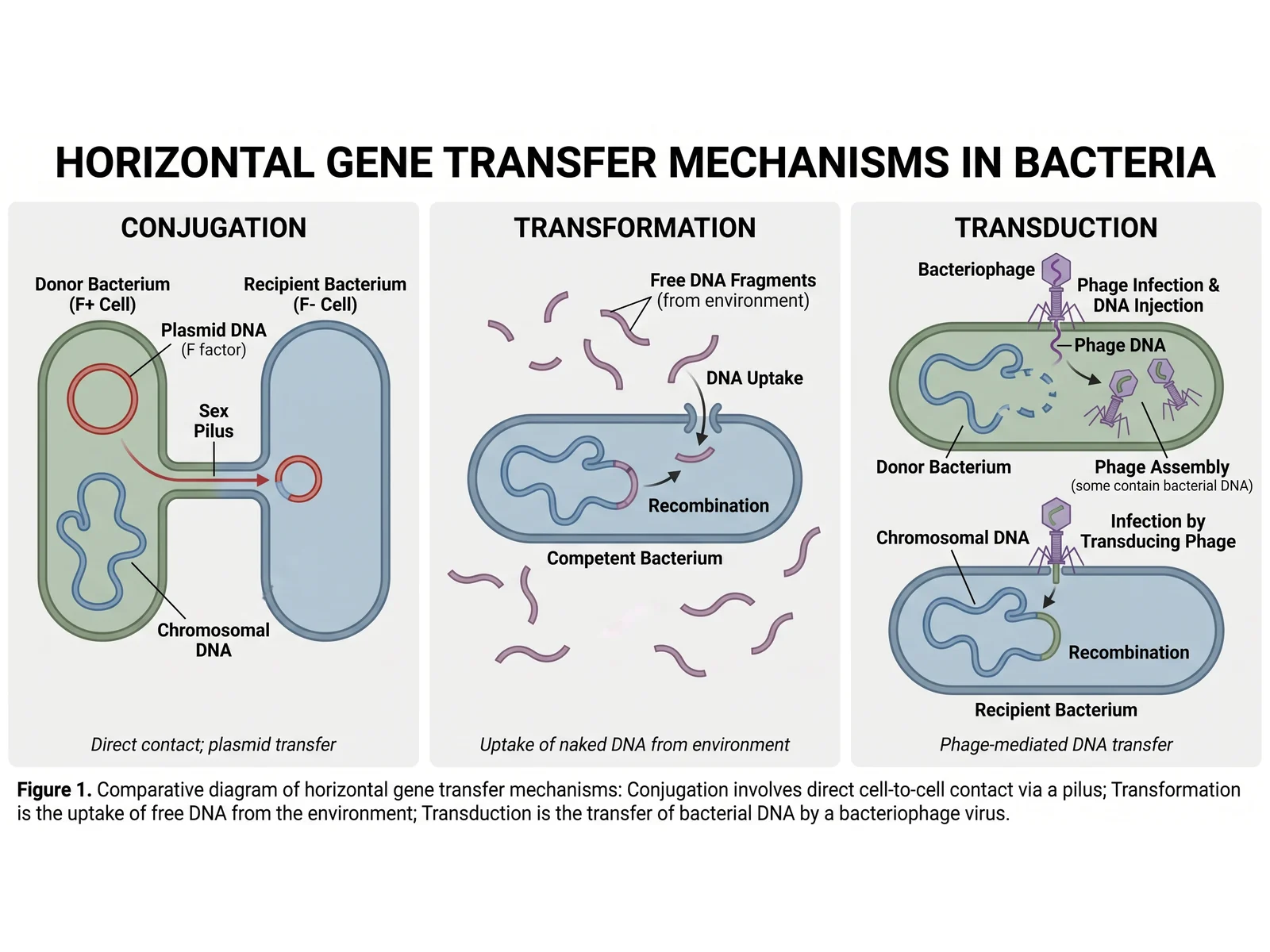
A three-panel comparison, F+ to F− conjugation and its rolling-circle close-up, transformation by a competent cell, a transducing phage delivering host DNA, and an overview of how one bacterium can gain genes by all three routes.
Prompt
Create a three-panel bacterial gene transfer diagram titled "Horizontal Gene Transfer Mechanisms in Bacteria" on a light grey background. Panel "Conjugation": a donor bacterium (F+ cell) with its chromosomal DNA and a red F plasmid (F factor), joined by a sex pilus to a recipient bacterium (F− cell); a red arrow carries plasmid DNA through the pilus to a new plasmid copy in the recipient; caption "Direct contact; plasmid transfer". Panel "Transformation": free DNA fragments from the environment around a competent bacterium; one fragment crosses the cell envelope ("DNA Uptake") and joins the chromosome ("Recombination"); caption "Uptake of naked DNA from environment". Panel "Transduction": a bacteriophage injects its DNA into a donor bacterium whose chromosome breaks into pieces; new phages assemble, some containing bacterial DNA; a transducing phage infects a recipient bacterium and the delivered DNA recombines into its chromosome; caption "Phage-mediated DNA transfer". Add a one-line figure legend summarising the three mechanisms.
Use this example
Prompt
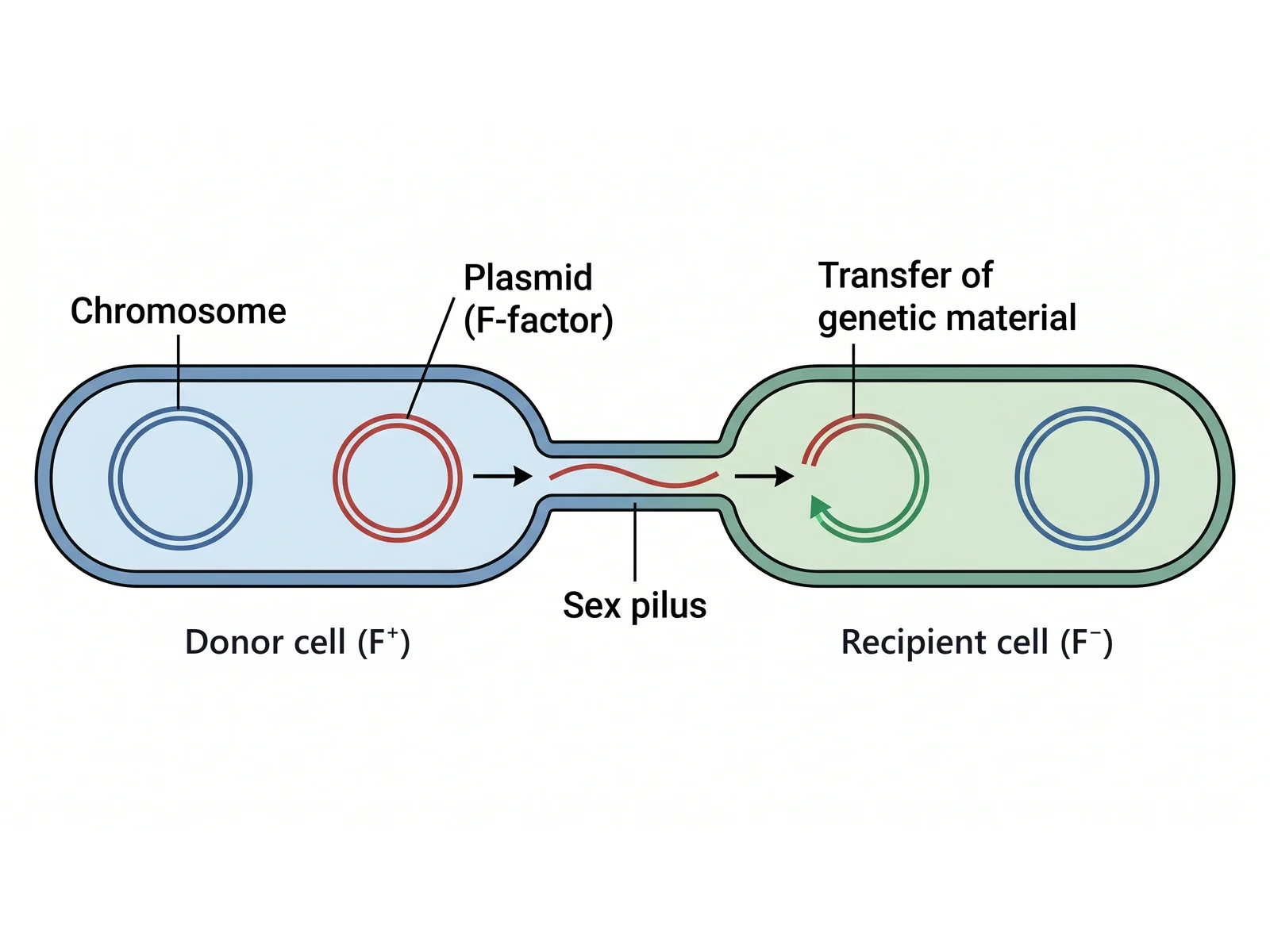
Create a diagram of bacterial conjugation on a white background: two rod-shaped cells side by side joined by a narrow sex pilus. Left, the "Donor cell (F+)", blue, with its circular chromosome and a red double-stranded plasmid labeled "Plasmid (F-factor)". A single red strand of plasmid DNA runs through the pilus, with black arrows pointing from donor to recipient. Right, the "Recipient cell (F−)", green, with its own chromosome and the arriving red strand curving into a circle while a green complementary strand is made along it, shown by a curved green arrow and labeled "Transfer of genetic material". Also label "Chromosome" and "Sex pilus".
Conjugation (F+ × F−)
Use this example
Prompt
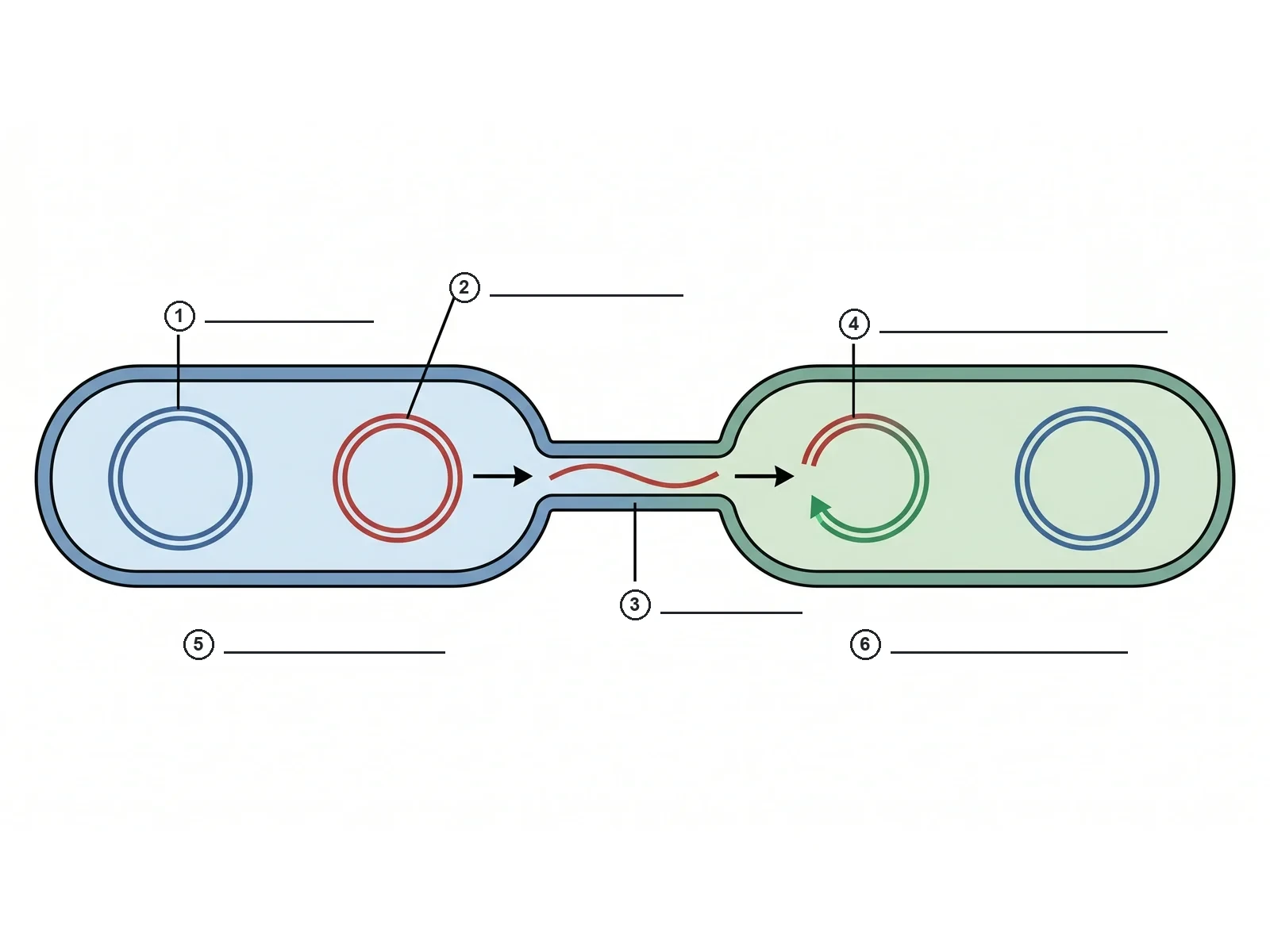
Create a blank bacterial conjugation worksheet for students to label, on a white background. Draw two rod-shaped cells side by side joined by a narrow sex pilus: a blue F+ donor on the left with its circular chromosome and a red double-stranded F plasmid, one red plasmid strand running through the pilus with black arrows from donor to recipient, and a green F− recipient on the right with its own chromosome and the arriving strand curving into a circle while a green complementary strand is made along it. Add exactly 6 numbered answer lines, each a small circled number with an empty ruled line, pointing at these targets: 1 = Chromosome, 2 = Plasmid (F factor), 3 = Sex pilus, 4 = Plasmid DNA transferred, 5 = Donor cell (F+), 6 = Recipient cell (F−). Print no names, no key and no word bank anywhere on the image.
Blank worksheetConjugation (F+ × F−)
Use this example
Prompt
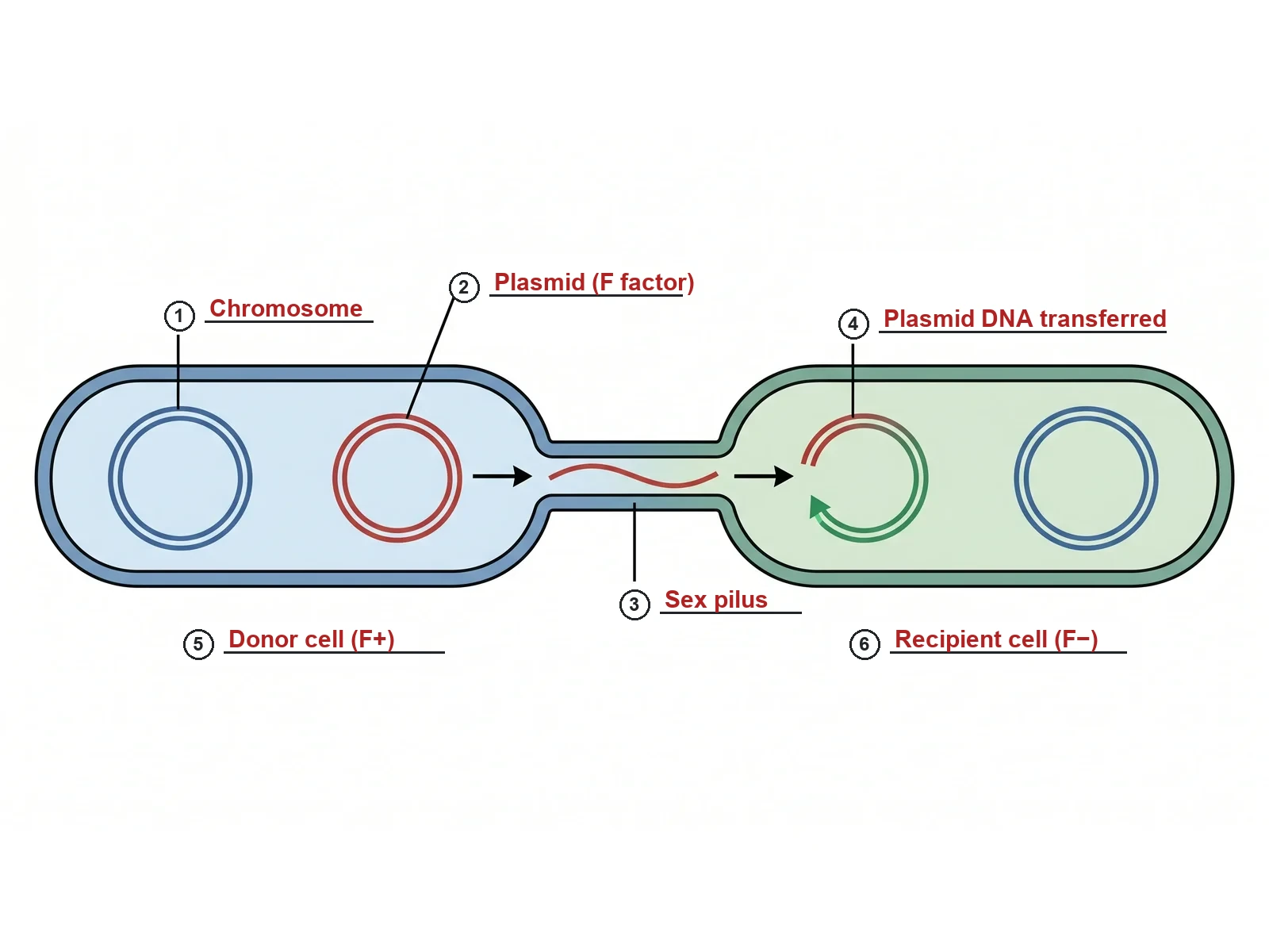
Create the answer key for a blank bacterial conjugation worksheet, on a white background. Draw two rod-shaped cells side by side joined by a narrow sex pilus: a blue F+ donor on the left with its circular chromosome and a red double-stranded F plasmid, one red plasmid strand running through the pilus with black arrows from donor to recipient, and a green F− recipient on the right with its own chromosome and the arriving strand curving into a circle while a green complementary strand is made along it. Add the same 6 circled numbers with ruled answer lines and write each answer on its line in red: 1 = Chromosome, 2 = Plasmid (F factor), 3 = Sex pilus, 4 = Plasmid DNA transferred, 5 = Donor cell (F+), 6 = Recipient cell (F−). Keep the numbering in this order so it matches the worksheet line for line.
Conjugation (F+ × F−)
Use this example
Prompt
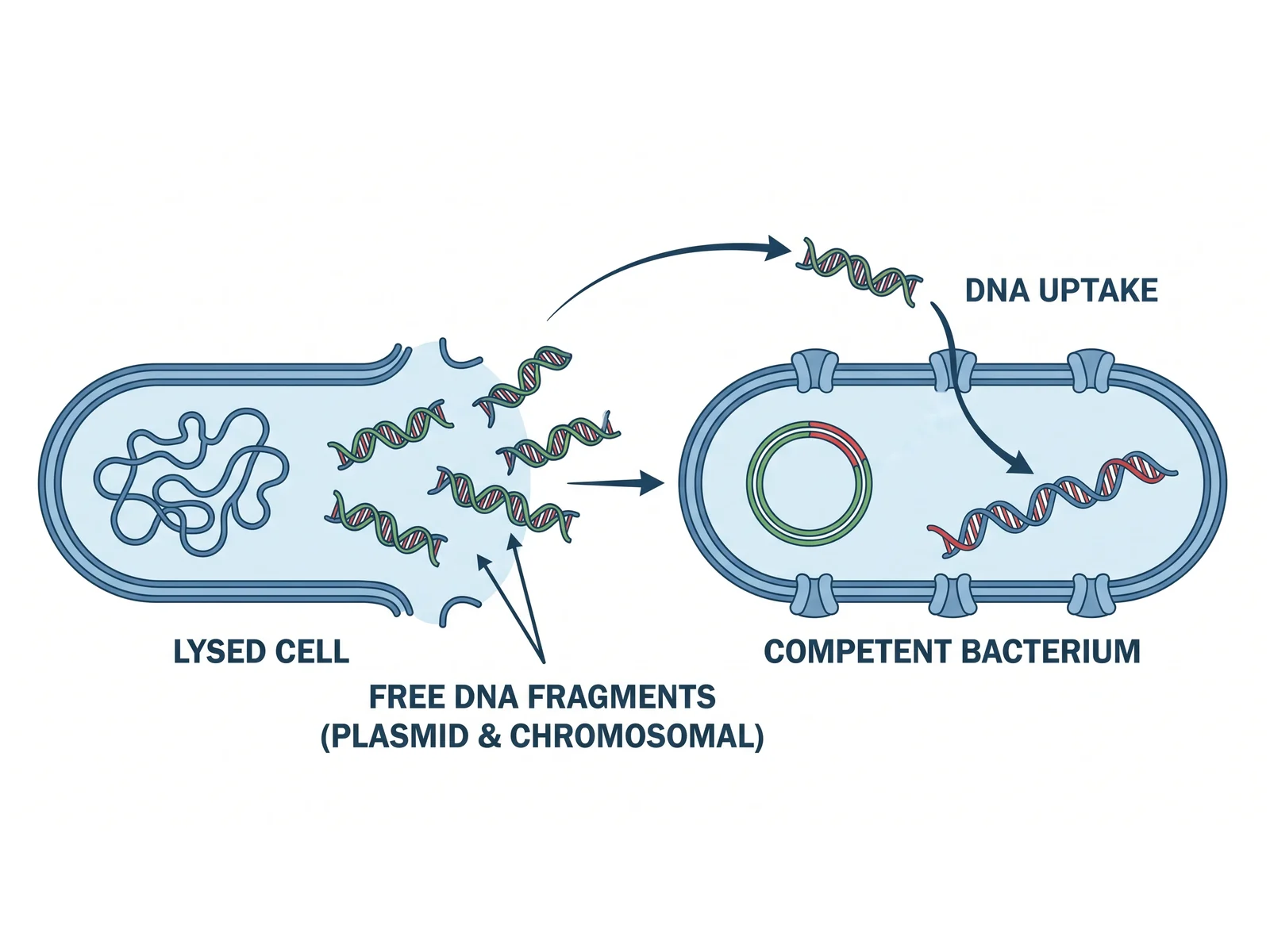
Create a bacterial transformation diagram on a white background in blue and green. Left, a "Lysed cell" with a burst envelope, its tangled chromosome still inside and short double-stranded DNA fragments spilling out, labeled "Free DNA fragments (plasmid & chromosomal)". Arrows carry one fragment to a "Competent bacterium" on the right whose envelope has uptake channels; label the step "DNA uptake". Inside the competent cell draw a circular plasmid in the donor’s green and red, and a stretch of the cell’s own blue DNA in which one strand is now red donor DNA, joined in by recombination.
Transformation
Use this example
Prompt
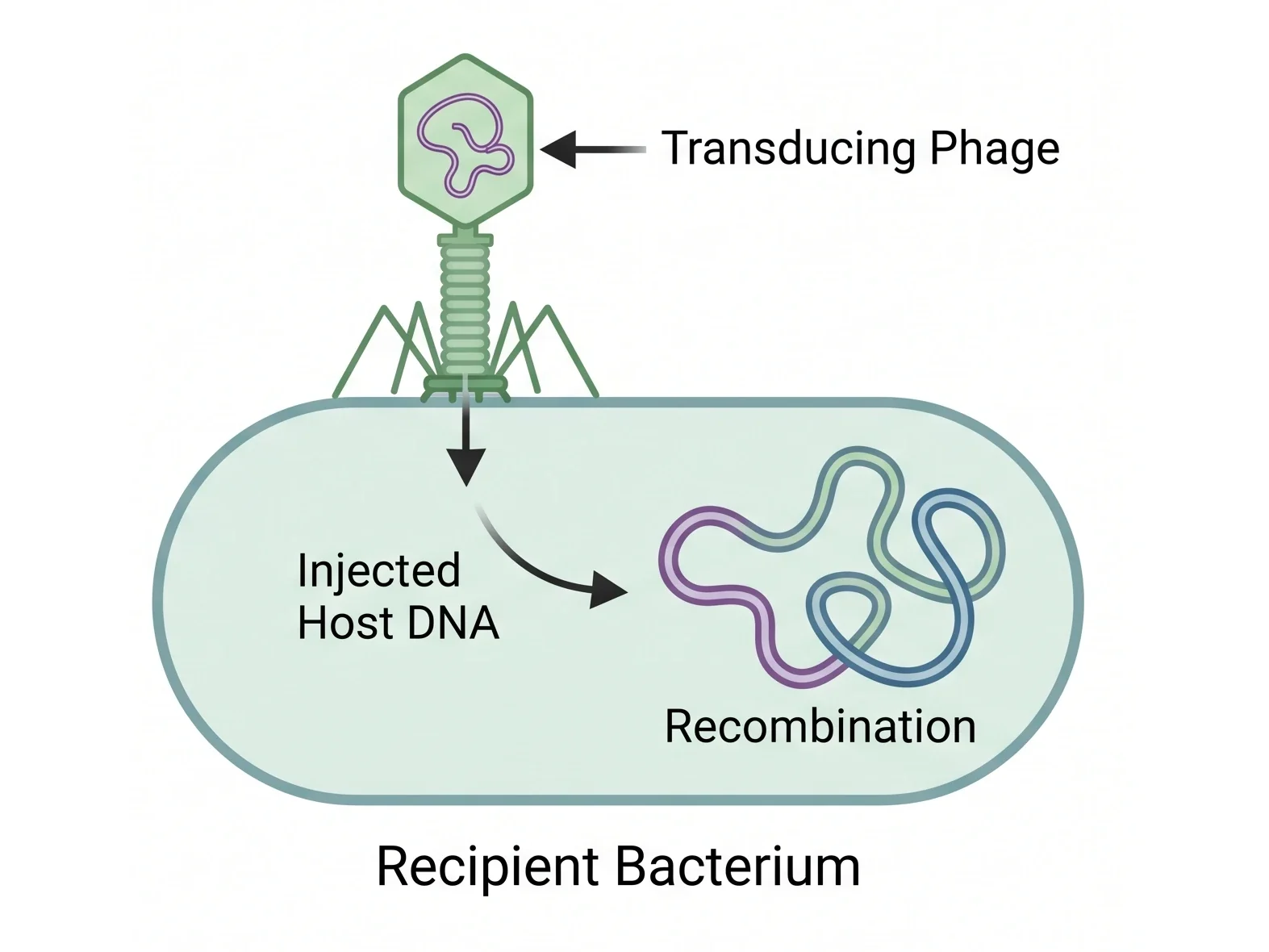
Create a transduction diagram on a white background: a bacteriophage labeled "Transducing Phage", its head holding a loop of bacterial DNA picked up from its previous host, attached by its tail fibres to the surface of a rod-shaped "Recipient Bacterium". An arrow shows the DNA going in through the tail, labeled "Injected Host DNA", and a curved arrow carries it to the recipient’s chromosome, where the incoming segment, drawn in purple, is joined into the chromosome; label it "Recombination".
Generalized transduction
Use this example
How to make a conjugation, transformation, and transduction diagram
1
Choose the mechanism, level, and labels
Compare all three, or pick F+ × F− conjugation, Hfr conjugation, transformation, generalized or specialized transduction, or add transposition. Set the level, then choose labeled, numbered, a blank worksheet, or a worksheet with a word bank.
2
Generate the figure
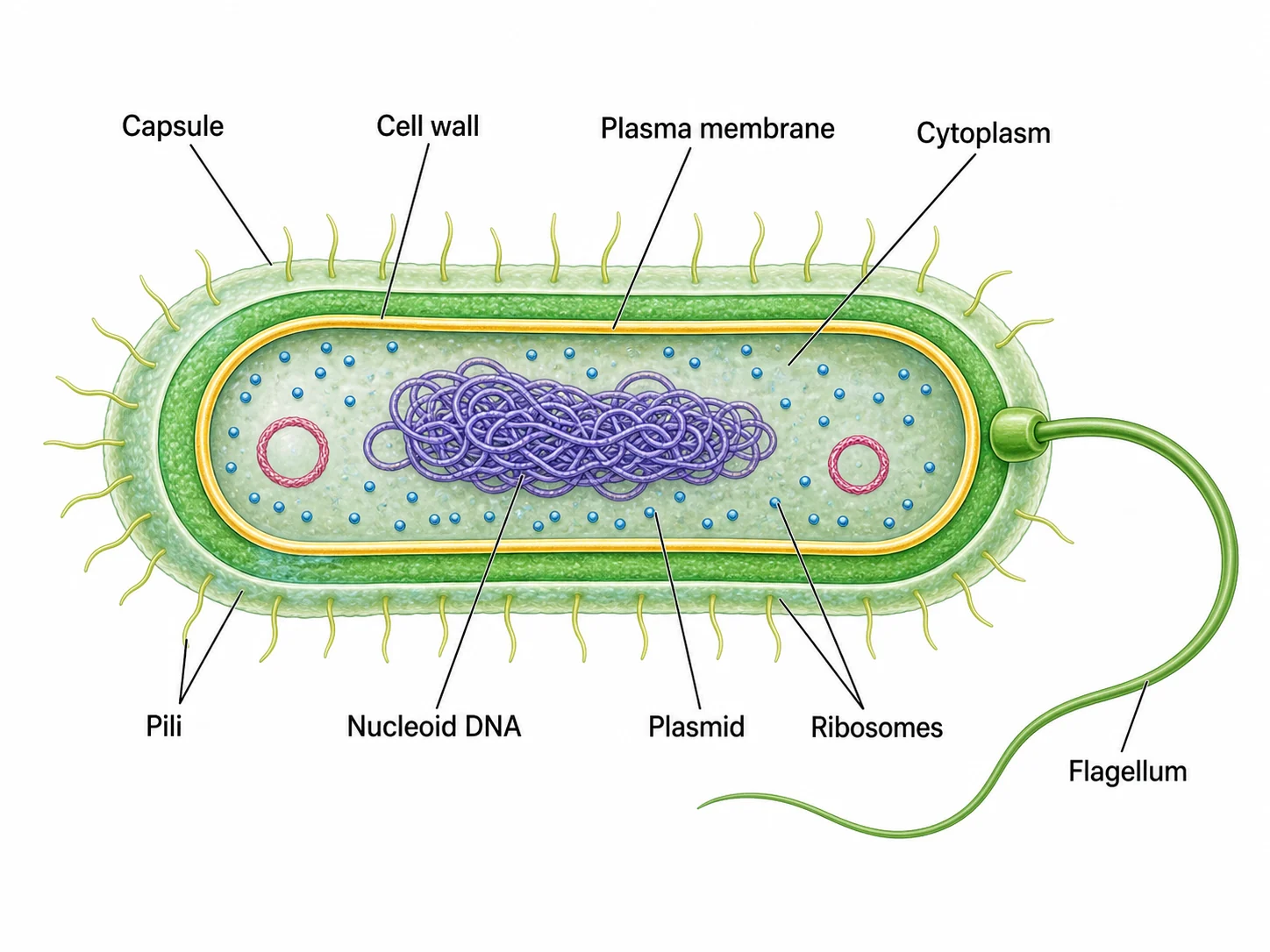
The generator draws bacteria with a nucleoid and no nucleus, sends a single plasmid strand across the pilus, keeps phages outside the cells they infect, and keeps the two kinds of transduction apart, then adds names, numbers, or empty blanks.
3
Make the answer key, check, and export
For a worksheet, make the answer key from the same result so the numbers match. Check the donor and recipient labels, the F status after mating, and the phage steps against the list below, then download the PNG.
Bacteria reproduce by binary fission, which copies one cell into two identical cells. Genes passed down that way move vertically, from parent to offspring. Bacteria also swap genes sideways, between cells that already exist and may belong to different strains or species. This horizontal gene transfer happens in three ways, and a bacterial gene transfer diagram puts them side by side: conjugation, transformation and transduction.
The figure works because the three panels can share a layout. Each has a donor, a recipient and a piece of DNA, and only the route between them changes: a pilus in conjugation, nothing at all in transformation, and a virus in transduction. Each panel ends the same way, with the new DNA kept in the recipient, either by joining its chromosome or by replicating as a plasmid.
Conjugation in bacteria, step by step
Conjugation is the one mechanism that needs two living cells in contact. The best-studied version uses the F (fertility) plasmid of Escherichia coli, and it is the one most courses ask students to describe.
Contact: the F+ donor makes a sex pilus, a thin retractable filament coded by genes on the F plasmid. The pilus tip binds an F− recipient, then retracts and draws the two cells together into a mating pair.
Nicking: an enzyme called relaxase cuts one strand of the plasmid at a site called the origin of transfer (oriT) and stays attached to its 5′ end.
Transfer: that single strand is fed into the recipient through a transfer machine in the membranes, a type IV secretion system, leading end first.
Replacement: in the donor, the strand left behind is copied by rolling-circle replication, so the donor never loses its plasmid. In the recipient, the incoming single strand is made double-stranded and closed into a circle.
Result: two F+ cells. The recipient, now called a transconjugant, can make its own pilus and pass the plasmid on.
F+, F−, Hfr and F′ cells
The same F factor can sit in a cell in three ways, and each one gives a different result when the cell conjugates.
F+ cells carry F as a free plasmid of about 100 kb. Crossed with F− cells they pass on the plasmid, but rarely any chromosomal genes.
F− cells have no F factor. They cannot make a pilus and act only as recipients.
Hfr cells have F integrated into the chromosome. Transfer starts at oriT inside F and drags the chromosome after it in a fixed order, so genes close to the start arrive first. Contact usually breaks long before the whole chromosome, about 100 minutes in E. coli, has crossed, which leaves the recipient with a linear fragment of donor chromosome. That fragment must recombine into the recipient chromosome to be kept, and the recipient usually stays F−.
F′ (F-prime) cells carry an F plasmid that left the chromosome imprecisely and took some neighbouring chromosomal genes with it. An F′ × F− cross passes those genes on with the plasmid, giving a recipient with two copies of them.
Conjugation in biology: the other meanings
Conjugation in biology means a pairing in which genetic material passes between two cells, so the word turns up outside bacteria too. In ciliates such as Paramecium, two cells join, each makes haploid micronuclei by meiosis, and they swap one micronucleus each before separating; both then have a new mix of genes. In the alga Spirogyra, cells of two filaments are joined by conjugation tubes and the contents of one cell move into the other to form a zygospore. Prokaryote conjugation differs from both: the transfer is one-way, only part of the genome moves, and no zygote forms. When an exam asks you to describe conjugation in bacteria, it means the F plasmid story above.
Transformation and competence
Transformation is gene transfer by uptake of naked DNA. When a bacterium dies and bursts, pieces of its chromosome and any plasmids it carried are released into the surroundings. A competent cell can bind that DNA and bring it inside.
Competence is a regulated state, not a constant feature. Species such as Streptococcus pneumoniae, Bacillus subtilis, Haemophilus influenzae and Neisseria switch on a set of competence genes, often when crowded or stressed.
Uptake: double-stranded DNA binds to the cell surface; in most species one strand is broken down while the other is pulled across the membrane.
Integration: the single strand pairs with the matching stretch of the recipient chromosome and replaces it by homologous recombination, which needs the RecA protein. A gene that differs from the recipient’s version changes its traits, making a transformant.
Plasmids: a whole plasmid that gets in can replicate on its own, without recombining.
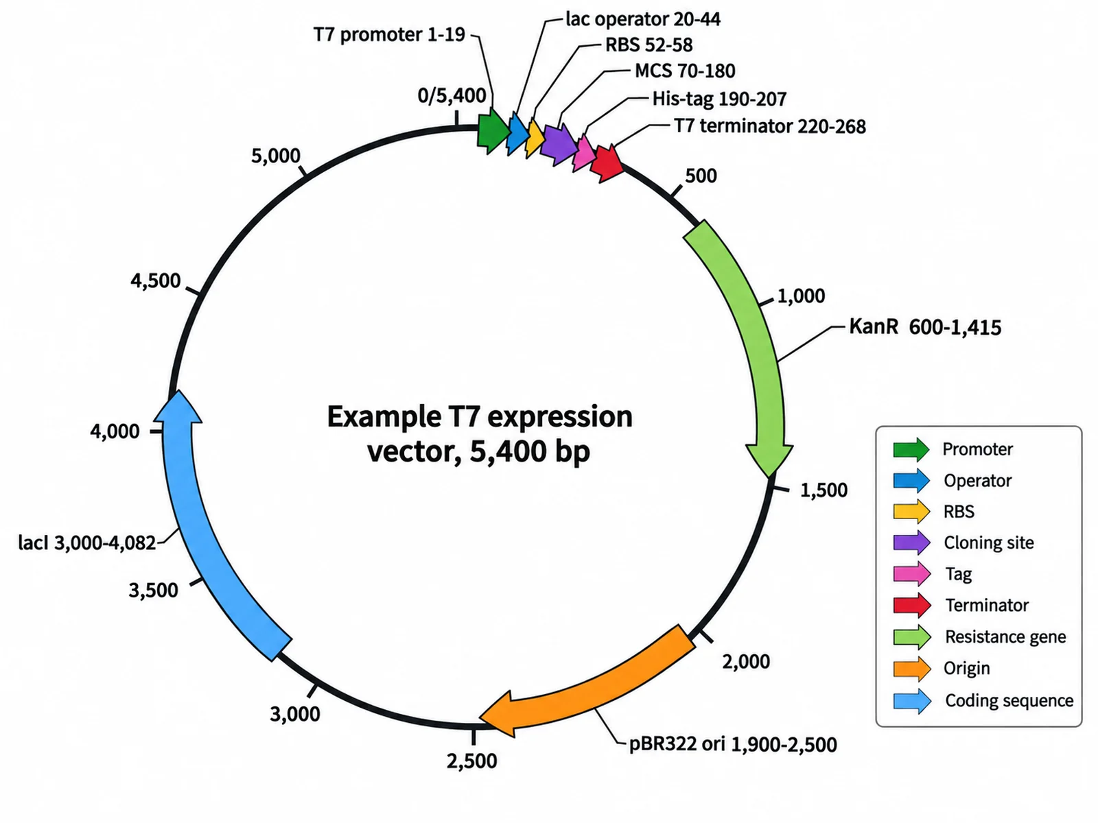
In the lab: Escherichia coli is not naturally competent, so it is made to take up plasmids by treatment with calcium chloride and a heat shock, or by electroporation. That is the transformation step in molecular cloning, and a plasmid map is the usual companion figure.
How transformation was discovered
In 1928 Frederick Griffith worked with two strains of Streptococcus pneumoniae: a smooth (S) strain with a capsule that killed mice, and a rough (R) strain without one that did not. Heat-killed S cells were harmless on their own, but mixed with live R cells they killed the mice, and live S bacteria were recovered from them. Something from the dead cells had transformed the living ones. In 1944 Oswald Avery, Colin MacLeod and Maclyn McCarty purified that transforming principle and showed that it was DNA: enzymes that destroyed protein or RNA left it active, while an enzyme that destroyed DNA stopped it. Transformation was the first evidence that genes are made of DNA, which is why it appears in genetics courses as well as microbiology.
Transduction: the horizontal transfer process that uses a virus
Transduction is gene transfer by a bacteriophage, a virus that infects bacteria. A phage has a protein head holding its genome, a tail and tail fibres. It lands on a specific receptor on the cell surface and injects its DNA, leaving the head and tail outside. Transduction happens when, by accident, a phage carries bacterial DNA instead of, or as well as, its own. Norton Zinder and Joshua Lederberg found it in Salmonella in 1952, while looking for conjugation: genes passed between strains kept apart by a filter that let only the virus through.
Generalized transduction vs specialized transduction
The two kinds of transduction differ in which phage life cycle is involved and in which genes can be carried. This is the comparison most often tested.
Life cycle: generalized transduction happens during the lytic cycle, when the phage kills its host. Specialized transduction needs a temperate phage and the lysogenic cycle, in which the phage first integrates into the chromosome as a prophage.
How the mistake happens: in generalized transduction, the host chromosome is broken up and a random fragment is packed into a phage head instead of phage DNA. In specialized transduction, the prophage excises imprecisely and takes the host DNA next to it.
Which genes move: generalized, any gene, as long as it fits in a head. Specialized, only the genes beside the integration site, such as gal or bio for phage lambda in Escherichia coli.
What the particle holds: a generalized transducing particle holds only host DNA; a specialized one holds a hybrid of phage and host DNA.
Examples: phages P22 in Salmonella and P1 in E. coli for generalized; lambda in E. coli for specialized.
Use in mapping: in generalized transduction, two genes close enough to fit in one head are often carried together. How often that cotransduction happens shows how close they are.
How to tell the three mechanisms apart
Cell contact: separate donor and recipient with a filter that bacteria cannot pass, as in the U-tube experiment. Conjugation stops; transformation and transduction can still happen.
DNase: add an enzyme that destroys free DNA. Transformation stops, because its DNA is exposed. Conjugation and transduction carry on, because the DNA is inside a mating pair or a phage head.
Living donor: conjugation needs a living F+ or Hfr donor. Transformation works with DNA from dead cells or with purified DNA. Transduction needs a phage that has grown on the donor.
Host range: conjugative plasmids can cross between distant species; transduction is limited to bacteria the phage can infect; transformation usually needs DNA similar enough to recombine.
After the DNA arrives: recombination, plasmids and barriers
Getting DNA into a cell is only half the process. A linear fragment of chromosome cannot replicate on its own, so unless it recombines into the recipient’s chromosome, swapping places with the matching stretch, it is broken down. A plasmid or a phage genome can replicate independently. Recipients also defend themselves: restriction enzymes cut DNA that lacks the cell’s own chemical marks, and CRISPR systems recognise and cut DNA from phages and plasmids met before. These barriers explain why gene transfer is most frequent between closely related bacteria. A diagram at university level can show them as gates on the recipient side.
Transposition and mobile genes
Many courses group transposition with the three transfer mechanisms, so students often draw it as a fourth one. Transposons, sometimes called jumping genes, are DNA segments that can move from one site to another, between the chromosome and a plasmid, or between two plasmids in the same cell. Transposition does not move DNA from one cell to another by itself, but it loads genes onto vehicles that can. A resistance gene on a transposon that jumps onto a conjugative plasmid can then reach many other bacteria. Choose Add transposition in the Mechanism menu for a fourth panel that shows this correctly, as movement within a genome.
Why gene transfer matters: resistance, toxins and biotechnology
Antibiotic resistance: R plasmids can carry several resistance genes at once and spread them by conjugation, including between species, which is a major reason resistance spreads quickly in hospitals and farms.
Toxins: some disease toxins are carried by phages. The genes for cholera toxin arrive in Vibrio cholerae with the CTX phage, and those for diphtheria toxin with a phage of Corynebacterium diphtheriae. Lysogenic phages that bring such genes change what their host can do.
Genome evolution: comparing bacterial genomes shows that a large share of their genes arrived by horizontal transfer rather than by descent, which blurs the branches of bacterial family trees.
Biotechnology: transformation puts recombinant plasmids into bacteria, conjugation moves genetic tools into hard-to-transform species, and transduction with phage P1 is a standard way to move mutations between E. coli strains.
Using a blank gene transfer worksheet in class
Start blank, end labeled: hand out the blank worksheet, then show the answer key made from the same result so every number matches.
Hide the headings: on a blank sheet the panel titles are left off, so students have to recognise each mechanism from the drawing.
One question per mechanism: ask what the recipient is called afterwards, transconjugant, transformant or transductant, and whether DNase would stop it.
Go deeper with the Mechanism menu: an Hfr sheet for gene order, a generalized vs specialized sheet for transduction, or a four-panel sheet with transposition.
Most common mistakes on a gene transfer diagram
These are the errors image models, and students, make most often when drawing how bacteria exchange genes. Check each one before a figure goes into graded work.
MistakeCorrect
MistakeA nucleus or a linear chromosome in a bacterium
CorrectNo nucleus: one circular chromosome in the nucleoid, plus small separate plasmids
MistakeA whole double-stranded plasmid sliding down the pilus
CorrectOne strand crosses; donor and recipient each make the complementary strand
MistakeThe recipient still labeled F− after an F+ × F− mating
CorrectBoth cells end up F+, each with a complete F plasmid
MistakeThe recipient of an Hfr mating turned into Hfr or F+
CorrectIt gets some chromosomal genes and usually stays F−
MistakeA whole phage drawn inside the cell it infects
CorrectOnly the DNA is injected; the head and tail stay outside
MistakeDonor and recipient joined in a transformation panel
CorrectNo contact and no phage: free DNA from a burst cell is taken up
MistakeGeneralized and specialized transduction swapped
CorrectGeneralized: any gene, lytic cycle. Specialized: only genes beside the prophage site
MistakeTransformation and transduction treated as the same thing
CorrectTransformation: naked DNA. Transduction: DNA carried inside a phage
MistakeConjugation called reproduction
CorrectNo new cell is made; it is one-way gene transfer
Conjugation vs transformation vs transduction
The three mechanisms side by side. Most exam questions turn on the first four rows.
Conjugation
Transformation
Transduction
How the DNA travels
Directly from donor to recipient
As free DNA in the surroundings
Inside a bacteriophage
Donor
A living F+ or Hfr cell
A cell that has died and burst, or added DNA
The cell the phage last infected
Cell-to-cell contact
Yes, through the sex pilus
No
No
Effect of DNase
None
Blocks it
None
What usually moves
A plasmid, or part of the chromosome from an Hfr cell
Short chromosomal fragments or a whole plasmid
A piece of chromosome that fits in a phage head
Recipient needs
No F plasmid (F−)
To be competent
A surface receptor the phage binds
Structures to label
Sex pilus, F plasmid, origin of transfer
Free DNA, uptake machinery, recombination
Phage head and tail, transducing particle, prophage
Recipient is called
Transconjugant
Transformant
Transductant
Discovered by
Lederberg and Tatum, 1946
Griffith, 1928; shown to be DNA by Avery, MacLeod and McCarty, 1944
Zinder and Lederberg, 1952
Conjugation, Transformation and Transduction FAQ
Conjugation is the transfer of DNA from one living bacterium to another through direct contact. In the classic Escherichia coli example, a donor cell carrying the F plasmid (an F+ cell) extends a sex pilus, attaches to a recipient without the plasmid (an F− cell), and pulls it close. One strand of the plasmid is then passed into the recipient while each cell makes the missing complementary strand. Both cells end up with a complete plasmid, so the recipient becomes F+ itself. In short, the conjugation definition in biology for bacteria is one-way, contact-dependent DNA transfer from donor to recipient.
Generate bacterial cell diagrams, plasmid maps, DNA and replication figures, blank labeling worksheets with matching answer keys, and lecture-ready biology illustrations with the same SciFigure AI workflow.