Urinary System Diagram AI

Urinary System Diagram Generator

Generate a labeled urinary system diagram following urine from the kidneys down the ureters to the bladder and out through the urethra, or a blank diagram of the urinary system for students to label. Describe the parts and the style you need, then download a clean figure for worksheets, revision notes, and slides.

Labeled and blankKidneys to urethraWorksheet-ready

AI Urinary System Diagram Generator

Describe which parts to label, or ask for a blank version to label in class.

Costs 5 credits557 / 2000 characters

Generate a publication-ready PNG from your description.

Preview

Urinary System Diagram Example Previews

Target aspect ratio: 3:4

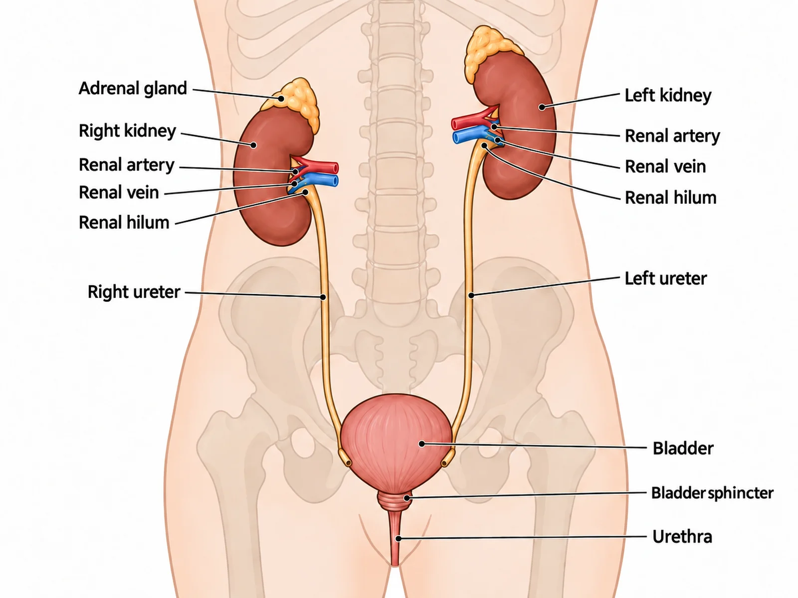
Labeled urinary system diagram naming both kidneys, both ureters, the bladder, the urethra, the renal artery and vein, and the adrenal glands
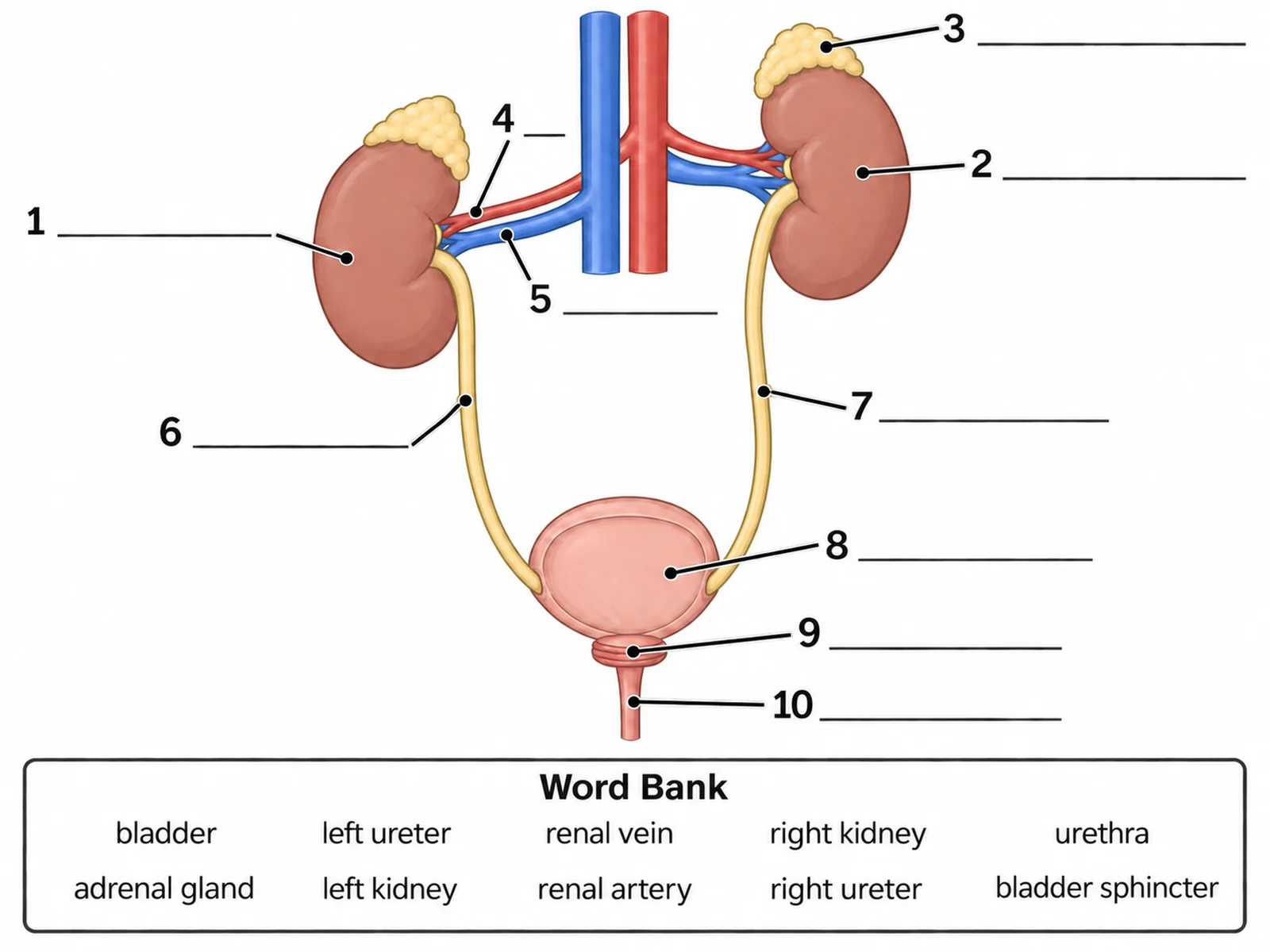
Blank unlabeled urinary system diagram with numbered empty label lines and a word bank for students to complete
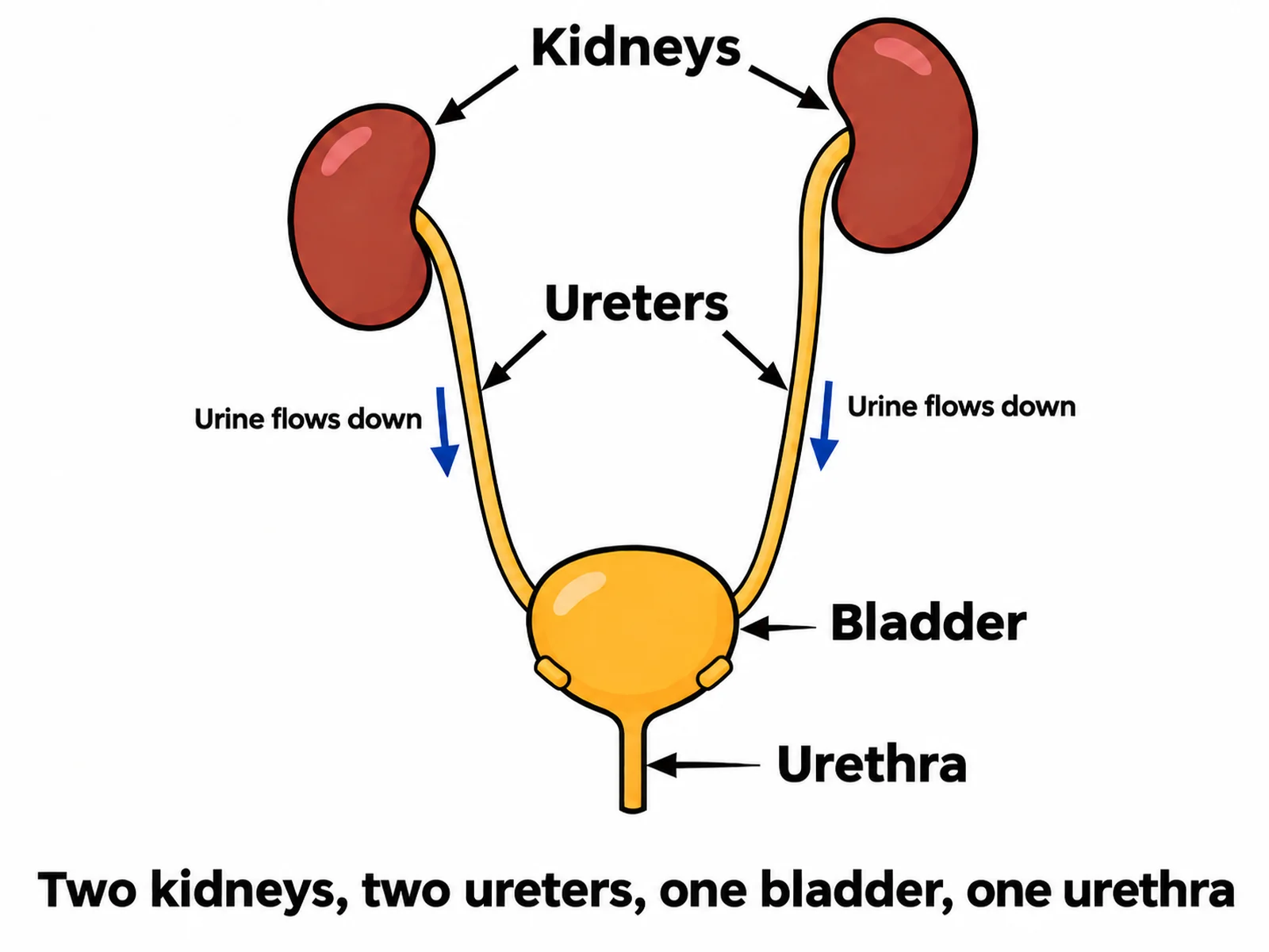
Simple urinary system drawing for younger students labeling only the kidneys, ureters, bladder, and urethra

Sample previews. Describe your diagram to generate your own urinary figure.

Urinary System Diagram Examples

Use these as visual directions for labeled anatomy, blank worksheets to label, simple versions for younger classes, kidney cross sections, and nephron close-ups.

Labeled urinary system diagram naming both kidneys, both ureters, the bladder, the urethra, the renal artery and vein, and the adrenal glands

Prompt

Create a fully labeled human urinary system diagram on a white background, drawn as a front-facing view of the abdomen and pelvis. Show exactly two bean-shaped kidneys high against the back wall, two ureters running down from them, one pear-shaped bladder in the pelvis, and one urethra leaving the bladder neck. Draw the patient’s right kidney on the VIEWER’S LEFT and clearly LOWER than the left kidney, because the liver sits above it. Cap each kidney with a separate adrenal gland, and show the renal artery and renal vein entering at the hilum. Run each ureter into the BACK of the bladder near its base at an oblique angle. Label exactly these targets with thin leader lines and readable dark sans-serif text: "Right kidney", "Left kidney", "Adrenal gland", "Renal artery", "Renal vein", "Renal hilum", "Right ureter", "Left ureter", "Bladder", "Bladder sphincter", and "Urethra".
Labeled Urinary System

Blank unlabeled urinary system diagram with numbered empty label lines and a word bank for students to complete

Prompt

Create a blank urinary system worksheet for students to label. Draw the same front-facing anatomy in clean line art with pale uniform fills and no answer labels: two kidneys with the one on the viewer’s left drawn slightly lower, an adrenal gland capping each kidney, the renal artery and renal vein at each hilum, two ureters running down the back wall, one bladder in the pelvis with both ureters entering at the back near its base, and one urethra below. Draw exactly ten thin leader lines from the ten targets to short horizontal ruled blanks, numbered 1 to 10. Add a word bank box underneath reading exactly "bladder, left ureter, renal vein, right kidney, urethra, adrenal gland, left kidney, renal artery, right ureter, bladder sphincter". Generous white space, no answer key.

Blank Urinary System to Label

Simple urinary system drawing for younger students labeling only the kidneys, ureters, bladder, and urethra

Prompt

Create a simple, easy-to-draw urinary system diagram for primary or lower-secondary students. Label only four things in large readable text: kidneys, ureters, bladder, and urethra. Use bold flat colours with thick dark outlines and plenty of white space, and omit the adrenal glands, the blood vessels, and the internal structure of the kidney. Keep the count and the geometry honest: two kidneys with the one on the viewer’s LEFT drawn a little lower than the other, two ureters, one bladder, one urethra. Add small downward arrows along both ureters labeled "Urine flows down" and a short caption reading "Two kidneys, two ureters, one bladder, one urethra".

Simple Urinary System Drawing

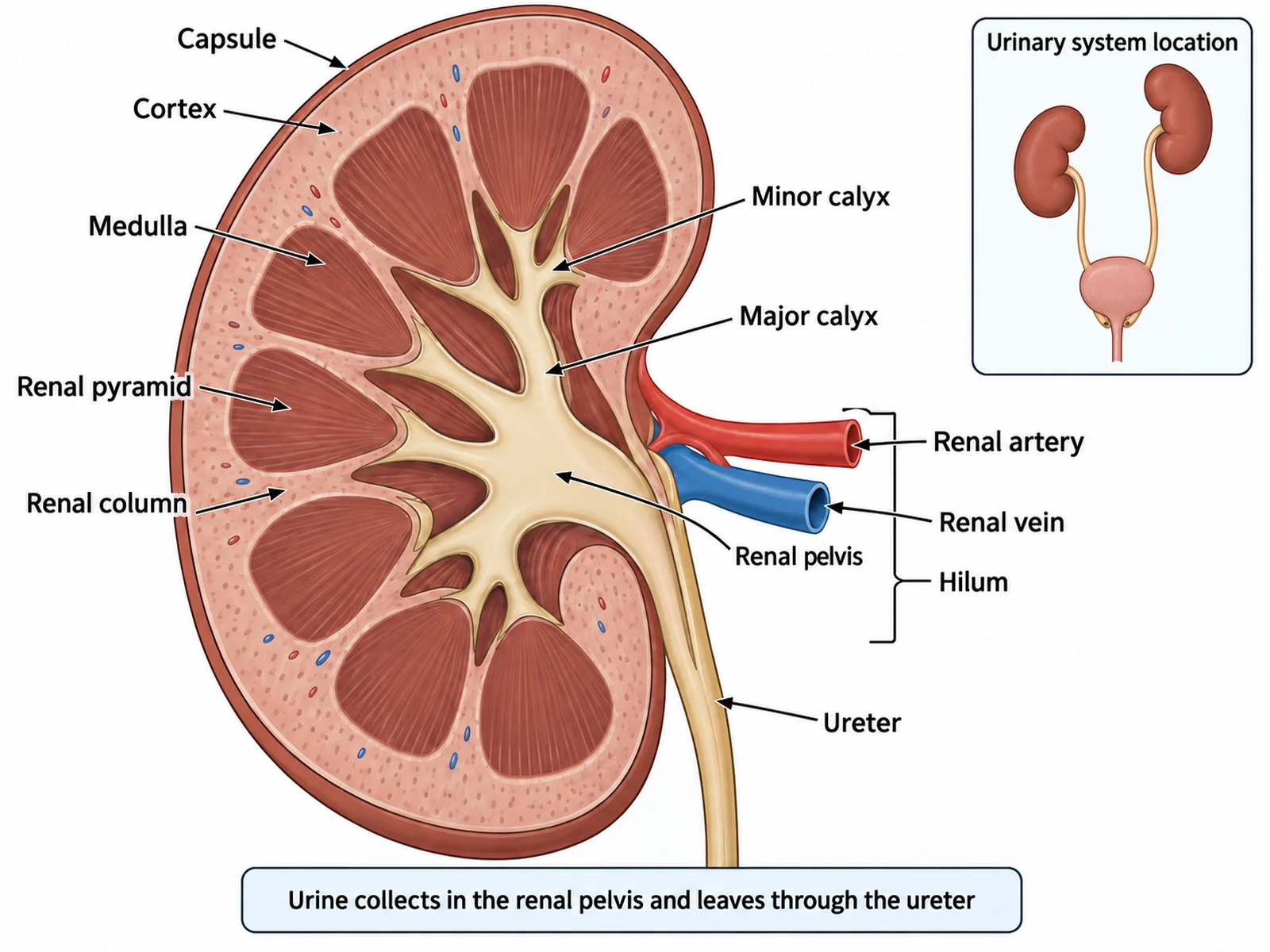
Labeled kidney cross section showing the cortex, medulla with renal pyramids, renal pelvis, and the renal artery, renal vein, and ureter at the hilum

Prompt

Create a large labeled cross section of a single human kidney on a white background, cut lengthwise so the interior is fully visible. Layer the structures correctly from the outside in: a thin fibrous capsule, then the pale outer CORTEX, then the darker MEDULLA containing several triangular renal pyramids whose tips point inward, then the funnel-shaped RENAL PELVIS collecting urine from the minor and major calyces. At the concave hilum, show the renal artery entering, the renal vein leaving, and the ureter leaving downward to carry urine away. Label capsule, cortex, medulla, renal pyramid, renal column, minor calyx, major calyx, renal pelvis, renal artery, renal vein, hilum, and ureter with thin leader lines, and add a caption reading "Urine collects in the renal pelvis and leaves through the ureter".

Kidney in Cross Section

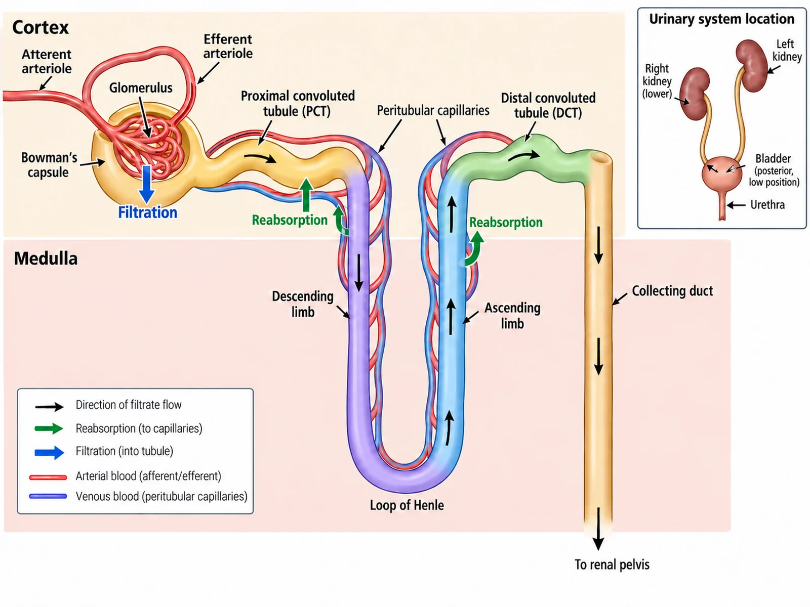
Labeled nephron diagram tracing filtrate from the glomerulus and Bowman’s capsule through the tubules and loop of Henle to the collecting duct

Prompt

Create a magnified, labeled diagram of a single nephron on a white background, with a pale band across the top marked "Cortex" and a pale band below it marked "Medulla" so the position of each part is obvious. Trace the filtrate in order: a GLOMERULUS of capillaries sitting inside Bowman’s capsule, then the proximal convoluted tubule, then the LOOP OF HENLE dipping down into the medulla with a descending limb and an ascending limb, then the distal convoluted tubule back in the cortex, then the collecting duct running down towards the renal pelvis. Keep the glomerulus, both convoluted tubules, and Bowman’s capsule in the cortex, and let only the loop of Henle reach into the medulla. Add one arrow at the glomerulus labeled "Filtration" and arrows along the tubule labeled "Reabsorption", and label the afferent and efferent arterioles and the surrounding capillaries.

The Nephron in Detail

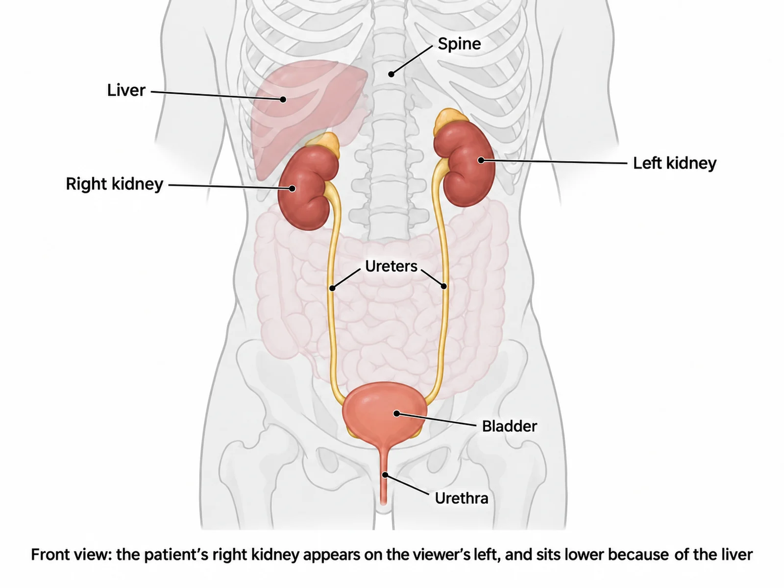
Front-facing torso diagram showing the kidneys retroperitoneal against the back wall behind the intestines, with the ureters running down to the bladder

Prompt

Create a labeled diagram showing the urinary system in position inside the human body. Draw a full front-facing torso outline in pale grey with the lower ribs, the spine, and a faint outline of the intestines sketched in, then render the urinary organs in saturated colour on top. Make the kidneys clearly RETROPERITONEAL — sitting against the back wall BEHIND the faint intestines, roughly level with the lowest ribs — and place the patient’s right kidney on the VIEWER’S LEFT of the frame, drawn lower than the left kidney with the liver sketched above it to show why. Run both ureters down the back wall into the bladder low in the pelvis. Label right kidney, left kidney, liver, ureters, bladder, urethra, and spine, and add a caption reading "Front view: the patient’s right kidney appears on the viewer’s left, and sits lower because of the liver".

Urinary System in the Body

How to make a labeled or blank urinary system diagram

1

Choose the view and the parts

A front-facing anatomical view of the whole system, a cross section through one kidney, or a magnified nephron. List the parts your syllabus requires, say whether the figure is male or female, and say whether you want the labels filled in or left blank.

2

Generate the diagram

The generator follows urine from two kidneys down two ureters into one bladder and out through one urethra, drops the right kidney below the left, and adds clean leader lines with readable text — or empty numbered lines for a worksheet.

3

Check the anatomy, then export

Confirm the lower kidney sits on the viewer’s left, the kidneys are behind the intestines, the ureters enter the back of the bladder near its base, and the urethra length matches the sex of the figure, then download the PNG for a worksheet, slide, or revision sheet.

What is a urinary system diagram?

A urinary system diagram is a labeled map of the organs that filter the blood and remove the resulting urine from the body. It is normally drawn from the front of the patient, following urine from the two kidneys high against the back wall, down the two ureters, into the single bladder in the pelvis, and out through one urethra. The same figure is used two ways in class: fully labeled as a reference, and as a blank diagram of the urinary system with numbered empty lines that students complete themselves.

Parts of the urinary system

  • Kidneys: two bean-shaped organs that filter the blood and make urine; they are retroperitoneal, lying against the back wall of the abdomen behind the intestines.
  • Adrenal glands: one caps each kidney, but they are endocrine organs — they sit on top of the kidney and are not part of it.
  • Renal artery and renal vein: blood arrives through the renal artery and leaves through the renal vein, both entering and leaving at the hilum alongside the ureter.
  • Ureters: two narrow muscular tubes, one from each kidney, that squeeze urine downward and enter the bladder at the back near its base.
  • Bladder: one muscular bag in the pelvis that stores urine; as it fills it pinches the angled ureter openings shut so urine cannot flow backwards.
  • Urethra: one tube out of the body, about 4 cm in a female and about 20 cm in a male, where it passes through the prostate below the bladder.
  • Sphincters: rings of muscle at the bladder neck that hold urine in until release.
  • Nephrons: about a million microscopic filtering units inside each kidney, which is where urine is actually made.

How urine is made and removed

  • Filtration: blood arriving through the renal artery is forced under pressure through the glomerulus into Bowman’s capsule, pushing water, salts, glucose, and urea out of the blood as filtrate.
  • Reabsorption: as the filtrate flows along the proximal convoluted tubule, the loop of Henle, and the distal convoluted tubule, glucose, most water, and useful salts are reabsorbed back into the surrounding capillaries.
  • Collection: what is left is urine; it drains through the collecting duct into the renal pelvis, which funnels it into the ureter at the hilum.
  • Transport and storage: muscular waves push urine down each ureter into the bladder, which stores it until the sphincters at its neck relax and it leaves through the urethra.

Common errors to check before you print

  • Both kidneys drawn at the same height. The right kidney sits slightly lower than the left because the liver occupies the space above it.
  • The kidneys mirrored. The diagram is a front view, so the patient’s right kidney — the lower one — belongs on the viewer’s left of the frame.
  • The kidneys placed in front of the intestines. They are retroperitoneal and belong behind the gut, against the back wall of the abdomen.
  • Ureters entering the top of the bladder. They enter at the back near the base, at an angle, and that angle is what prevents urine flowing back to the kidneys.
  • Adrenal glands drawn as part of the kidney. They sit on top of each kidney with their own outline and belong to the endocrine system.
  • A female diagram given a male-length urethra. The female urethra is about 4 cm; the male is about 20 cm and passes through the prostate.
  • More than one bladder or urethra. The count is always two kidneys, two ureters, one bladder, one urethra.

Urinary System Diagram Generator FAQ

The organs in the order urine travels: two kidneys, two ureters, one bladder, and one urethra. Around them, label the renal artery and renal vein at each kidney, the adrenal gland sitting on top of each kidney, and the sphincter at the neck of the bladder. Most syllabuses also expect the inside of the kidney to be named — cortex, medulla with its renal pyramids, and the renal pelvis where the ureter begins.

Need other anatomy figures?

Generate body system diagrams, organ close-ups, cell diagrams, and worksheet-ready biology illustrations with the same SciFigure AI workflow.