Animal Cell Diagram AI

Animal Cell Diagram Generator

Generate a labeled animal cell diagram with every organelle named, or a blank version for students to label themselves. Describe the parts and the style you need, then download a clean figure for worksheets, revision notes, and slides.

Labeled and unlabeledEvery organelle namedWorksheet-ready

AI Animal Cell Diagram Generator

Describe which animal cell parts to label and the style you need.

Costs 5 credits359 / 2000 characters

Generate a publication-ready PNG from your description.

Preview

Animal Cell Diagram Example Previews

Target aspect ratio: 4:3

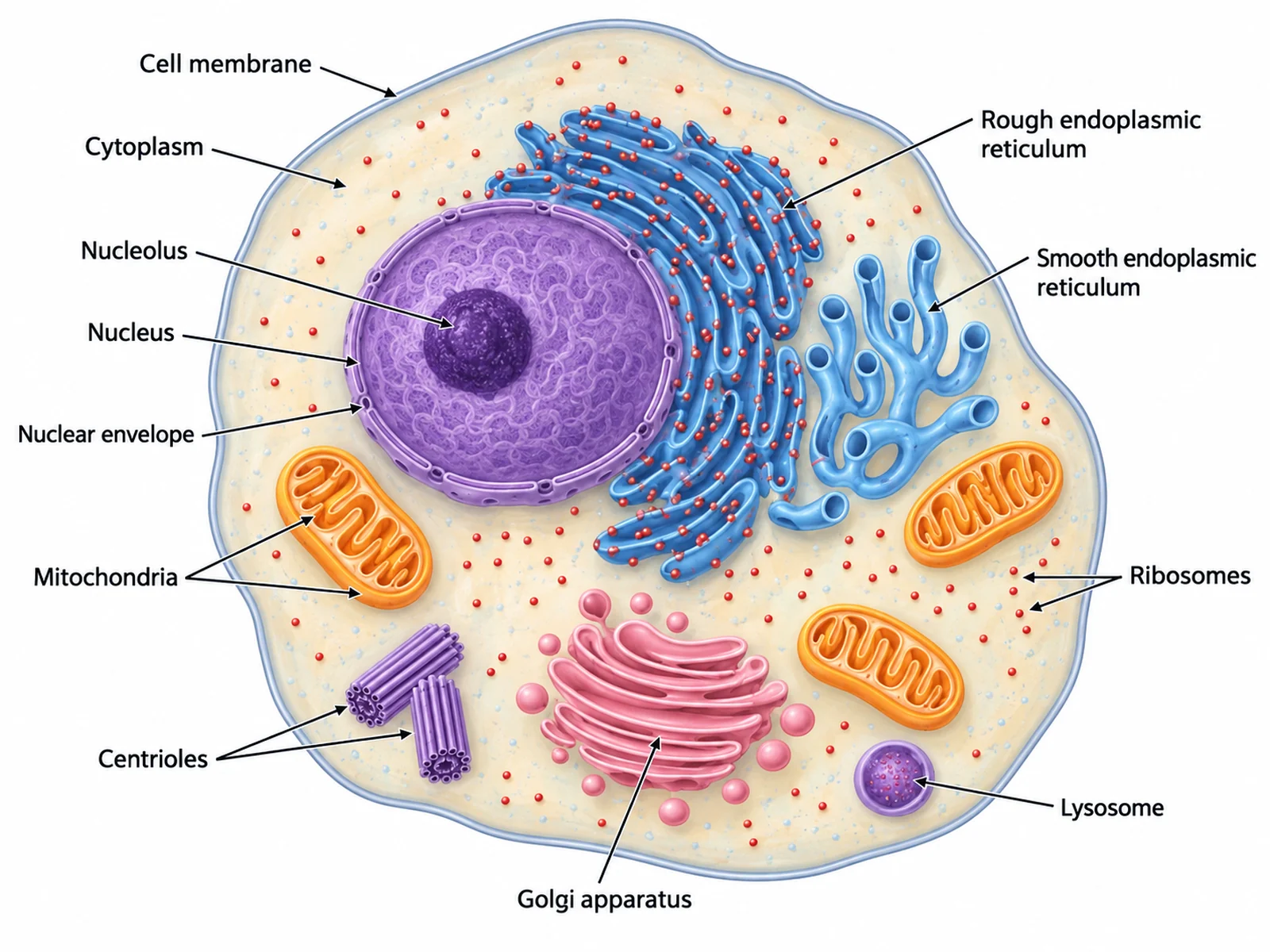
Labeled animal cell diagram naming the cell membrane, cytoplasm, nucleus, nucleolus, mitochondria, rough and smooth endoplasmic reticulum, ribosomes, Golgi apparatus, lysosome, and centrioles
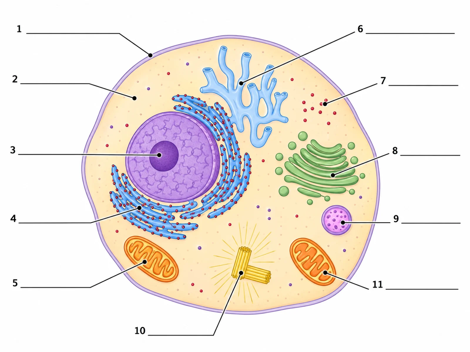
Blank unlabeled animal cell diagram with numbered empty label lines for students to complete
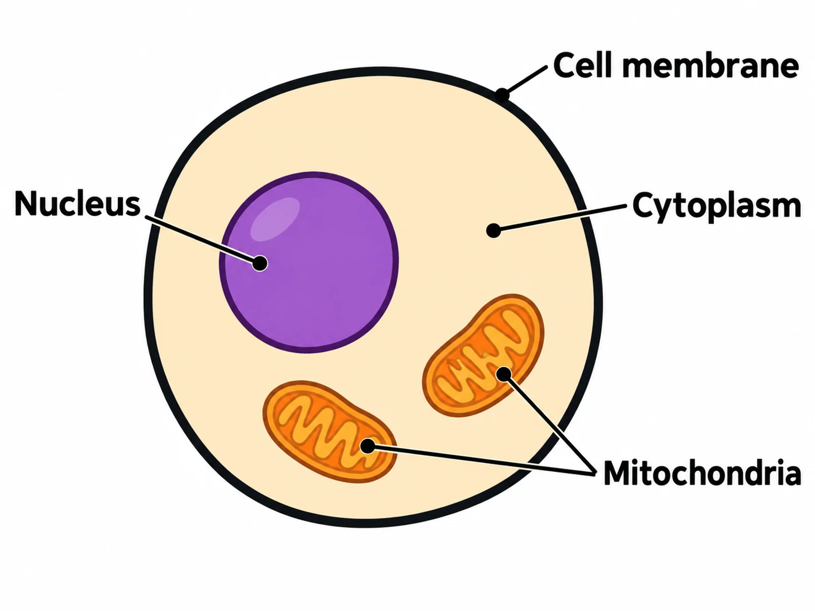
Simple animal cell diagram for younger students labeling only the cell membrane, cytoplasm, nucleus, and mitochondria

Sample previews. Describe your diagram to generate your own animal cell figure.

Labeled Animal Cell Diagram Examples

Use these as visual directions for labeled diagrams, blank worksheets, simple versions for younger classes, and animal versus plant cell comparisons.

Labeled animal cell diagram naming the cell membrane, cytoplasm, nucleus, nucleolus, mitochondria, rough and smooth endoplasmic reticulum, ribosomes, Golgi apparatus, lysosome, and centrioles

Prompt

Create a fully labeled animal cell diagram on a white background. Draw a rounded cell with a single thin cell membrane and no cell wall. Include a large round purple nucleus with a darker nucleolus and a visible nuclear envelope, two or three orange bean-shaped mitochondria with internal cristae, blue folded membrane stacks studded with small red ribosome dots labeled rough endoplasmic reticulum, a separate smooth tubular network with no dots labeled smooth endoplasmic reticulum, stacked pink flattened cisternae labeled Golgi apparatus, a small purple lysosome, and a pair of centrioles. Label every structure with a thin leader line and readable dark sans-serif text.
Labeled Animal Cell

Blank unlabeled animal cell diagram with numbered empty label lines for students to complete

Prompt

Create a blank animal cell worksheet. Draw the same animal cell outline and organelles in clean line art with soft flat fills, but leave every label empty: draw thin leader lines from each structure out to a short horizontal ruled blank line with no text on it. Number the blanks 1 to 11. Include generous white space and no answer key.

Blank Animal Cell Worksheet

Simple animal cell diagram for younger students labeling only the cell membrane, cytoplasm, nucleus, and mitochondria

Prompt

Create a simple animal cell diagram for primary or lower-secondary students. Show only four labeled parts: cell membrane, cytoplasm, nucleus, and mitochondria. Use bold flat colors, thick clean outlines, large readable text, and plenty of white space. Omit all other organelles.

Simple Animal Cell

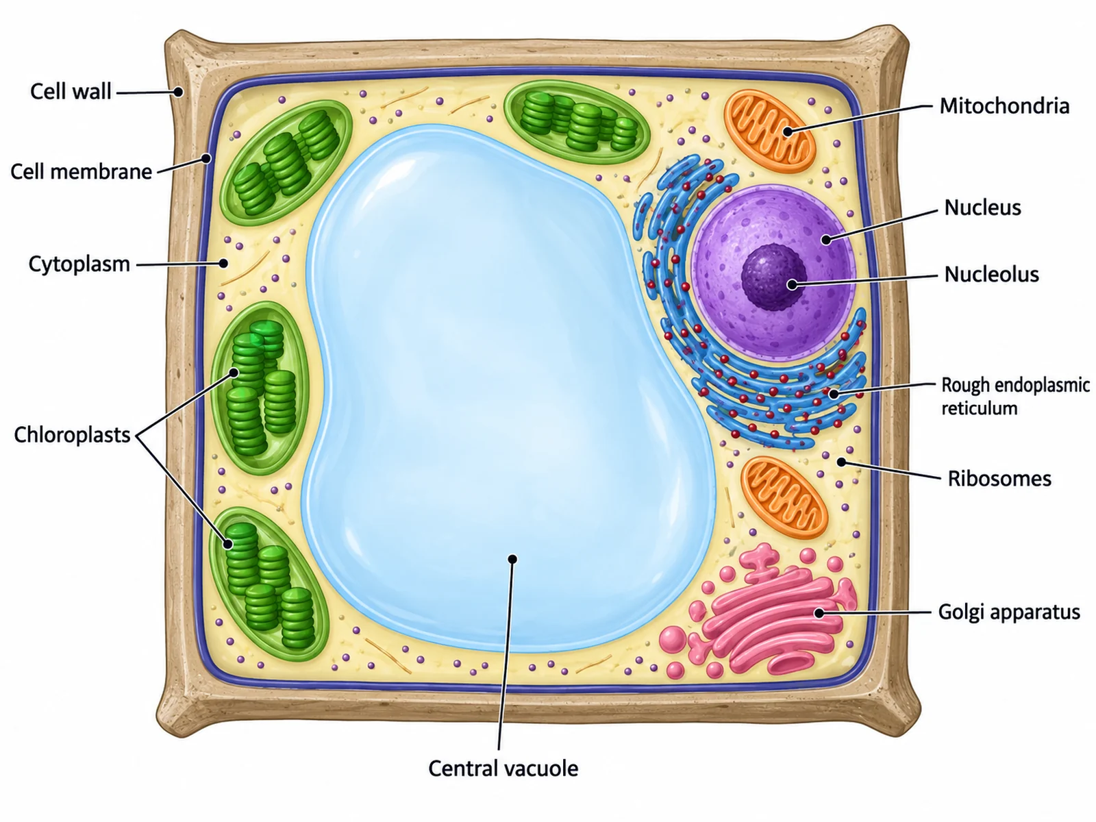
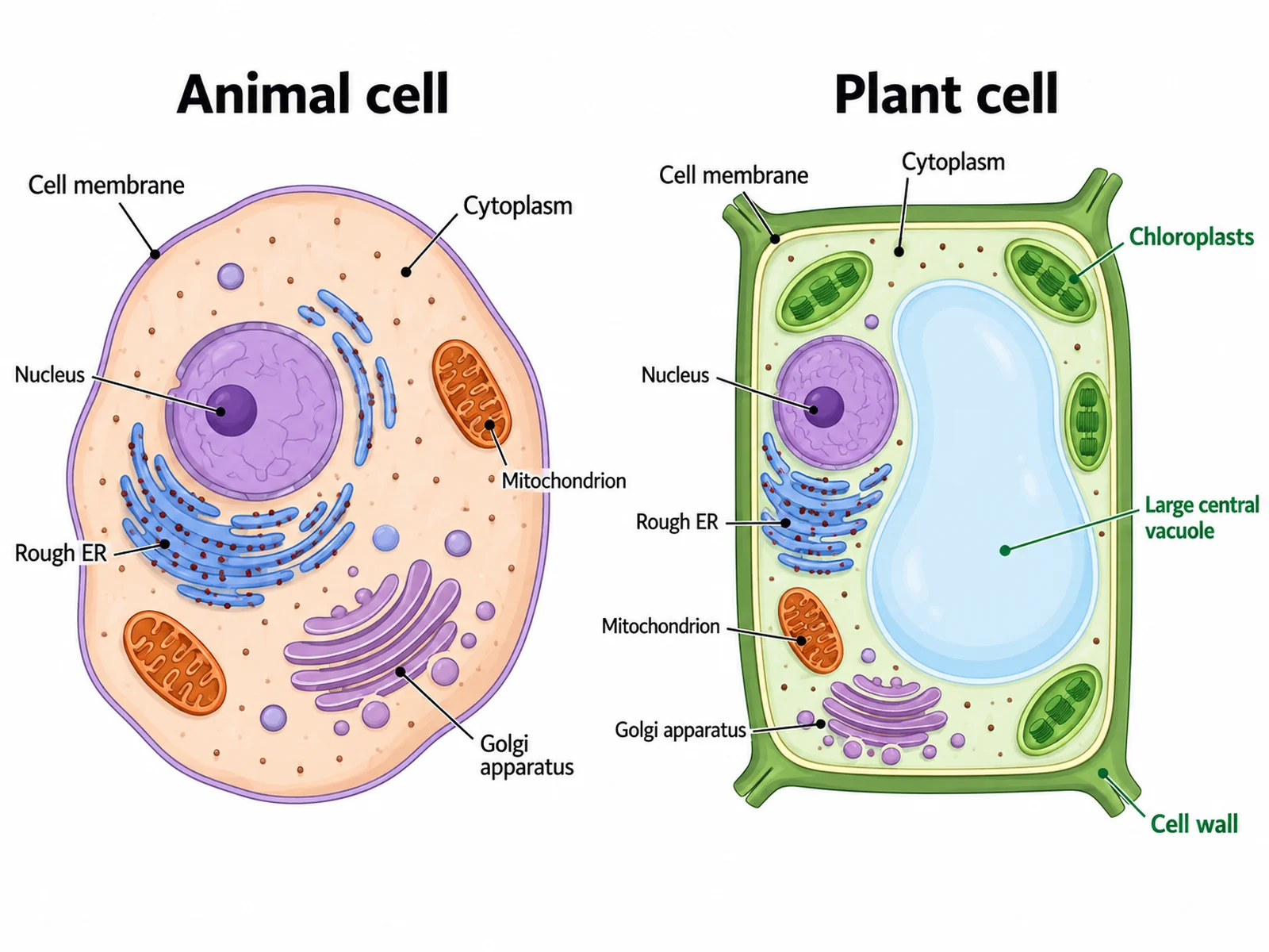
Side-by-side comparison of a labeled animal cell and a labeled plant cell highlighting the cell wall, chloroplasts, and central vacuole as plant-only structures

Prompt

Create a side-by-side comparison diagram of an animal cell and a plant cell on a white background. Draw the rounded animal cell on the left and the boxy plant cell on the right, each labeled. Highlight the three structures the animal cell lacks with a distinct accent color and a short callout on the plant side: cell wall, chloroplasts, and large central vacuole. Add a heading over each cell reading "Animal cell" and "Plant cell".

Animal vs Plant Cell

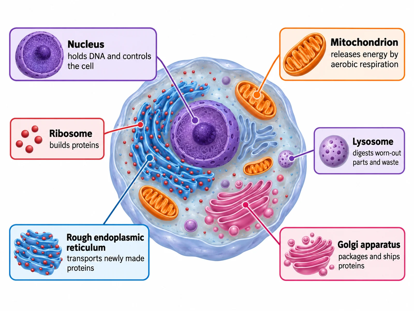
Animal cell diagram with callout cards describing the function of each labeled organelle

Prompt

Create an animal cell diagram with function callouts. Draw the labeled animal cell in the centre and surround it with small rounded cards connected by thin leader lines, each card naming one organelle in bold with a one-line function beneath it: nucleus "holds DNA and controls the cell", mitochondrion "releases energy by aerobic respiration", ribosome "builds proteins", rough endoplasmic reticulum "transports newly made proteins", Golgi apparatus "packages and ships proteins", and lysosome "digests worn-out parts and waste".

Animal Cell Organelle Functions

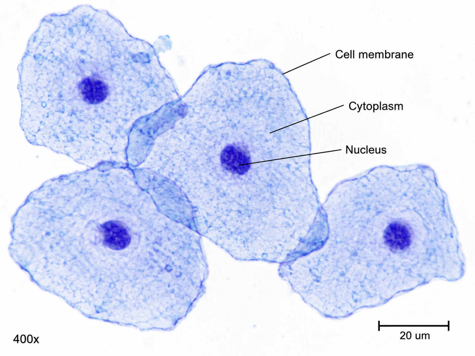
Labeled diagram of cheek cells as seen under a light microscope, showing the cell membrane, cytoplasm, and stained nucleus

Prompt

Create a labeled diagram showing human cheek cells as seen through a light microscope at 400x after methylene blue staining, in the style of a biology practical guide. Draw several flat, irregular, overlapping cells with a thin cell membrane, pale blue cytoplasm, and one darkly stained round nucleus each. Label cell membrane, cytoplasm, and nucleus, and add a scale bar reading "20 um".

Animal Cell Under the Microscope

How to make a labeled animal cell diagram

1

List the parts to label

Name the organelles your syllabus requires, such as cell membrane, cytoplasm, nucleus, mitochondria, ribosomes, and Golgi apparatus. Say whether you want a labeled version, a blank worksheet, or both.

2

Generate the diagram

The generator draws the cell with clean leader lines and readable text, keeping the membrane as the only boundary and the nucleus clearly central.

3

Check every label, then export

Confirm each leader line points at the right structure, especially rough versus smooth endoplasmic reticulum, then download the PNG for a worksheet, slide, or revision sheet.

What is an animal cell diagram?

An animal cell diagram is a labeled cross-section of a typical animal cell showing where each organelle sits and what it is called. Because an animal cell has no rigid wall, it is drawn as a rounded, slightly irregular shape bounded only by the cell membrane, with the nucleus near the centre and the other organelles spread through the cytoplasm. A labeled animal cell diagram is how school biology introduces organelles; the same drawing with the labels removed is how it tests them.

Parts of an animal cell and what each one does

  • Cell membrane: a partially permeable phospholipid layer that encloses the cell and controls what enters and leaves.
  • Cytoplasm: the jelly-like fluid where most chemical reactions of the cell take place.
  • Nucleus: contains the DNA and controls the cell; the dense nucleolus inside it builds ribosomes.
  • Mitochondria: bean-shaped organelles with folded inner cristae that release energy by aerobic respiration.
  • Ribosomes: tiny particles, free in the cytoplasm or attached to membrane, that build proteins.
  • Rough endoplasmic reticulum: ribosome-studded membrane stacks near the nucleus that transport new proteins.
  • Smooth endoplasmic reticulum: tubular membrane without ribosomes that makes lipids and stores calcium.
  • Golgi apparatus: stacked flattened sacs that modify, package, and ship proteins out of the cell.
  • Lysosomes: small sacs of digestive enzymes that break down worn-out organelles and waste.
  • Centrioles: paired cylindrical structures that organise the spindle fibres during cell division.

Labeled or blank: which one do you need?

  • Labeled animal cell diagram: use it to teach the organelles, as a revision sheet, or as the answer key for a worksheet.
  • Blank or unlabeled animal cell diagram: use it to test recall. The same drawing with empty numbered lines becomes the worksheet.
  • Simple animal cell diagram: four parts and larger text, for younger students or for a slide seen from the back of a room.
  • Animal versus plant cell: the fastest way to show that the cell wall, chloroplasts, and central vacuole are absent from animal cells.

Animal Cell Diagram Generator FAQ

A complete labeled animal cell diagram names the cell membrane, cytoplasm, nucleus, nucleolus, nuclear envelope, mitochondria, rough and smooth endoplasmic reticulum, ribosomes, Golgi apparatus, lysosomes, centrioles, and vesicles. Most school syllabuses require at least the cell membrane, cytoplasm, nucleus, and mitochondria.

Need other biology figures?

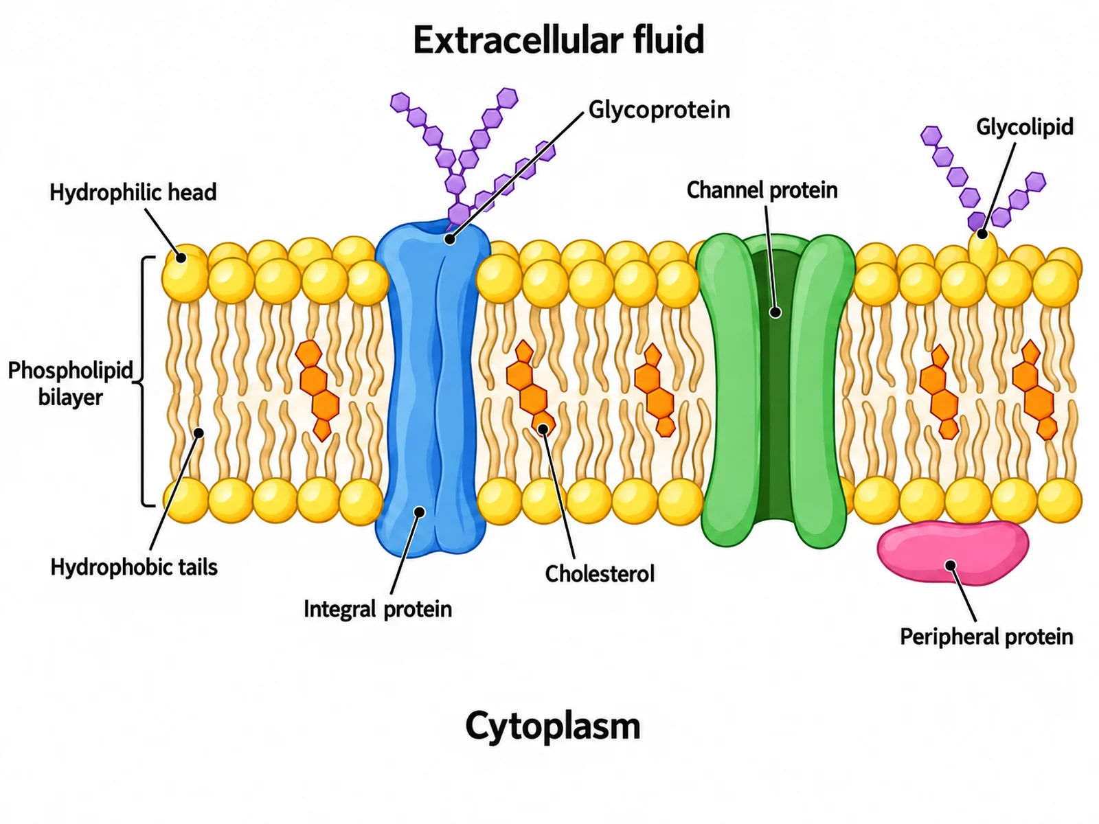
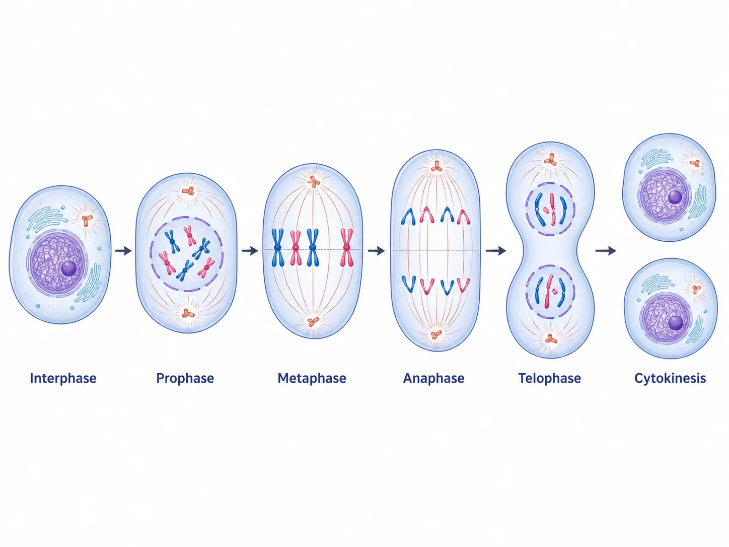
Generate plant cell diagrams, cell membrane figures, mitosis sequences, DNA replication visuals, and worksheet-ready biology illustrations with the same SciFigure AI workflow.