Ear Diagram AI

Ear Diagram Generator

Generate an ear diagram to label, or a fully labeled cross-section covering the outer, middle, and inner ear. Download a clean figure for worksheets, revision notes, and slides.

To label or labeledOuter, middle, inner earWorksheet-ready

AI Ear Diagram Generator

Describe which ear structures to label and the style you need.

Costs 5 credits310 / 2000 characters

Generate a publication-ready PNG from your description.

Preview

Ear Diagram Example Previews

Target aspect ratio: 4:3

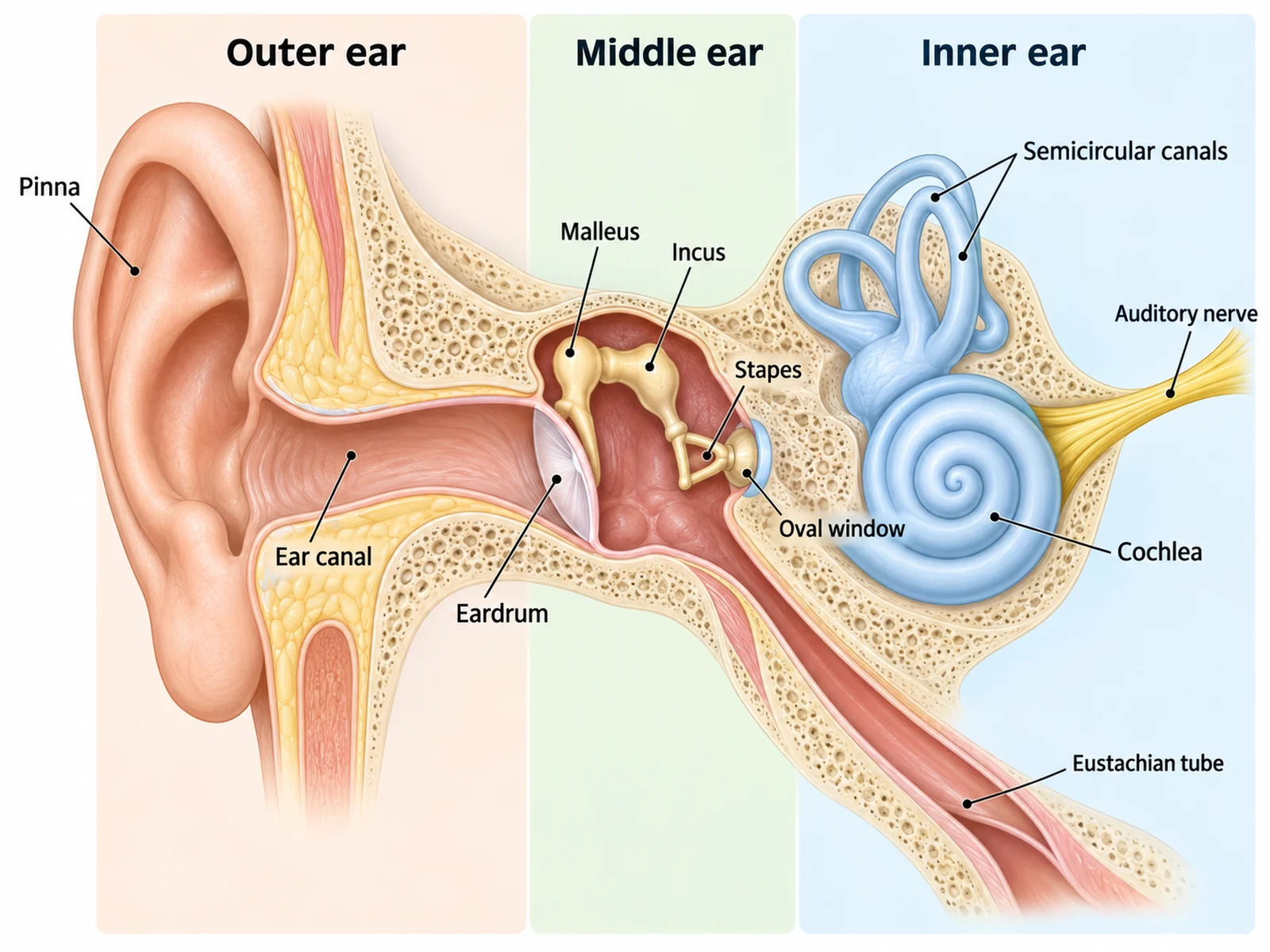
Labeled human ear cross-section naming the pinna, ear canal, eardrum, malleus, incus, stapes, oval window, Eustachian tube, cochlea, semicircular canals, and auditory nerve
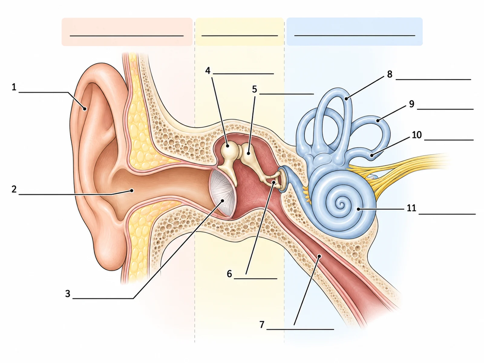
Blank unlabeled ear diagram with numbered empty label lines for students to complete
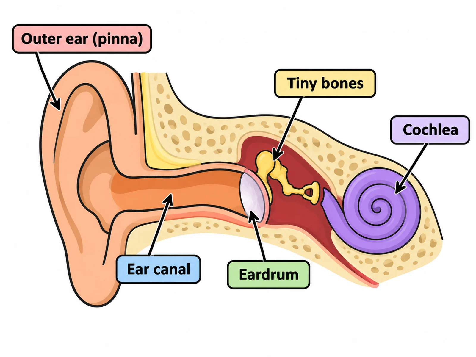
Simple ear diagram for younger students labeling only the pinna, ear canal, eardrum, ossicles, and cochlea

Sample previews. Describe your diagram to generate your own ear figure.

Ear Diagram Examples

Use these as visual directions for labeled cross-sections, blank worksheets, simple versions, ossicle close-ups, and sound pathway figures.

Labeled human ear cross-section naming the pinna, ear canal, eardrum, malleus, incus, stapes, oval window, Eustachian tube, cochlea, semicircular canals, and auditory nerve

Prompt

Create a fully labeled human ear diagram on a white background, drawn as a cross-section facing right. From the outside in, show a skin-toned pinna, an ear canal tunnelling inward, a taut eardrum membrane, then the three ossicles in order — malleus attached to the eardrum, incus in the middle, stapes with its footplate in the oval window — then a spiral cochlea of about two and a half turns in pale blue with the auditory nerve leaving it toward the left, three semicircular canals arching above the cochlea, and the Eustachian tube running down and forward from the middle ear cavity. Label every structure with a thin dark leader line and readable sans-serif text, and add three soft background tints with headings "Outer ear", "Middle ear", and "Inner ear".
Labeled Ear Diagram

Blank unlabeled ear diagram with numbered empty label lines for students to complete

Prompt

Create a blank ear diagram worksheet. Draw the same ear cross-section in clean line art with soft flat fills, but leave every label empty: draw thin leader lines from each structure out to a short horizontal ruled blank line with no text on it. Number the blanks 1 to 11. Keep the three region tints but leave their headings blank too, with a ruled line above each. Generous white space, no answer key.

Ear Diagram to Label

Simple ear diagram for younger students labeling only the pinna, ear canal, eardrum, ossicles, and cochlea

Prompt

Create a simple ear diagram for primary or lower-secondary students. Show only five labeled parts in everyday wording: "Outer ear (pinna)", "Ear canal", "Eardrum", "Tiny bones", and "Cochlea". Use bold flat colors, thick clean outlines, large readable text, and plenty of white space. Omit the Eustachian tube, oval window, and individual ossicle names.

Simple Ear Diagram

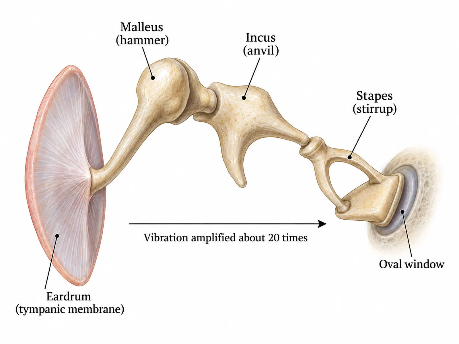
Magnified diagram of the malleus, incus, and stapes showing how they connect the eardrum to the oval window

Prompt

Create a magnified labeled diagram of the middle ear ossicles on a white background. Show the eardrum on the left as a taut membrane, then the malleus with its handle attached to the eardrum, the incus articulating with it, and the stapes with its stirrup shape and footplate seated in the oval window on the right. Label each bone with both its Latin and common name: "Malleus (hammer)", "Incus (anvil)", "Stapes (stirrup)". Add a thin arrow along the chain labeled "Vibration amplified about 20 times" and small labels for "Eardrum (tympanic membrane)" and "Oval window".

The Three Ossicles

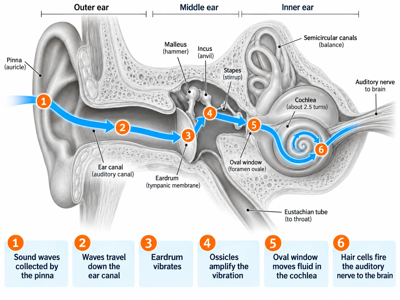
Ear diagram tracing the path of sound from the pinna through the ossicles to the cochlea and auditory nerve

Prompt

Create an ear diagram that traces the path of sound. Draw the labeled ear cross-section in muted grey, then overlay a single bright numbered pathway with six stops, each with a short caption: "1 Sound waves collected by the pinna", "2 Waves travel down the ear canal", "3 Eardrum vibrates", "4 Ossicles amplify the vibration", "5 Oval window moves fluid in the cochlea", "6 Hair cells fire the auditory nerve to the brain". Draw the path as a thick curved arrow following the real anatomy.

How Sound Travels

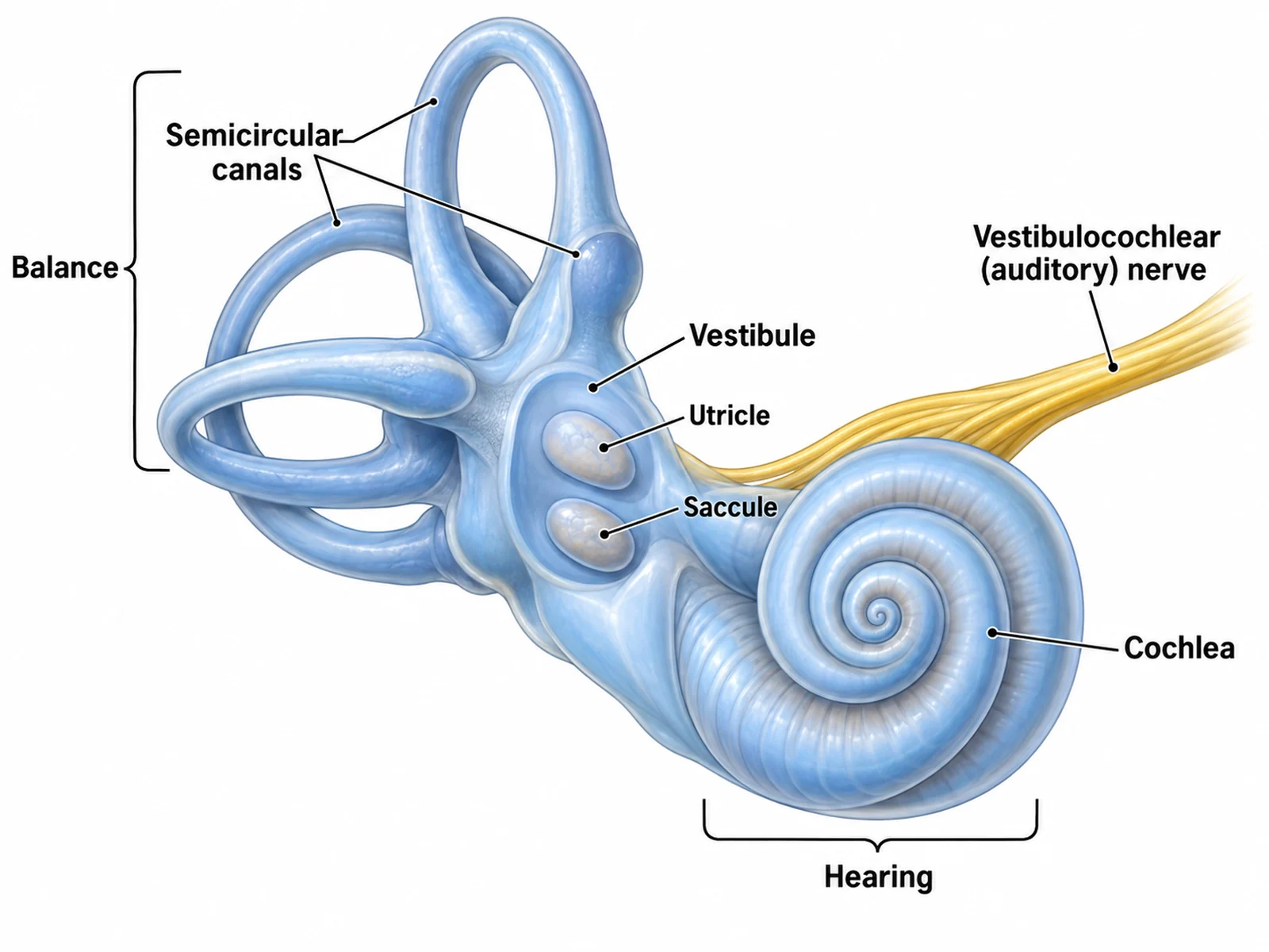
Labeled inner ear diagram showing the cochlea, three semicircular canals, vestibule, and their roles in hearing and balance

Prompt

Create a labeled inner ear diagram on a white background, magnified from the rest of the ear. Show the spiral cochlea of about two and a half turns, the vestibule, and the three semicircular canals arranged at right angles to one another, with the vestibulocochlear nerve leaving toward the brain. Label "Cochlea", "Semicircular canals", "Vestibule", "Utricle", "Saccule", and "Vestibulocochlear (auditory) nerve". Add two short captions with leader brackets: "Hearing" pointing to the cochlea and "Balance" pointing to the semicircular canals and vestibule.

Inner Ear and Balance

How to make a labeled ear diagram

1

Choose the scope

The whole ear in cross-section, a close-up of the three ossicles, or the inner ear alone. Say whether you want labels filled in or blank lines to complete.

2

Generate the diagram

The generator keeps the structures in anatomical order from outer to inner ear and adds clean leader lines.

3

Check the anatomy, then export

Confirm the ossicle order is malleus, incus, stapes and that the cochlea spirals correctly, then download the PNG for a worksheet, slide, or revision sheet.

What is an ear diagram?

An ear diagram is a labeled cross-section through the human ear, cut so that structures normally hidden inside the skull become visible. It is almost always drawn in three regions — outer, middle, and inner ear — because that division explains the job each part does: collecting sound, amplifying it, and converting it into nerve impulses. The blank version of the same drawing is the standard way the topic is tested.

Parts of the ear and what each one does

  • Pinna (auricle): the visible flap that collects sound waves and funnels them into the canal.
  • Ear canal: carries sound to the eardrum and produces wax that traps dust and bacteria.
  • Eardrum (tympanic membrane): a thin taut membrane that vibrates when sound waves strike it.
  • Malleus, incus, stapes: three tiny bones that amplify the vibration roughly twentyfold.
  • Oval window: the membrane the stapes footplate pushes against, passing vibration into the inner ear.
  • Eustachian tube: connects the middle ear to the throat and equalises pressure on both sides of the eardrum.
  • Cochlea: a fluid-filled spiral lined with hair cells that convert vibration into nerve impulses.
  • Semicircular canals: three fluid-filled loops at right angles that detect head rotation for balance.
  • Auditory (vestibulocochlear) nerve: carries hearing and balance signals to the brain.

Common errors to check before you print

  • Ossicles out of order. From the eardrum inward it is always malleus, then incus, then stapes.
  • Cochlea drawn as a flat circle. It is a spiral of about two and a half turns.
  • Eustachian tube pointing upward. It runs down and forward toward the throat.
  • Semicircular canals drawn in one plane. The three loops sit at right angles to one another.

Ear Diagram Generator FAQ

The outer ear: pinna (auricle), ear canal (external auditory canal), and eardrum (tympanic membrane). The middle ear: malleus (hammer), incus (anvil), stapes (stirrup), oval window, and Eustachian tube. The inner ear: cochlea, semicircular canals, vestibule, and the auditory (vestibulocochlear) nerve.

Need other anatomy figures?

Generate sense organ diagrams, body system figures, cell diagrams, and worksheet-ready illustrations with the same SciFigure AI workflow.