Tutorials
Labeled Brain Diagram: How to Label the Lobes, Cerebellum, Brainstem and More
How to label a brain diagram: the lobes, cerebellum and brainstem on a side view, the midline parts on a sagittal section, what each does, and a worksheet.
 SciFigure AI
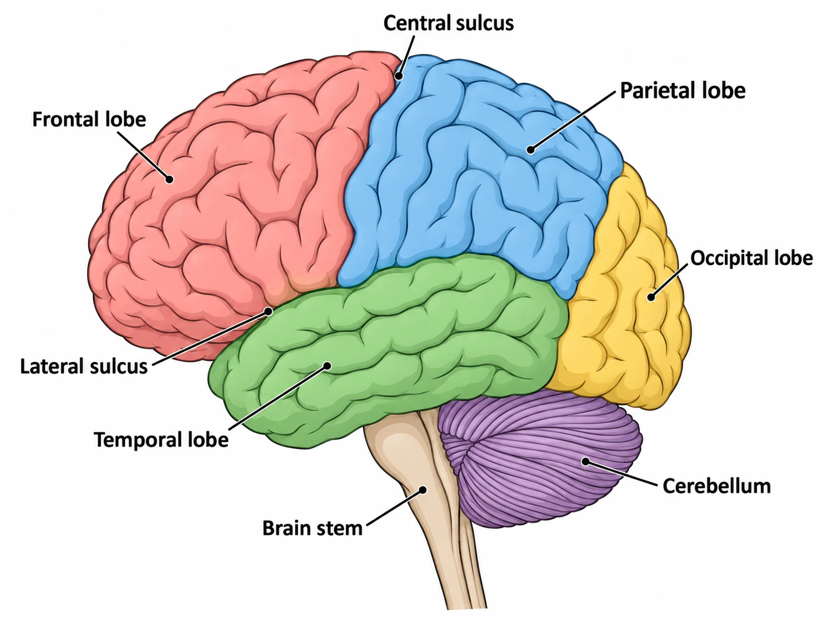
The single most useful thing to know before labeling a brain diagram is which view you are looking at. A side view of the brain shows the surface: the four lobes, the cerebellum and the brainstem. It cannot show the corpus callosum, the thalamus or the hypothalamus, because those are buried in the middle. To label them you need a brain cut down the midline. A surprising number of lost marks come from labeling a structure on a view where it cannot be seen.
This guide covers what to label at each level, the two standard views and what belongs on each, every structure with what it looks like and what it does, the labeling conventions that gain marks, the mistakes that lose them, and a numbered worksheet with an answer key. If you need a figure, the brain diagram generator draws a labeled side view, a blank brain diagram to label, a sagittal section and a plain brain outline.
The short answer: what to label on a brain diagram
On a side (lateral) view: frontal lobe, parietal lobe, temporal lobe, occipital lobe, cerebellum, brainstem, and the two grooves that divide the lobes, the central sulcus and the lateral sulcus.
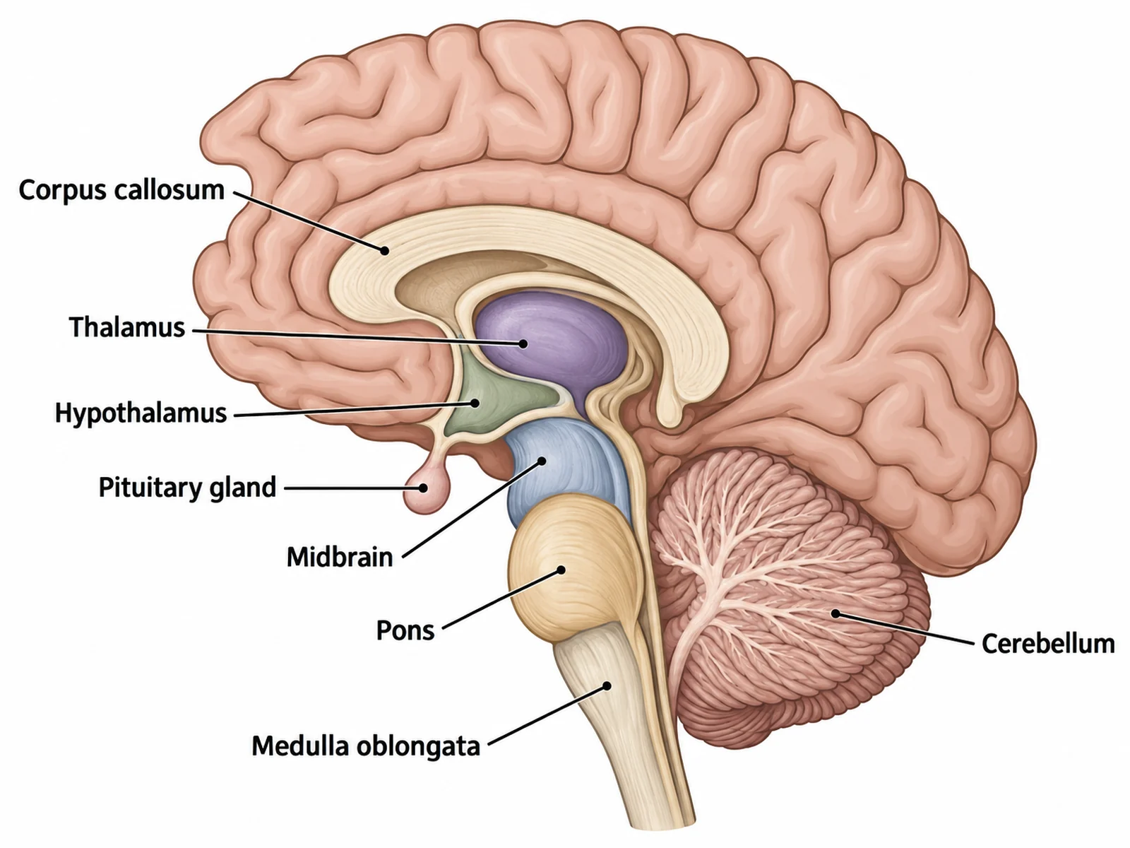
On a midline (sagittal) section: corpus callosum, thalamus, hypothalamus, pituitary gland, midbrain, pons, medulla oblongata and cerebellum, with the cerebral cortex around the outside.
The four lobes are all part of the cerebrum, the large folded upper part of the brain made of two hemispheres. The cerebellum is a separate structure, not a fifth lobe.
What to label at each level
| Level | View | Labels expected |
|---|---|---|
| Middle school, KS3, grades 6–8 | Side view | Cerebrum, cerebellum, brainstem (or medulla), often the four lobes |
| GCSE, high school biology | Side view, sometimes a section | Cerebral cortex, cerebellum and medulla, with their functions; the hypothalamus and pituitary gland appear in the hormones topic |
| AP Psychology, A-level Psychology, IB | Side view and sagittal section | The four lobes, motor and somatosensory cortex, Broca's and Wernicke's areas, corpus callosum, thalamus, hypothalamus, pituitary gland, hippocampus, amygdala, medulla, pons, cerebellum |
| College anatomy and neuroscience | All views, including coronal sections | The above plus precentral and postcentral gyri, longitudinal fissure, insula, midbrain, pineal gland, fornix, ventricles, arbor vitae, and the gray and white matter |
AQA's GCSE Biology specification is the clearest example of the second row: students must identify the cerebral cortex, cerebellum and medulla on a diagram of the brain and describe their functions. That makes those three words, not "cerebrum", "little brain" or "brain stem", the safest labels for a GCSE answer.
The two views, and what can be labeled on each
| View | How it is drawn | Can show | Cannot show |
|---|---|---|---|
| Lateral (side) view | The brain seen from the side, usually the left hemisphere facing left | The four lobes, central and lateral sulci, motor and sensory strips, Broca's and Wernicke's areas, cerebellum, brainstem | Corpus callosum, thalamus, hypothalamus, pineal gland, ventricles |
| Midsagittal section | The brain cut down the middle, between the two hemispheres, cut face toward you | Corpus callosum, thalamus, hypothalamus, pituitary gland, pineal gland, midbrain, pons, medulla, cerebellum with arbor vitae, ventricles | Most of the lateral surface, the insula, and the hippocampus and amygdala, which lie off the midline inside the temporal lobe |
| Coronal section | The brain sliced from ear to ear | Gray and white matter, lateral ventricles, basal nuclei, insula, hippocampus | The full outline of each lobe |
If a question asks you to label the hippocampus or the amygdala, expect a coronal section or a cut-away "limbic system" figure. They are not visible on a plain side view or a midline cut.
Labeling the side view: lobes, cerebellum and brainstem
| Part | Where it is on the diagram | What it does |
|---|---|---|
| Frontal lobe | The front of the cerebrum, in front of the central sulcus | Planning, decision-making, personality and voluntary movement. The primary motor cortex runs down its back edge (the precentral gyrus) |
| Parietal lobe | Top of the brain, behind the central sulcus | Processes touch, temperature, pain and body position. The primary somatosensory cortex runs down its front edge (the postcentral gyrus); also spatial awareness |
| Temporal lobe | Low on the side, below the lateral sulcus | Hearing (primary auditory cortex), understanding language (Wernicke's area, usually in the left hemisphere) and, on its inner side, memory |
| Occipital lobe | The back of the cerebrum | Vision: the primary visual cortex |
| Central sulcus | The groove running down the side from the top, between the frontal and parietal lobes | Boundary between movement (in front) and body sensation (behind) |
| Lateral sulcus (lateral fissure) | The deep groove running back along the side, above the temporal lobe | Separates the temporal lobe from the frontal and parietal lobes; hides the insula |
| Broca's area | Lower part of the frontal lobe, usually on the left | Producing speech |
| Cerebellum | Below the occipital lobe at the back, with much finer, tighter folds | Coordination, balance, posture and the learning of movements |
| Brainstem | The stalk descending in front of the cerebellum into the spinal cord | Automatic life-support functions: heart rate, breathing, blood pressure, swallowing; carries every signal between the brain and the spinal cord |
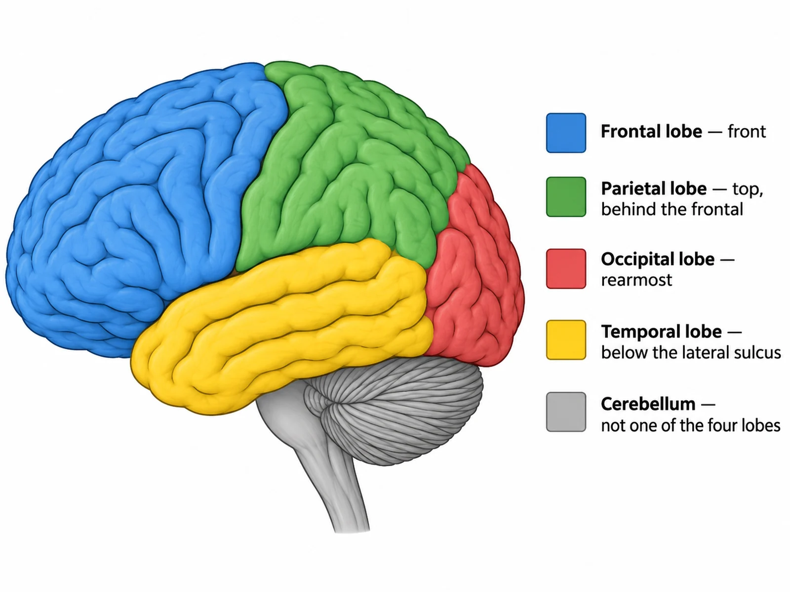
How to find the lobe boundaries. The central sulcus is the long groove that runs from the top of the brain down and slightly forward toward the lateral sulcus; everything in front of it is frontal lobe. The lateral sulcus is the deep groove running back along the side; the temporal lobe is everything below it. The occipital lobe is the rearmost part, and its front boundary is mostly seen on the inner surface, so on a side view it is drawn as a line running down from near the top of the back of the brain toward the cerebellum.
Labeling the sagittal section: the midline structures
| Part | Where it is on the diagram | What it does |
|---|---|---|
| Corpus callosum | The thick C-shaped band arching above the center | The largest bundle of nerve fibers joining the left and right hemispheres, so they can share information |
| Thalamus | The rounded mass just below the corpus callosum, in the center | Relay station: passes sensory information (every sense except smell) on to the cerebral cortex |
| Hypothalamus | Just below and in front of the thalamus | Homeostasis: body temperature, hunger, thirst and water balance; controls the pituitary gland |
| Pituitary gland | A small pea-shaped gland hanging below the hypothalamus on a stalk | Releases hormones that control many other endocrine glands |
| Pineal gland | A tiny gland behind the thalamus, above the midbrain | Releases melatonin, which helps regulate the sleep-wake cycle |
| Midbrain | The top part of the brainstem, below the thalamus | Visual and auditory reflexes and eye movement; a relay for motor and sensory pathways |
| Pons | The rounded bulge at the front of the brainstem, below the midbrain | Relays signals between the cerebrum and the cerebellum; helps control breathing |
| Medulla oblongata | The lowest part of the brainstem, joining the spinal cord | Controls heart rate, breathing rate, blood pressure, and reflexes such as swallowing and vomiting |
| Cerebellum | Behind the pons and medulla; its cut surface shows a branching pattern | Coordination and balance; the branching white matter is called the arbor vitae ("tree of life") |
Cerebrum, cerebral cortex and brainstem: getting the words right
Three pairs of terms are easily confused, and markers notice.
- Cerebrum vs cerebral cortex. The cerebrum is the whole upper part of the brain, both hemispheres. The cerebral cortex is its outer layer of gray matter, only a few millimeters thick and folded into gyri (ridges) and sulci (grooves). GCSE mark schemes ask for the cortex; when you label it, end the line on the outer surface.
- Brainstem vs medulla. The brainstem is the midbrain, pons and medulla oblongata together. The medulla is the lowest part. On a GCSE diagram labeled for the cortex, cerebellum and medulla, the "medulla" line goes to the lower brainstem, just above the spinal cord.
- Cerebrum vs cerebellum. One letter apart and easy to swap in a hurry. Cerebrum is the big folded top; cerebellum is the small finely ridged structure at the back.
Labeling conventions that gain marks
- Say which view it is, or check the one you are given before you label anything midline.
- Label lobes with the line ending in the middle of the lobe, not on a boundary, so there is no doubt which region you mean.
- Label sulci with the line ending in the groove itself.
- Keep a consistent orientation. Most diagrams show the left hemisphere from the left, frontal lobe on the viewer's left. If yours faces the other way, the lobes still run front to back in the same order.
- Use straight, ruled lines that do not cross, with labels outside the outline in columns.
- Write "lobe" after each lobe name, and capitalize Broca's and Wernicke's areas, which are named after people.
- Pair functions with structures in a short note under each label if the question asks "label and describe", for example "medulla: controls breathing and heart rate".
How to label a brain diagram, step by step
- Identify the view. Folded surface and a stalk underneath means a side view; a C-shaped band in the middle means a sagittal section.
- Find the front. On a side view, the frontal lobe is the large rounded end opposite the cerebellum; the temporal lobe points toward the front.
- Find the two grooves. The lateral sulcus runs back along the side; the central sulcus runs down from the top. Label them first: they define the lobes.
- Label the lobes: frontal in front of the central sulcus, parietal behind it on top, temporal below the lateral sulcus, occipital at the back.
- Label the cerebellum at the back underneath, and the brainstem as the stalk in front of it.
- On a sagittal section, work from top to bottom: corpus callosum, thalamus, hypothalamus, pituitary, then midbrain, pons and medulla down the brainstem, and the cerebellum behind.
- Check that nothing midline is labeled on a side view, the cerebellum is not called a lobe, and the parietal and occipital lobes are not swapped.
Mistakes that cost marks
- The cerebellum labeled as a lobe, or counted as part of the cerebrum.
- Parietal and occipital swapped. Parietal is on top toward the back; occipital is the rearmost lobe.
- The temporal lobe placed above the lateral sulcus. It is below it.
- The brainstem drawn behind the cerebellum. It runs down in front of it.
- Corpus callosum, thalamus or hypothalamus labeled on a side view. They are only visible in a section.
- The hypothalamus placed above the thalamus. It is below it, as the name says ("hypo" means under).
- The pituitary gland labeled as part of the brainstem. It hangs from the hypothalamus.
- Cerebrum and cerebellum swapped, in spelling or in position.
- "Cerebrum" written where the specification asks for the cerebral cortex.
- The medulla line ending on the pons. The medulla is the lowest part of the brainstem, joining the spinal cord.
- Broca's area placed in the temporal lobe or Wernicke's in the frontal. Broca's is frontal (speech production); Wernicke's is temporal (comprehension).
- Hippocampus or amygdala labeled on a midline section. They lie inside the temporal lobe, off the midline.
Printable brain diagram worksheet with answer key
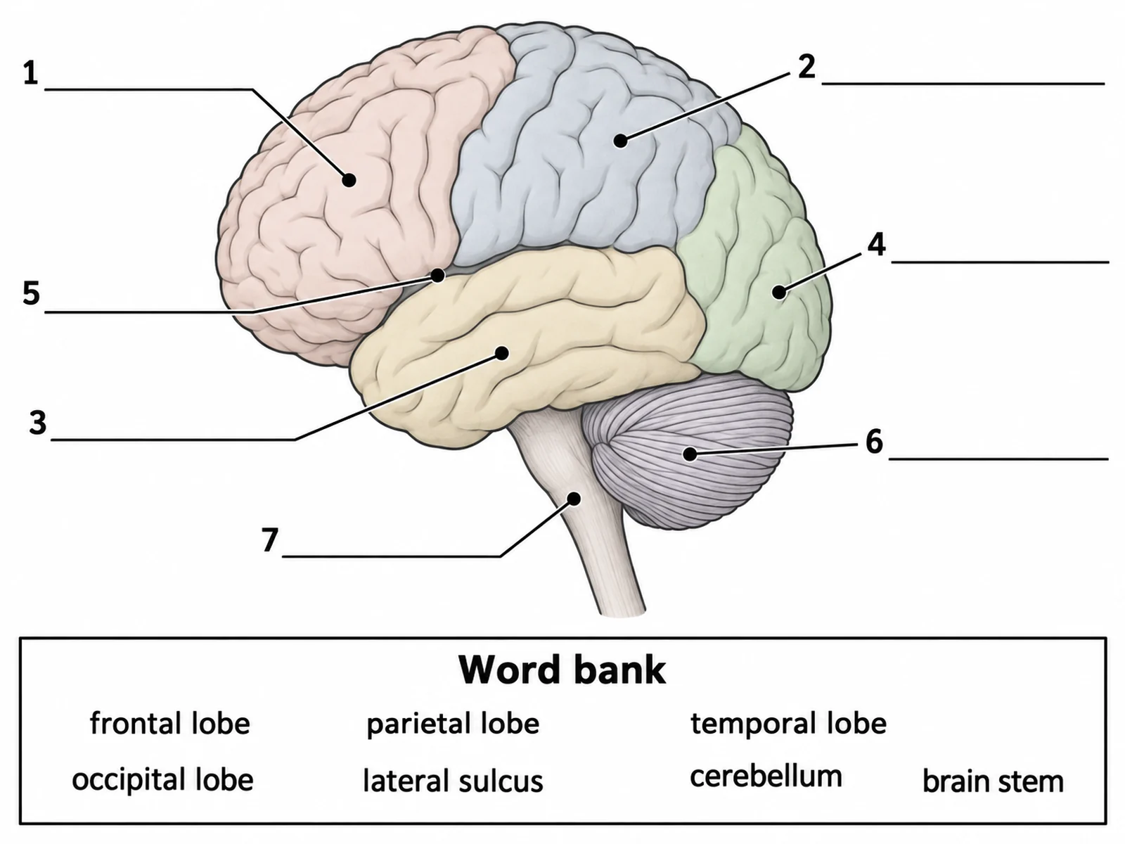
Label the seven numbered parts of this side view. The word bank is on the sheet; the answers follow.
Answer key
- Frontal lobe
- Parietal lobe
- Temporal lobe
- Occipital lobe
- Lateral sulcus
- Cerebellum
- Brainstem
For a harder sheet, cover the word bank, or make a sagittal section worksheet with the corpus callosum, thalamus, hypothalamus, pituitary and brainstem parts on the brain diagram generator. Generate the labeled and the blank version from the same description and you have the answer key too.
Frequently asked questions
What are the main parts of the brain to label?
The cerebrum with its four lobes (frontal, parietal, temporal, occipital), the cerebellum and the brainstem (midbrain, pons, medulla oblongata). A midline section adds the corpus callosum, thalamus, hypothalamus and pituitary gland.
What are the four lobes of the brain and what do they do?
The frontal lobe (planning, decisions, personality, voluntary movement, speech production), parietal lobe (touch, body position, spatial awareness), temporal lobe (hearing, language comprehension, memory) and occipital lobe (vision).
Is the cerebellum a lobe?
No. The four lobes belong to the cerebrum. The cerebellum is a separate structure below the occipital lobe at the back of the brain, with much finer folds, and it coordinates movement and balance.
Which part of the brain controls breathing and heart rate?
The medulla oblongata, the lowest part of the brainstem. The pons helps regulate breathing as well.
What does the corpus callosum do?
It is a thick band of nerve fibers joining the left and right hemispheres so they can exchange information. It can only be seen when the brain is cut down the midline.
What is the difference between the cerebrum and the cerebral cortex?
The cerebrum is the whole upper brain: both hemispheres. The cerebral cortex is its folded outer layer of gray matter, a few millimeters thick, where most higher processing happens.
Where are the thalamus and hypothalamus?
In the center of the brain, below the corpus callosum. The thalamus is the larger rounded mass; the hypothalamus is just below it, with the pituitary gland hanging underneath.
What is the difference between a lateral view and a sagittal view of the brain?
A lateral view shows the outer surface from the side: lobes, cerebellum and brainstem. A sagittal section shows the brain cut down the middle, exposing structures such as the corpus callosum and thalamus.
What are the brain's folds called?
The ridges are gyri (singular gyrus) and the grooves are sulci (singular sulcus); the deepest grooves are called fissures. Folding packs a large area of cortex inside the skull.
Where can I get a blank brain diagram to label?
Use the worksheet above, or make one with the brain diagram generator, which draws labeled and blank side views, sagittal sections and plain brain outlines.
Related guides and tools
- How to label an ear diagram: the hearing pathway that ends in the temporal lobe.
- How to label the digestive system: another anatomy worksheet, labeled organ by organ.
- Neuron diagram and synapse diagram: the cells the brain is made of.
- Endocrine system diagram: the hypothalamus and pituitary as hormone glands.
- Dermatome chart and anatomy diagram generator for the rest of the nervous system and body.
- More guides in this series: labeling a plant cell, drawing the stages of mitosis, labeling a cell membrane.
Sources
- OpenStax, Anatomy and Physiology 2e, 13.2 The Central Nervous System: cerebrum, lobes, diencephalon, brainstem and cerebellum.
- OpenStax, Biology 2e, 35.3 The Central Nervous System: functions of the lobes and the midline structures.
- Javed K, Reddy V, Lui F. Neuroanatomy, Cerebral Cortex. StatPearls, NCBI Bookshelf.
- Basinger H, Hogg JP. Neuroanatomy, Brainstem. StatPearls, NCBI Bookshelf.
- Jimsheleishvili S, Dididze M. Neuroanatomy, Cerebellum. StatPearls, NCBI Bookshelf.
- AQA, GCSE Biology (8461) specification, Homeostasis and response: the brain structures required at GCSE.