Tutorials
How to Label the Digestive System: Every Organ From Mouth to Anus
How to label a digestive system diagram: every organ from mouth to anus and its job, where each one sits, what to label at each level, and a worksheet.
 SciFigure AI
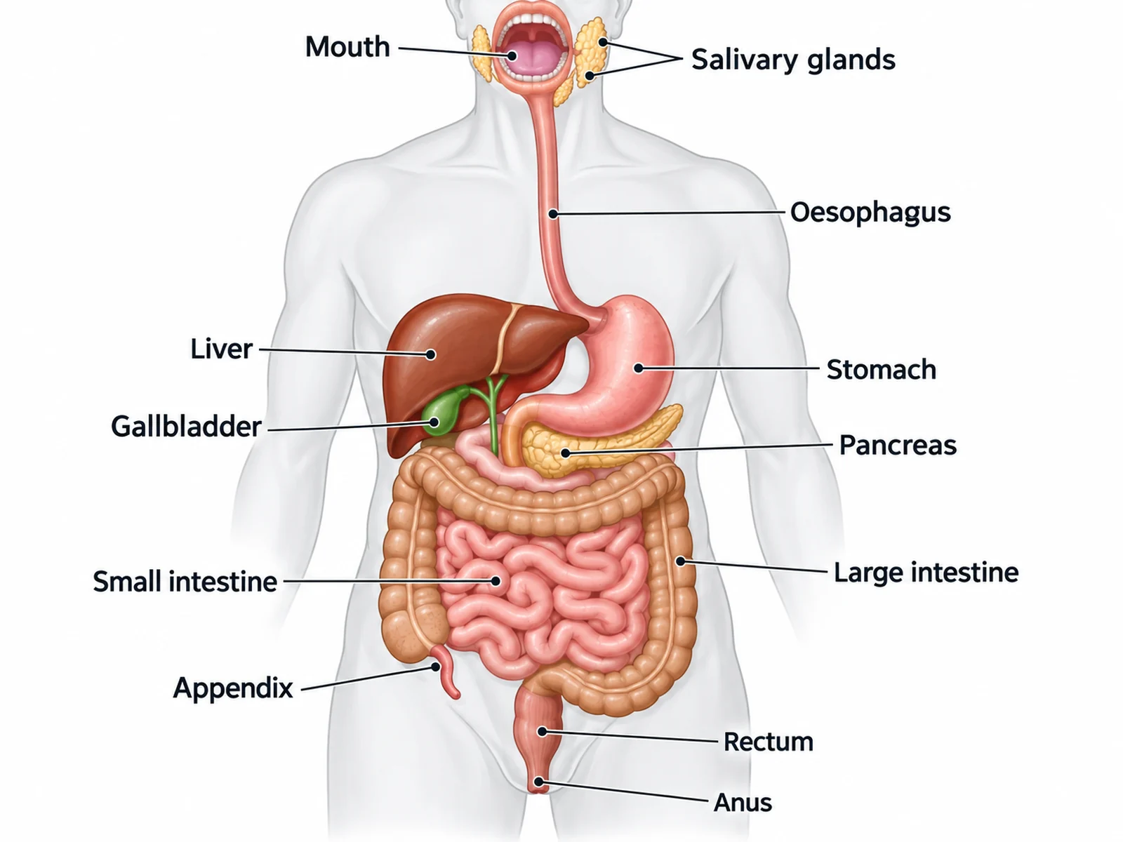
The digestive system is two things drawn as one. There is a tube that food passes through, from mouth to anus, and there are organs beside the tube that food never enters but which pour their secretions into it: the salivary glands, the liver, the gallbladder and the pancreas. A labeled digestive system diagram that keeps those two groups straight, and puts each organ on the correct side of the body, is most of the way to full marks.
This guide covers what to label at each level, every organ with what it looks like and what it does, where each one sits in the body, close-ups of the stomach and the small intestine, the labeling conventions that gain marks, the mistakes that lose them, and a numbered worksheet with an answer key. If you need a figure, the digestive system diagram generator draws a labeled version, a blank version to label, a simple version for younger students and a flow chart of the food route.
The short answer: what to label on a digestive system diagram
The alimentary canal (the tube), in the order food passes through it: mouth, pharynx, esophagus, stomach, small intestine (duodenum, jejunum, ileum), large intestine (cecum, colon, rectum), anus.
The accessory organs (beside the tube): salivary glands, liver, gallbladder, pancreas.
Most school diagrams also expect the appendix, a small closed tube attached to the cecum. US and UK spellings differ for several of these, and both are accepted: esophagus or oesophagus, cecum or caecum, feces or faeces.
What to label at each level
| Level | Labels expected | Usually also asked |
|---|---|---|
| Middle school, KS3, grades 6–8 | Mouth, esophagus, stomach, small intestine, large intestine, rectum, anus; often liver and pancreas | The order food travels in |
| GCSE, high school biology | The above plus salivary glands, gallbladder and appendix | Which enzymes are made where; what bile does; where nutrients and water are absorbed |
| A-level, AP Biology, IB | The above plus duodenum and ileum, the stomach's sphincters, villi and microvilli, and the layers of the gut wall | How villi are adapted for absorption; how the structure of the ileum wall relates to its function |
| College anatomy and physiology | The above plus pharynx, jejunum, cecum, ascending, transverse, descending and sigmoid colon, hepatic and splenic flexures, ileocecal valve, common bile duct, pancreatic duct, hepatopancreatic ampulla, and the internal and external anal sphincters | Peritoneum and mesentery, blood supply via the hepatic portal vein, and the histology of each region |
Every organ and what it does
The alimentary canal
| Organ | What it looks like on a diagram | What it does |
|---|---|---|
| Mouth | The opening at the top, with teeth and tongue | Chewing breaks food into smaller pieces (mechanical digestion); saliva moistens it, and salivary amylase starts breaking starch down |
| Pharynx | The throat, behind the mouth | Shared by food and air; swallowing pushes food into the esophagus while the epiglottis covers the airway |
| Esophagus | A straight muscular tube running down the chest, behind the windpipe | Moves food to the stomach by peristalsis, waves of muscle contraction. No digestion happens here |
| Stomach | A J-shaped bag under the diaphragm, on the body's left | Stores food and churns it with acid and enzymes into a thick liquid called chyme. Hydrochloric acid kills most microbes and provides the low pH at which pepsin starts digesting protein |
| Duodenum | The first, C-shaped part of the small intestine, curving round the head of the pancreas | Receives chyme from the stomach plus bile and pancreatic juice; most chemical digestion is completed here and further along the small intestine |
| Jejunum and ileum | The long coiled tube filling the center of the abdomen | Finish digestion and absorb nutrients into the blood through millions of villi. The ileum also absorbs vitamin B12 and bile salts |
| Cecum | A pouch at the start of the large intestine, low on the body's right | Receives material from the ileum; the appendix hangs from it |
| Appendix | A narrow, closed, finger-like tube about 7.5 cm long | Contains lymphoid tissue; no role in digestion in humans |
| Colon | The wide tube framing the small intestine: up the body's right, across the top, down the left | Absorbs water and salts, turning liquid waste into solid feces; houses gut bacteria |
| Rectum | The last straight section, in the midline of the pelvis | Stores feces until they are passed |
| Anus | The opening at the end of the canal, with an internal (involuntary) and external (voluntary) sphincter | Egestion: removal of feces from the body |
The accessory organs
| Organ | Where it is | What it does |
|---|---|---|
| Salivary glands | Around the mouth: in front of the ears, under the jaw and under the tongue | Make saliva, which contains amylase and mucus |
| Liver | The large organ in the upper right of the abdomen, under the diaphragm; the largest gland in the body | Makes bile. It also processes the nutrients absorbed from the intestine, stores glycogen and breaks down toxins |
| Gallbladder | A small green sac on the underside of the liver | Stores and concentrates bile, and releases it into the duodenum after a meal |
| Pancreas | A long, flat gland behind the stomach, its head in the curve of the duodenum | Secretes pancreatic juice into the duodenum: amylase, proteases and lipase, plus bicarbonate to neutralize stomach acid. Separately, it makes the hormones insulin and glucagon |
Two points from these tables come up again and again.
Bile is not an enzyme. It is made in the liver, stored in the gallbladder, and does two jobs in the small intestine: it emulsifies fats, breaking large droplets into smaller ones so lipase has more surface to work on, and it helps neutralize the acid in chyme. A diagram that labels the gallbladder "makes bile" or "makes lipase" loses the mark.
Food never enters the accessory organs. The liver, gallbladder and pancreas connect to the duodenum by ducts. Arrows on your diagram should show secretions flowing into the small intestine, not food flowing out to these organs.
Where each organ sits: the left-right rule
Digestive system diagrams are drawn as if you are facing the person, so the body's right side is on the viewer's left. Almost every position mistake comes from forgetting this mirror.
| Organ | Body side | Where it appears on a front-view diagram |
|---|---|---|
| Liver | Upper right | Upper left of the drawing, the largest organ there |
| Gallbladder | Under the liver | Small sac tucked under the liver, on the viewer's left |
| Stomach | Upper left | Upper right of the drawing, just below where the esophagus ends |
| Pancreas | Behind the stomach, across the midline | Below and behind the stomach, often partly hidden |
| Small intestine | Center | The coiled mass in the middle |
| Ascending colon, cecum and appendix | Right | Running up the viewer's left, with the appendix at the bottom left |
| Descending colon | Left | Running down the viewer's right |
| Rectum and anus | Midline | Bottom center |
A quick check: the liver and the appendix are on the same side of the body (the right), and the stomach is on the other. On a front-view diagram that puts the liver and the appendix both on your left as you look at the page.
The stomach in close-up
Many A-level and college questions ask for the stomach on its own.
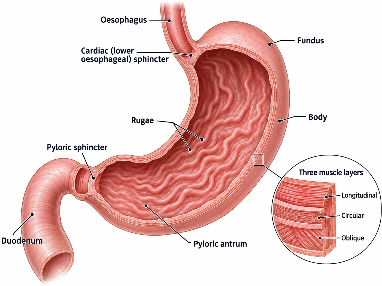
Label it from top to bottom:
- Lower esophageal (cardiac) sphincter: where the esophagus joins the stomach; it stops acid flowing back up.
- Fundus: the dome that rises above the entrance from the esophagus.
- Body: the large main part of the stomach.
- Rugae: folds in the lining that flatten out as the stomach fills.
- Pyloric antrum and pyloric sphincter: the narrowing lower end and the ring of muscle that lets chyme into the duodenum a little at a time.
- Muscle layers: most of the gut has two layers, longitudinal and circular; the stomach adds a third, inner oblique layer, which is why it can churn food so thoroughly.
The small intestine and villi
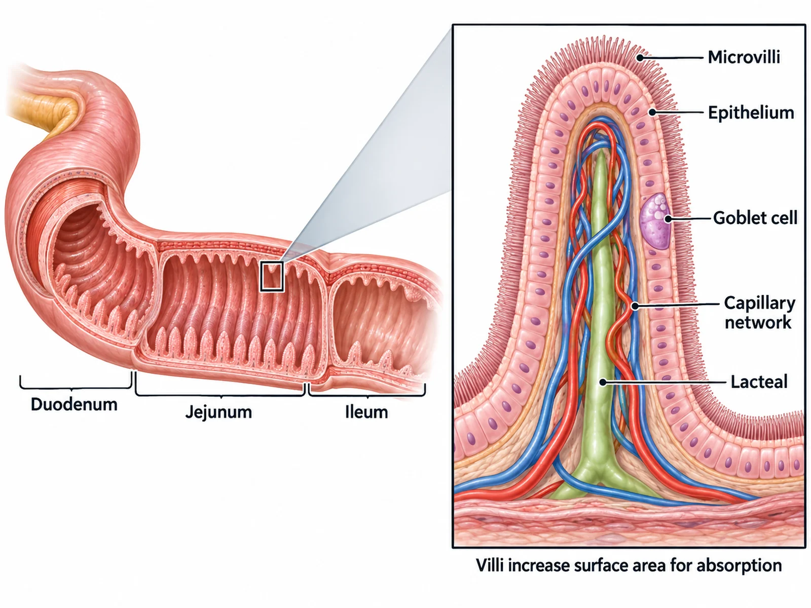
Absorption is the small intestine's job, and its structure is built around surface area. In a living person it is about 3 m long, and its inner surface is folded three times over: circular folds of the lining, villi on the folds, and microvilli on the cells of the villi. Together they give an absorptive surface of around 200 m².
| Villus structure | Why it matters |
|---|---|
| Epithelium one cell thick | Short diffusion distance into the blood |
| Microvilli (brush border) | Multiply the surface area of each cell; carry digestive enzymes |
| Capillary network | Absorbs glucose and amino acids, which are carried to the liver in the hepatic portal vein |
| Lacteal | A lymph vessel that absorbs fats |
| Goblet cells | Secrete mucus that protects the lining |
| Good blood supply | Carries absorbed nutrients away, keeping the concentration gradient steep |
Where digestion happens: enzymes and secretions
| Food | Enzyme | Made by | Works in | Products |
|---|---|---|---|---|
| Starch | Amylase | Salivary glands, pancreas | Mouth, small intestine | Maltose (split into glucose by maltase) |
| Protein | Pepsin | Stomach | Stomach | Shorter polypeptides |
| Protein | Trypsin and other proteases | Pancreas | Small intestine | Shorter peptides |
| Fats (lipids) | Lipase | Pancreas | Small intestine | Fatty acids and glycerol |
| Disaccharides and peptides | Maltase, sucrase, lactase, peptidases | Cells of the small intestine lining | Surface of the small intestine | Monosaccharides and amino acids |
| Fat droplets | Bile (not an enzyme) | Liver, stored in the gallbladder | Small intestine | Smaller droplets (emulsified fat) |
Labeling conventions that gain marks
- Label the tube in order, top to bottom, then the accessory organs. It keeps the lines tidy and shows you know the route.
- Rule straight label lines that end exactly on the organ named, and never cross.
- Keep left-side organs labeled on the left and right-side organs on the right: liver, gallbladder, ascending colon and appendix on one side; stomach, pancreas and descending colon on the other.
- Point the "small intestine" line at the coils in the middle, and the "large intestine" line at the wide, pouched tube framing them.
- Use one spelling system consistently: esophagus, cecum, feces; or oesophagus, caecum, faeces.
- Do not label "intestine" alone. Say small or large.
- Spell ileum with an e. The ilium, with an i, is part of the hip bone.
How to label a digestive system diagram, step by step
- Find the mouth and follow the tube down: mouth, esophagus, stomach. Label each as you reach it.
- Find the stomach's exit and follow it into the duodenum, then the coils of the small intestine.
- Find the cecum at the lower body-right (the viewer's lower left) where the small intestine meets the large, and label the appendix hanging from it.
- Trace the large intestine up, across and down, then the rectum and anus at the bottom center.
- Now label the accessory organs: salivary glands at the top, the liver above the stomach on the body's right, the gallbladder under the liver, the pancreas behind the stomach.
- Check the sides: liver and appendix on the body's right, stomach on the left.
- Check the words: gallbladder stores bile, the liver makes it, the pancreas makes enzymes, and nothing is labeled "intestine" without "small" or "large".
Mistakes that cost marks
- Liver and stomach on the wrong sides because the diagram was not mirrored.
- Appendix on the wrong side. It is on the body's right, at the cecum.
- Small and large intestine swapped. The large intestine is wider and shorter and frames the coils of the small intestine.
- The gallbladder said to make bile. It stores and concentrates it; the liver makes it.
- Bile called an enzyme. It emulsifies fats and neutralizes acid but digests nothing.
- Food drawn passing through the liver or pancreas. They are accessory organs connected by ducts.
- The pancreas drawn in front of the stomach. It lies behind it.
- The esophagus labeled "trachea", or drawn joining the airway.
- Digestion placed in the esophagus. It only transports food.
- Egestion called excretion. Removing undigested food as feces is egestion; excretion is removing waste made by the body's own metabolism, such as urea and carbon dioxide.
- "Ilium" written for ileum.
- The rectum and anus labeled as the same structure. The rectum stores feces; the anus is the opening.
Printable digestive system worksheet with answer key
Label the twelve numbered structures, then add the mouth, salivary glands and anus yourself if your worksheet requires them. The answers follow.
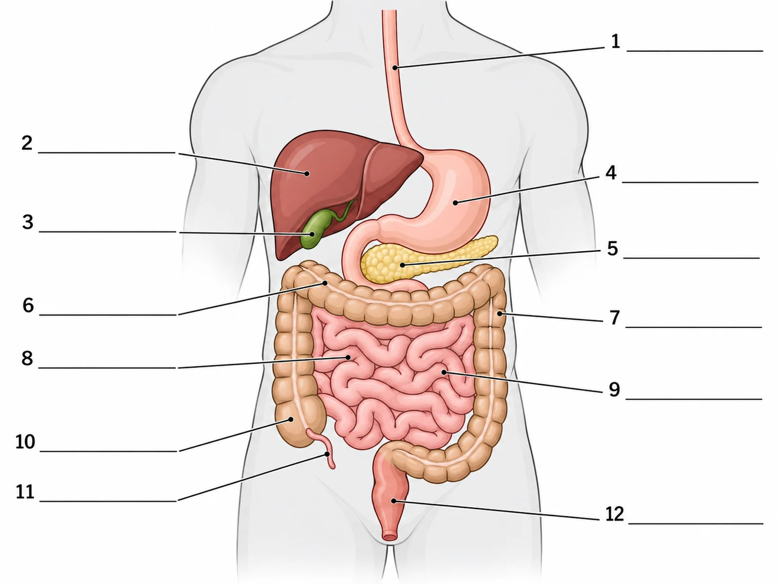
Answer key
- Esophagus (oesophagus)
- Liver
- Gallbladder
- Stomach
- Pancreas
- Large intestine (transverse colon)
- Large intestine (descending colon)
- Small intestine
- Small intestine (8 and 9 both point to the coiled small intestine; a schematic like this does not show where the jejunum ends and the ileum begins, so "small intestine" is the safe answer for both)
- Cecum, the start of the large intestine
- Appendix
- Rectum
To match your own syllabus, for example a simple five-organ diagram for younger classes, or a version with the duodenum and bile duct labeled, describe it on the digestive system diagram generator and ask for the labeled and blank versions of the same figure.
Frequently asked questions
What are the organs of the digestive system in order?
Mouth, pharynx, esophagus, stomach, small intestine (duodenum, jejunum, ileum), large intestine (cecum, colon, rectum) and anus. The salivary glands, liver, gallbladder and pancreas are accessory organs beside the tube.
What are the accessory organs of digestion?
The salivary glands, liver, gallbladder and pancreas (and, in some textbooks, the teeth and tongue). They help digestion by producing or storing secretions, but food does not pass through them.
Which side of the body is the liver on?
The right, in the upper abdomen under the diaphragm. On a front-view diagram it appears on the viewer's left. The stomach is on the body's left.
Why is it called the small intestine if it is longer?
The names refer to the width of the tube, not its length. The small intestine is about 3 m long in a living person but narrow; the large intestine is about half as long but more than twice as wide.
What does the large intestine do?
It absorbs water and salts from the material left after digestion, forming feces, and it houses gut bacteria. Very little digestion happens there.
What does bile do, and is it an enzyme?
Bile emulsifies fats, splitting large fat droplets into small ones so lipase can digest them faster, and helps neutralize acid from the stomach. It is not an enzyme. It is made by the liver and stored in the gallbladder.
Where does most absorption happen?
In the small intestine, through the villi. Glucose and amino acids enter the capillaries; fats enter the lacteals. The large intestine absorbs mainly water and salts.
Is the pancreas part of the digestive system?
Yes, as an accessory organ: it makes amylase, proteases, lipase and bicarbonate, which flow into the duodenum. It also makes insulin and glucagon, which belong to the endocrine system.
What is the difference between digestion, absorption and egestion?
Digestion breaks large food molecules into small ones. Absorption moves those small molecules into the blood or lymph. Egestion removes the undigested remainder from the body as feces.
Where can I get a blank digestive system diagram?
Use the worksheet above, or make one for your syllabus with the digestive system diagram generator, which draws labeled and blank versions, a simple version and a flow chart of the food route.
Related guides and tools
- How to label an ear diagram and how to label a brain diagram: two more body-system worksheets, region by region.
- How to label a cell membrane diagram: the transport processes that absorb nutrients across the villus.
- Anatomy diagram generator for the whole body, and the endocrine system diagram for the pancreas's hormone role.
- Urinary system diagram: the excretion system, for comparing excretion with egestion.
- Respiratory system diagram: the airway that the esophagus runs behind.
- More guides in this series: labeling a plant cell, drawing the stages of mitosis.
Sources
- OpenStax, Anatomy and Physiology 2e, 23.1 Overview of the Digestive System, 23.3 The Mouth, Pharynx, and Esophagus, 23.4 The Stomach, 23.5 The Small and Large Intestines and 23.6 Accessory Organs in Digestion.
- OpenStax, Biology 2e, 34.1 Digestive Systems.
- Patricia JJ, Dhamoon AS. Physiology, Digestion. StatPearls, NCBI Bookshelf.
- Chaudhry SR, Liman MNP, Omole AE, Peterson DC. Anatomy, Abdomen and Pelvis: Stomach. StatPearls, NCBI Bookshelf.
- Collins JT, Nguyen A, Omole AE, Badireddy M. Anatomy, Abdomen and Pelvis, Small Intestine. StatPearls, NCBI Bookshelf.