Tutorials
Labeled Plant Cell Diagram: Every Part, What It Does, and How to Label It
How to label a plant cell diagram: the parts to label at each level, what every organelle looks like and does, the mistakes that cost marks, and a worksheet.
 SciFigure AI
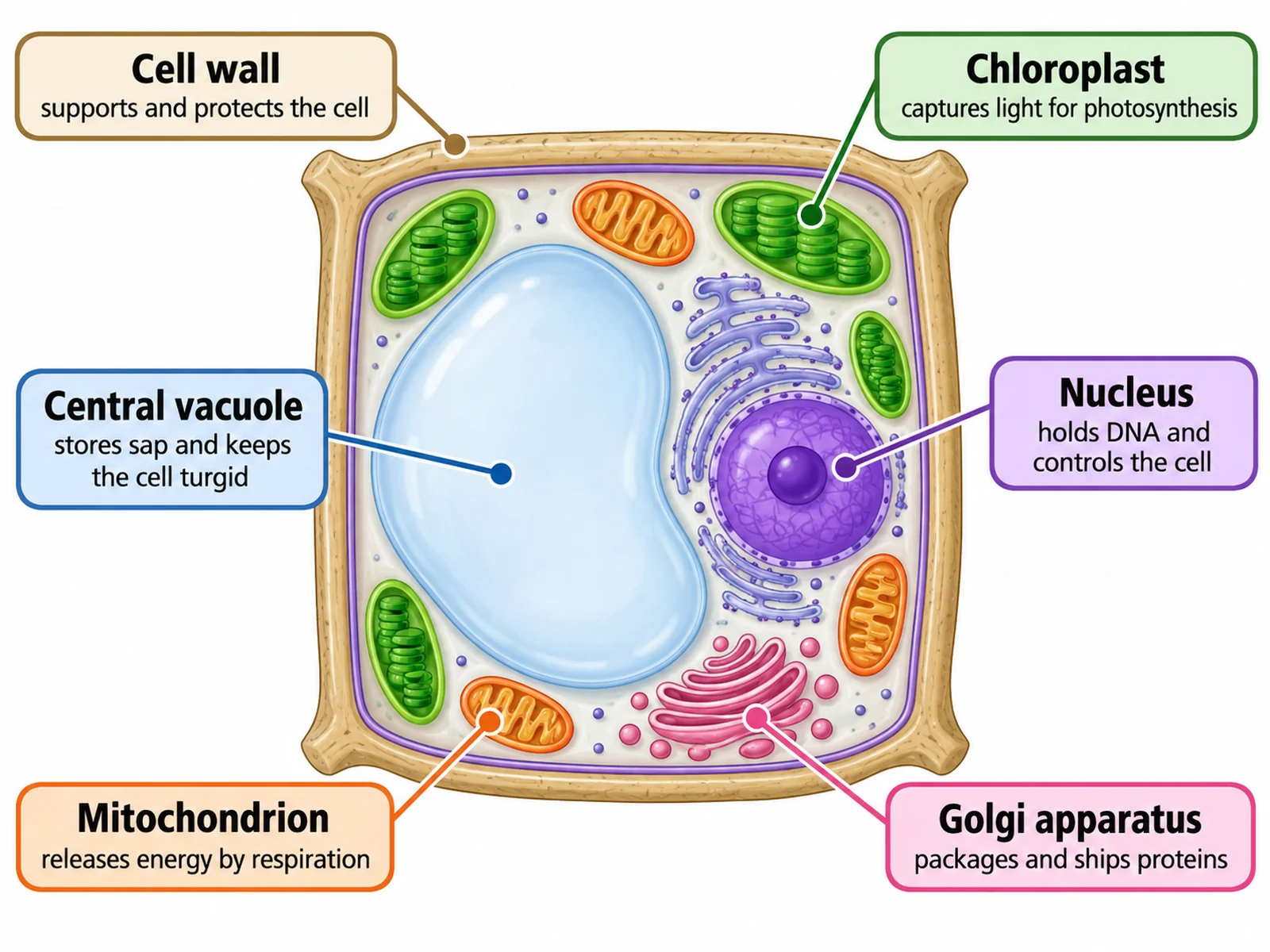
A plant cell diagram looks easy because the shape is easy: a box with a big bubble in it. Marks are lost elsewhere. The cell wall and the cell membrane get one label line between them, the nucleus ends up floating in the vacuole, mitochondria are left out because "plants have chloroplasts instead", and a drawing made from an onion slide somehow has chloroplasts in it.
This guide covers what to label at each level from middle school to college, what every structure looks like on the page and what it does, the labeling conventions examiners look for, a step-by-step method, the mistakes that cost marks, and a numbered worksheet with an answer key. If you need a figure to work on, the plant cell diagram generator draws a labeled version, a blank version and a simplified one from the same description.
The short answer: what to label on a plant cell
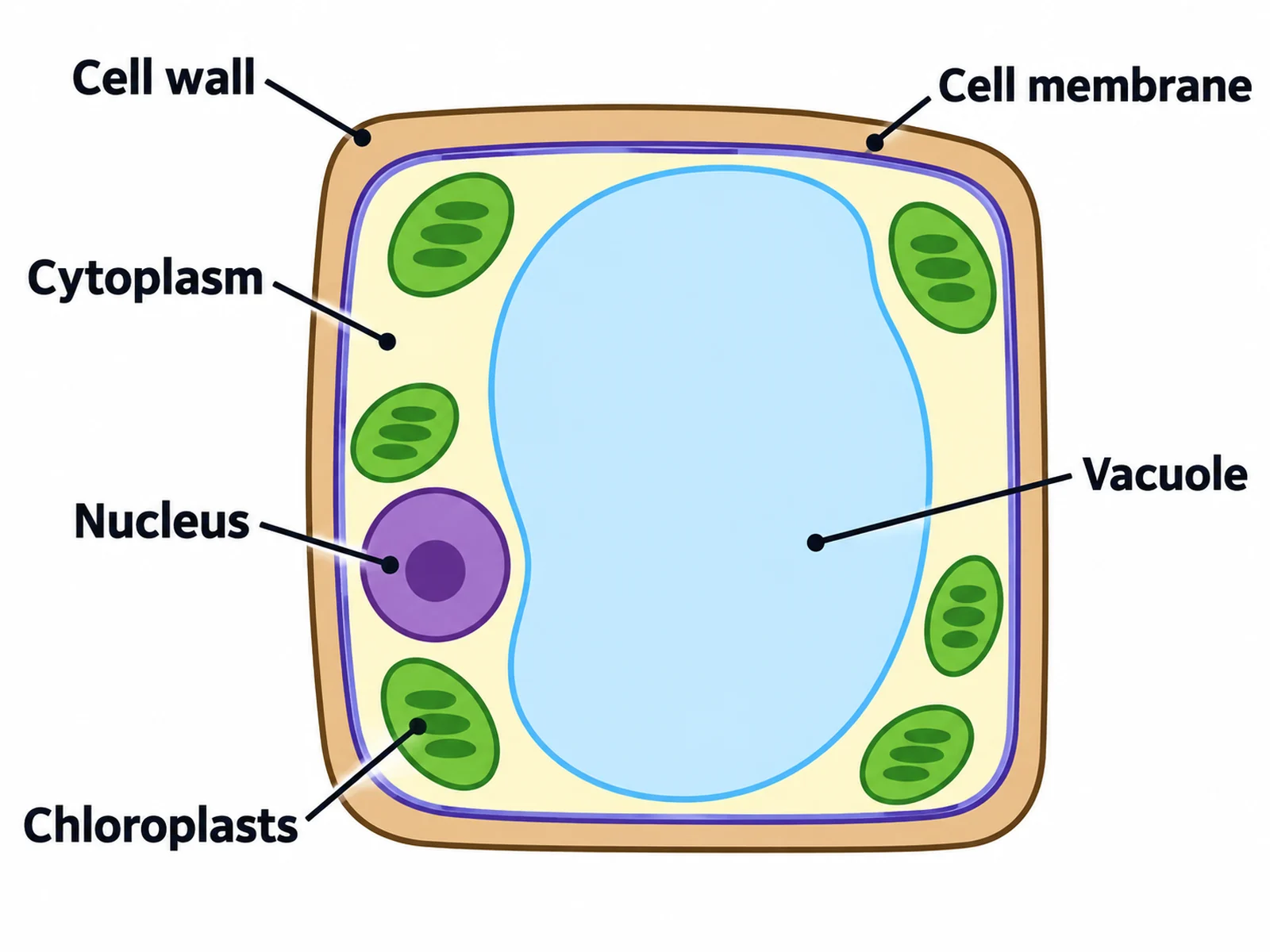
Every plant cell diagram, at any level, needs these six: cell wall, cell membrane, cytoplasm, nucleus, chloroplasts, and the large central vacuole. Three of them (the wall, chloroplasts and the permanent vacuole) are what make it a plant cell rather than an animal cell, so leaving any of them off is the most expensive omission you can make.
From there, the list grows with the course:
| Level | Labels expected | What examiners are really checking |
|---|---|---|
| Middle school, KS3, grades 6–8 | Cell wall, cell membrane, cytoplasm, nucleus, chloroplasts, vacuole (often mitochondria too) | That you can tell a plant cell from an animal cell and know the wall is outside the membrane |
| GCSE, IGCSE, high school biology | The above, always with mitochondria, plus ribosomes; the vacuole described as permanent and filled with cell sap; the wall described as cellulose | Function statements in the right words: the wall strengthens the cell, the membrane controls what enters and leaves, the vacuole holds cell sap |
| A-level, AP Biology, IB | The above plus nuclear envelope and pores, nucleolus, rough and smooth ER, Golgi apparatus, vesicles, tonoplast, plasmodesmata, and the internal structure of the chloroplast | Ultrastructure: what you would see in an electron micrograph, and which structures plant and animal cells share |
| College cell biology, botany | The above plus middle lamella, primary and secondary wall, amyloplasts (starch grains), peroxisomes, cytoskeleton, and the 70S ribosomes and DNA inside chloroplasts and mitochondria | Correct relationships: which membranes are continuous, which organelles have double membranes, where the wall is built |
AQA's GCSE specification is a useful benchmark for the middle row: it lists the nucleus, cytoplasm, cell membrane, mitochondria and ribosomes as the parts animal cells have, and says plant cells often also have chloroplasts and a permanent vacuole filled with cell sap, plus a cellulose cell wall. Note the word often. Not every plant cell has chloroplasts, which matters later.
Every part of a plant cell and what it does
| Part | What it looks like on a diagram | What it does |
|---|---|---|
| Cell wall | A thick band or double line forming the outer box | Made mainly of cellulose. Gives the cell strength and a fixed shape, and stops it bursting when water rushes in. It is fully permeable, not selective |
| Cell membrane (plasma membrane) | A thin single line pressed tight against the inside of the wall | Partially permeable: controls which substances enter and leave the cell |
| Cytoplasm | The space between the membrane and the vacuole, often only a thin layer | Where most of the cell's chemical reactions take place; organelles sit in it |
| Nucleus | A round body, usually pushed against one side of the cell | Contains the DNA as chromosomes and controls the cell's activities |
| Nuclear envelope | The double membrane around the nucleus, drawn with small gaps (pores) | Separates DNA from cytoplasm; the pores let mRNA and ribosome subunits out |
| Nucleolus | A darker spot inside the nucleus | Makes ribosomal RNA and assembles ribosome subunits |
| Chloroplast | Green oval or lens shape, with stacked discs (grana) drawn inside | Photosynthesis: absorbs light with chlorophyll and makes glucose from carbon dioxide and water |
| Large central vacuole | One big sac, usually the largest thing in the cell | Stores cell sap (water, sugars, salts, pigments); pushes the cytoplasm outward so the cell is turgid and firm |
| Tonoplast | The membrane forming the edge of the vacuole | Controls what moves in and out of the vacuole |
| Mitochondrion | Small oval with a folded inner membrane (cristae) | Aerobic respiration: releases energy from glucose as ATP. Plant cells need these as much as animal cells do |
| Ribosomes | Tiny dots, free or on the rough ER | Protein synthesis |
| Rough endoplasmic reticulum | Folded membrane sheets dotted with ribosomes, near the nucleus | Carries and folds the proteins its ribosomes make |
| Smooth endoplasmic reticulum | Tubular membranes without ribosomes | Makes lipids |
| Golgi apparatus | A stack of curved flattened sacs with small vesicles budding off | Modifies and packages proteins; in plant cells it also makes the pectins and hemicelluloses of the cell wall |
| Plasmodesmata | Narrow channels drawn as gaps crossing the wall between two cells | Connect the cytoplasm of neighboring cells so water, ions and small molecules can pass directly |
| Middle lamella | A thin layer between the walls of two adjacent cells | Cements neighboring cells together; rich in pectin |
| Amyloplast (starch grain) | Pale oval grain, common in storage cells such as potato | Stores starch |
Two structures in that table carry most of the subtlety.
The cell wall and the membrane are two separate things, drawn as two separate lines. In a healthy, well-watered cell the membrane is pressed so firmly against the wall that the two can look like one boundary, but a labeled diagram has to show both, and each label line must end on its own line. If you draw a visible gap between them, you have drawn a plasmolyzed cell, one that has lost water by osmosis, which is a different diagram with a different meaning.
The vacuole is the reason the nucleus is at the edge. In a mature plant cell the central vacuole can take up most of the cell's volume, which squeezes the cytoplasm into a thin layer against the wall and pushes the nucleus to one side. A nucleus drawn in the middle of the vacuole is not a style choice; it is a nucleus inside a sac of cell sap, which is wrong.
Inside the chloroplast (A-level and AP)
At A-level, AP and IB you are often asked to label the chloroplast itself, either as a zoom-in from the cell or as a separate diagram:
- Outer and inner membrane (the chloroplast envelope): two membranes, so draw two lines.
- Thylakoids: flattened membrane sacs where the light-dependent reactions happen.
- Granum (plural grana): a stack of thylakoids, the coin-stack shape most diagrams show.
- Stroma: the fluid around the grana, where the Calvin cycle (the light-independent reactions) happens.
- Intergranal lamellae: thylakoid membranes that link one granum to the next.
- Starch grains and lipid droplets: stores. Add 70S ribosomes and a loop of DNA when the question is about endosymbiotic theory: they are the evidence that chloroplasts descend from bacteria.
The chloroplast diagram page draws this organelle on its own, and the photosynthesis diagram shows where each stage happens inside it.
Plant cell vs animal cell: what to leave on, what to leave off
About half of the marks on a plant cell question come from knowing what not to draw. This is the comparison to revise from:
| Feature | Plant cell | Animal cell |
|---|---|---|
| Cell wall | Present, made of cellulose | Absent |
| Shape | Regular, often box-shaped, fixed by the wall | Irregular or rounded |
| Chloroplasts | In green, photosynthetic cells | Never |
| Vacuole | One large permanent central vacuole with a tonoplast | Small, temporary vacuoles if any |
| Centrioles | Absent in flowering plants | Present, a pair near the nucleus |
| Lysosomes | Rarely drawn: the vacuole does much of the digestive work | Present |
| Connections between cells | Plasmodesmata through the walls | Gap junctions and other junctions in the membrane |
| Carbohydrate store | Starch | Glycogen |
| Mitochondria, nucleus, ribosomes, ER, Golgi | Present | Present |
| Division of the cytoplasm | A cell plate forms across the middle | A cleavage furrow pinches the cell in two |
The last row of shared structures is the one students forget. Plant cells respire around the clock, so they have mitochondria; chloroplasts make the glucose, mitochondria release its energy. For the other half of the comparison, see how to label an animal cell diagram, and the plant vs animal cell diagram page draws both cells side by side with the plant-only structures highlighted.
What you actually see under a light microscope
A textbook plant cell and a plant cell under the school microscope are not the same picture, and a biological drawing must show what is on the slide, not what is in the book.
A light microscope can resolve detail down to roughly 0.2 µm. That is enough to see the cell wall, the nucleus, chloroplasts and the outline of the vacuole, but not ribosomes (about 25 nm across), the ER, or the internal membranes of a chloroplast. Those come from electron micrographs. So:
- A drawing of a leaf cell or pondweed (Elodea) cell from a light microscope shows the wall, chloroplasts around the edge, cytoplasm and sometimes the nucleus. Chloroplasts may appear to move round the cell: that is cytoplasmic streaming.
- A drawing of an onion epidermis cell shows no chloroplasts at all. Onion bulb cells grow underground and do not photosynthesize. Adding green ovals because "plant cells have chloroplasts" is a classic lost mark.
- A root hair cell also has no chloroplasts, for the same reason.
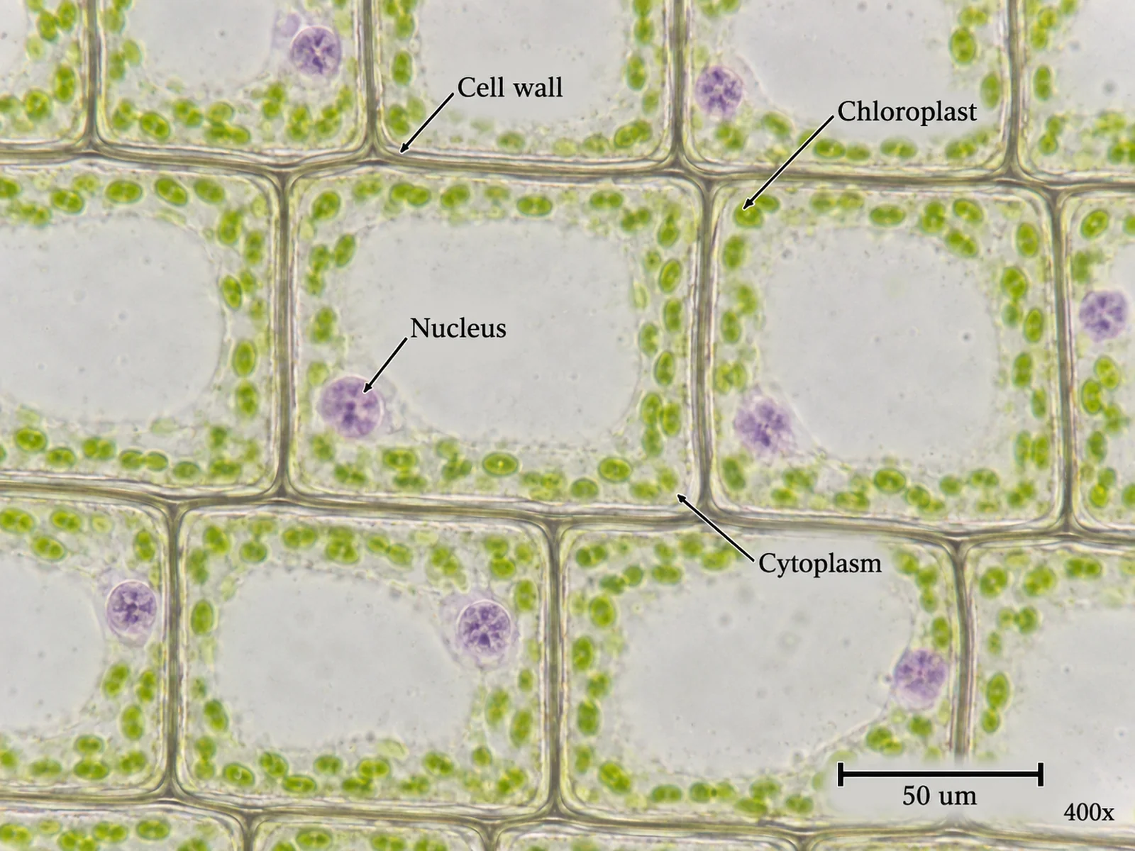
If the question gives you a micrograph, add a title, the magnification, and a scale bar or the magnification calculation. Magnification is image size divided by actual size: a cell drawn 60 mm long that is really 60 µm long (0.06 mm) has been drawn at ×1000. The microscope diagram page covers the parts of the microscope itself if that is the next question on the sheet.
Labeling conventions that gain marks
- Draw label lines with a ruler, as single straight lines with no arrowheads.
- End each line exactly on the structure. On a plant cell the commonest failure is a cell wall line that stops on the membrane, or a membrane line that stops on the wall. Make the wall thick enough that both lines have something distinct to touch.
- Never cross label lines. If two would cross, move one label to the other side of the cell.
- Keep the labels outside the cell, lined up in neat columns. Text written inside the vacuole covers the structure you are labeling.
- Write labels horizontally, even where the line comes in at an angle.
- Match singular and plural to what the line touches. One line to one green oval is chloroplast; one bracket or several lines to several is chloroplasts. The same goes for mitochondrion and mitochondria.
- Use the expected term. "Cell membrane" or "plasma membrane", not "cellulose membrane"; "permanent vacuole" or "central vacuole" at GCSE and above, not just "bubble"; "Golgi apparatus" with a capital G.
- Use clean single lines and no shading in a biological drawing. If you use color on a printed diagram, use it to carry meaning (green for chloroplasts is fine) and keep it consistent.
- Give the drawing a title and, for anything from a microscope, the magnification.
- Label a repeated structure once, on one clear example, unless the question asks you to show all of them.
How to label a plant cell diagram, step by step
- Decide the level and write your list first. Six labels for an introductory diagram, eight or nine for GCSE, fifteen or more for A-level. Counting them now tells you how much space to leave.
- Draw the box with room around it. Draw the wall as a thick band and the membrane as a thin line just inside it, touching. Leave a wide margin on both sides for labels.
- Draw the vacuole next. Make it big, roughly central, with its own outline (the tonoplast). Everything else goes in the cytoplasm around it.
- Place the nucleus against one side, in the cytoplasm, with the nucleolus inside it.
- Add chloroplasts around the edge if the cell is photosynthetic, then mitochondria, ER near the nucleus, and the Golgi stack.
- Label from the outside in. Wall first, membrane second, then cytoplasm and vacuole. These lines are short and set up clean columns on both sides.
- Label everything else by position. Structures on the left of the cell get labels on the left, structures on the right get labels on the right. This single habit prevents nearly every crossed line.
- Check every line ends on its structure, every label is spelled correctly, and nothing is on the cell that should not be: no centrioles, no chloroplasts in a root or onion cell, no nucleus in the vacuole.
Twelve mistakes that cost marks
- One label line used for both the cell wall and the cell membrane.
- A gap drawn between wall and membrane in a cell that is meant to be normal and turgid.
- The nucleus drawn inside the vacuole instead of in the cytoplasm.
- Mitochondria left out because the cell has chloroplasts.
- Chloroplasts drawn in an onion epidermis or root cell.
- Several small vacuoles instead of one large permanent one.
- Centrioles drawn in a plant cell.
- The cell wall described as partially permeable. It is the membrane that is selective; the wall lets water and dissolved substances through freely.
- "Chloroplasts" written on a line pointing at a single chloroplast, or "mitochondria" on one mitochondrion.
- Ribosomes, ER or Golgi drawn on a drawing that is supposed to show a light-microscope slide.
- The nucleolus labeled as the nucleus. On many diagrams a line drawn into the nucleus ends on the dark nucleolus, so check where yours lands.
- Labels written inside the cell, or lines that cross.
Printable plant cell worksheet with answer key
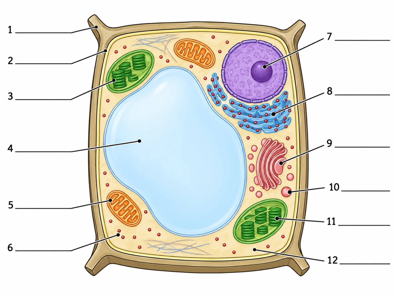
The fastest way to learn the labels is to fill in a blank, check it, and do it again a day later. Here is a numbered blank plant cell to label. The answers are below it.
Answer key
- Cell wall
- Cell membrane
- Chloroplast
- Central (permanent) vacuole
- Mitochondrion
- Ribosome
- Nucleolus (the line ends on the dark body inside the nucleus, not on the nucleus itself)
- Rough endoplasmic reticulum
- Golgi apparatus
- Vesicle
- Granum, a stack of thylakoids inside a chloroplast (at GCSE, "chloroplast" is the expected answer)
- Cytoplasm
To make a worksheet that matches your own syllabus exactly, describe the labels you want on the plant cell diagram generator and ask for a blank version with numbered lines. Generate the labeled version from the same description and you have the answer key. Check every label against your textbook before handing it out.
Frequently asked questions
How many parts should a labeled plant cell diagram have?
Six for an introductory diagram (cell wall, cell membrane, cytoplasm, nucleus, chloroplasts, vacuole). Eight to ten at GCSE and high-school level, adding mitochondria, ribosomes, and the vacuole's cell sap. Fifteen or more at A-level, AP and IB, adding the nucleolus, nuclear envelope, rough and smooth ER, Golgi apparatus, tonoplast and plasmodesmata.
What three parts does a plant cell have that an animal cell does not?
A cellulose cell wall, chloroplasts, and a large permanent vacuole filled with cell sap. At higher levels plasmodesmata are a fourth. Animal cells, in turn, have centrioles, which flowering plant cells lack.
Do plant cells have mitochondria?
Yes. Plant cells carry out aerobic respiration all the time, day and night, so they have mitochondria just as animal cells do. Chloroplasts make glucose by photosynthesis; mitochondria release the energy stored in it.
Why is the nucleus at the side of a plant cell?
Because the large central vacuole fills most of the cell and pushes the cytoplasm, and everything in it, into a thin layer against the wall. The nucleus sits in that layer.
What is the difference between the cell wall and the cell membrane?
The wall is a rigid layer of cellulose outside the membrane. It supports the cell and is fully permeable. The membrane is a thin phospholipid bilayer just inside the wall, and it is partially permeable: it decides what enters and leaves. The cell membrane labeling guide covers the membrane's own structure.
Does every plant cell have chloroplasts?
No. Only cells that photosynthesize have them, mainly in leaves and green stems. Root cells, onion bulb cells, and many storage cells have none. Mature xylem vessels have no living contents at all.
What is the tonoplast?
The membrane around the central vacuole. It controls what moves between the cytoplasm and the cell sap. It is usually expected from A-level and AP upward.
Do plant cells have lysosomes and centrioles?
Flowering plant cells have no centrioles; they build their spindle without them. Lysosomes are seldom drawn in plant cells because the central vacuole carries out much of the same digestive work, which is how most introductory textbooks treat it.
How do you draw a plant cell from a microscope slide?
Draw only what you can see, with clear continuous lines and no shading. A leaf or pondweed cell shows the wall, cytoplasm, chloroplasts and sometimes the nucleus; an onion cell shows the wall, cytoplasm and nucleus, with no chloroplasts. Add a title and the magnification.
Where can I get a blank plant cell diagram to label?
Use the worksheet above, or make one that matches your syllabus with the plant cell diagram generator, which draws a labeled version, a blank version with numbered lines, and a simple version for younger students.
Related guides and tools
- How to label an animal cell diagram: the other half of the plant vs animal comparison.
- How to label a cell membrane diagram: the fluid mosaic model in detail.
- How to draw the stages of mitosis: what happens when the cell divides, including the plant cell plate.
- Chloroplast diagram, leaf cross-section diagram and Golgi apparatus diagram for the organelles and tissues around the cell.
- Cell diagram generator for bacterial, fungal and specialized cells.
- More guides in this series: labeling an ear diagram, labeling the digestive system, labeling a brain diagram.
Sources
- OpenStax, Biology 2e, 4.3 Eukaryotic Cells: plant cell structures, chloroplast structure, the central vacuole, the absence of centrosomes in plant cells, and digestion in plant vacuoles.
- OpenStax, Biology 2e, 4.6 Connections between Cells and Cellular Activities: plasmodesmata.
- OpenStax, Biology 2e, 4.4 The Endomembrane System and Proteins: ER, Golgi apparatus and vesicles.
- OpenStax, Biology 2e, 10.2 The Cell Cycle: cell plate formation in plant cytokinesis.
- AQA, GCSE Biology (8461) specification, Cell biology: required plant and animal cell structures, microscopy and magnification.