Tutorials
How to Draw and Label the Stages of Mitosis (Prophase to Cytokinesis)
How to draw and label the stages of mitosis: what each phase looks like, the structures to label, chromosome counts that add up, and a worksheet with answers.
 SciFigure AI
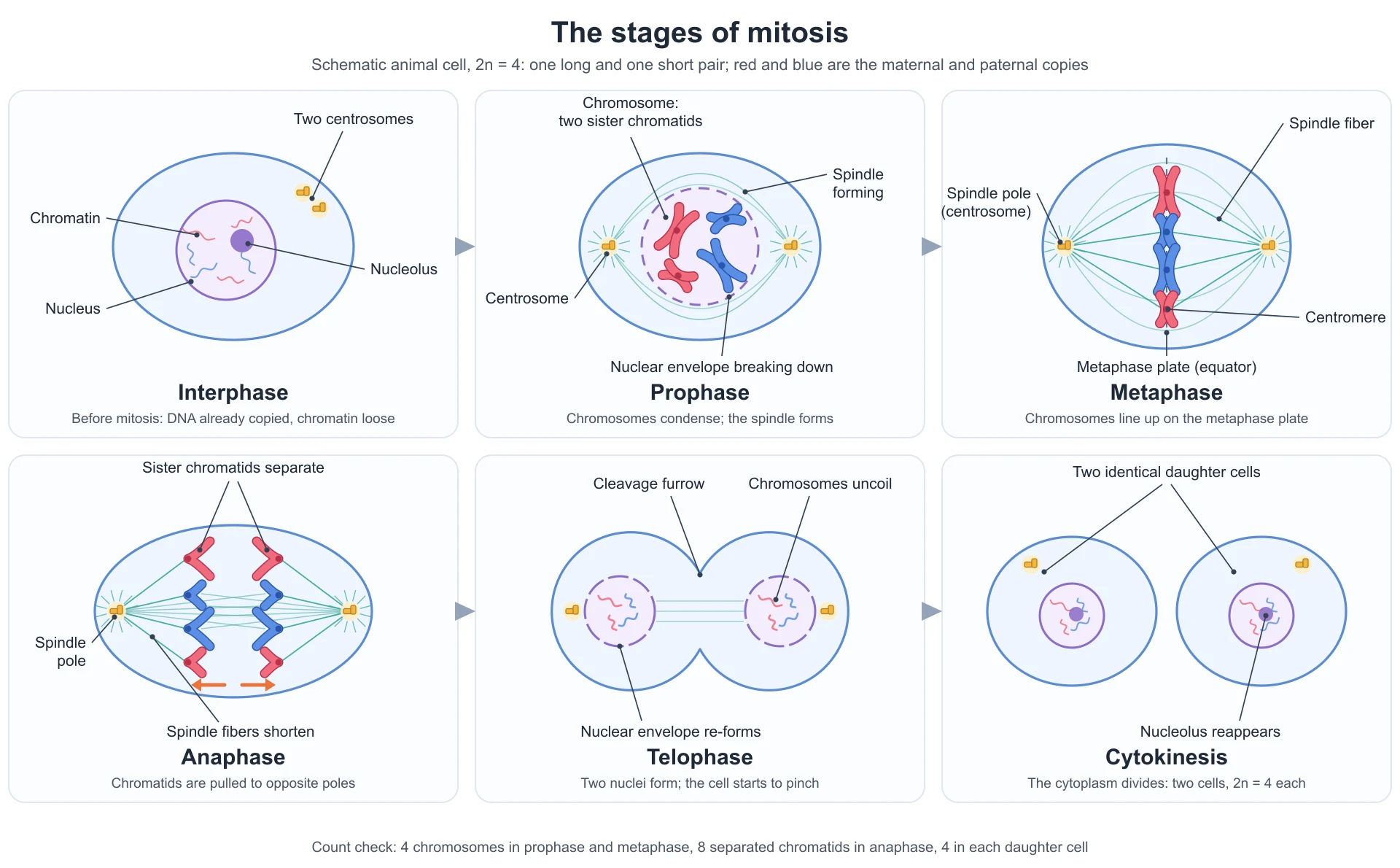
Most mitosis diagrams that lose marks are not badly drawn. They are inconsistent. Six chromosomes in prophase become four in metaphase. The metaphase plate is drawn running from pole to pole instead of across the spindle. Anaphase shows X-shaped chromosomes moving apart, which would mean nothing has actually separated. Each panel looks plausible on its own; together they describe a cell that could not exist.
This guide shows how to draw and label each stage so the whole sequence holds together: the order of the stages, what to label at each level, what every phase looks like and how to tell it apart, the chromosome counts that must add up, plant versus animal cells, the mistakes that cost marks, and a printable worksheet with an answer key. The figure above was drawn for this guide with a diploid number of four, so you can check every count against it. To make your own version for a worksheet or slide, use the mitosis diagram generator.
The stages of mitosis in order
Mitosis has four stages, usually remembered as PMAT: prophase, metaphase, anaphase, telophase. Two more names appear on almost every diagram:
- Interphase comes before mitosis. It is not a stage of mitosis, and it is not a resting phase: the cell grows and copies its DNA (in S phase) during it.
- Cytokinesis is the division of the cytoplasm. It usually begins during late anaphase or telophase, but strictly it is a separate process from mitosis, which is the division of the nucleus.
College and AP courses add a fifth stage, prometaphase, between prophase and metaphase, when the nuclear envelope fragments and spindle fibers attach to the chromosomes. If your course uses it, draw it; if not, its events belong to late prophase.
What to label at each level
| Level | Stages to show | Structures to label |
|---|---|---|
| Middle school, KS3 | The sequence in order, ending in two cells | Nucleus, chromosomes, cell membrane, two daughter cells |
| GCSE, high school biology | Interphase, prophase, metaphase, anaphase, telophase, cytokinesis | Chromosomes, nuclear membrane, spindle fibers, poles, the equator of the cell |
| A-level, AP Biology, IB | All of the above, often with prometaphase | Sister chromatids, centromere, kinetochore, centrosome and centrioles (animal cells), metaphase plate, nucleolus, cleavage furrow or cell plate |
| College cell biology | All six stages including prometaphase | Kinetochore, polar and astral microtubules, condensed chromatin, contractile ring, phragmoplast, and the two parts of anaphase |
Check your own specification before you learn the stage names. AQA's GCSE Biology specification, for example, says students do not need to know the different phases of mitosis, only that the chromosomes are copied, one set is pulled to each end of the cell and the nucleus divides, then the cytoplasm and membrane divide. Many other GCSE, IGCSE and US high-school courses do expect the four names.
Every stage: what to draw, what to label, how to recognize it
| Stage | What the chromosomes look like | Nuclear envelope | Spindle | The one feature that identifies it |
|---|---|---|---|---|
| Interphase | Not visible as separate chromosomes; loose chromatin | Intact, with a nucleolus | None | A normal-looking nucleus |
| Prophase | Condensed, each one two sister chromatids joined at the centromere | Breaking down; nucleolus disappears | Forming between centrosomes moving apart | Visible X-shaped chromosomes still in a nucleus-shaped cluster |
| Prometaphase | Condensed, being moved by spindle fibers | Fragmented | Fibers attaching to kinetochores | Chromosomes scattered, no envelope, not yet lined up |
| Metaphase | Lined up in a single row across the middle of the cell | Gone | Fully formed, fibers from both poles attached to each centromere | One line of chromosomes on the equator |
| Anaphase | Sister chromatids separated, each pulled centromere-first toward a pole | Gone | Kinetochore fibers shortening; cell elongating | Two sets of V-shaped chromatids moving apart |
| Telophase | Arrived at the poles and starting to uncoil | Re-forming around each set; nucleoli reappear | Breaking down | Two nuclei in one (often pinching) cell |
| Cytokinesis | Uncoiled again as chromatin | Intact in each new nucleus | Gone | Two separate daughter cells |
Interphase
Draw: a normal cell with a round nucleus, a nucleolus, and thin squiggly chromatin. In an animal cell, add two centrosomes near the nucleus: the centrosome is copied during interphase, ready to form the two spindle poles.
Label: nucleus, nucleolus, chromatin, centrosomes, cell membrane.
Remember: by the end of interphase the DNA has already been copied, so every chromosome is already two sister chromatids. You just cannot see them yet.
Prophase
Draw: the chromosomes as short, thick X shapes: two chromatids joined at a centromere. Draw the nuclear envelope as a broken (dashed) line, leave out the nucleolus, and show the two centrosomes moving to opposite sides of the nucleus with the first spindle fibers between them.
Label: chromosome (or "two sister chromatids"), centromere, centrosome, spindle fibers forming, nuclear envelope breaking down.
Prometaphase (college and AP)
Draw: chromosomes scattered through the cell with no envelope around them, and spindle fibers from both poles reaching in to attach at the centromeres.
Label: kinetochore (the protein structure at the centromere where fibers attach), kinetochore microtubules.
Metaphase
Draw: every chromosome lined up in a single row across the middle of the cell, at right angles to the line joining the two poles. Each centromere has a fiber from one pole attached to one chromatid and a fiber from the other pole attached to its sister. Chromosomes are at their most condensed in metaphase, which is why karyotypes are made from metaphase cells.
Label: metaphase plate (or equator), spindle fibers, spindle pole or centrosome, centromere.
The metaphase plate is not a structure. It is the imaginary plane halfway between the poles, the line your chromosomes sit on. If you draw it as a dashed line, it must cross the spindle, passing through the centromeres, never run from pole to pole.
Anaphase
Draw: the sister chromatids of every chromosome pulled apart, the two copies moving to opposite poles. Each separated chromatid is drawn as a V (or J or rod, depending on where its centromere is) with the centromere leading and the arms trailing behind toward the middle. The cell is longer than in metaphase.
Label: sister chromatids separating (once separated, each is counted as a chromosome), spindle fibers shortening, spindle poles.
Telophase
Draw: a complete set of chromosomes at each pole, uncoiling into thinner lines, with a new nuclear envelope forming around each set and the nucleoli reappearing. In an animal cell the cell membrane is already pinching in at the middle.
Label: nuclear envelope re-forming, chromosomes uncoiling, cleavage furrow (animal) or cell plate (plant).
Cytokinesis
Draw: two separate daughter cells, the same size, each with a normal interphase-looking nucleus.
Label: daughter cells, cleavage furrow (animal) or new cell wall (plant). For a full mark, state that the two cells are genetically identical to each other and to the parent cell.
The structures to label, and what each one is
| Structure | What it is | Where it appears |
|---|---|---|
| Chromatin | DNA wound around histone proteins, loosely packed | Interphase, late telophase |
| Chromosome | One DNA molecule with its proteins; after replication, two identical sister chromatids joined at a centromere | Visible as a distinct body from prophase to telophase |
| Sister chromatids | The two identical copies of a replicated chromosome, joined at the centromere | Prophase and metaphase; they separate in anaphase |
| Centromere | The constricted region joining two sister chromatids | Prophase to metaphase |
| Kinetochore | A protein complex on each side of the centromere where spindle fibers attach | Prometaphase and metaphase |
| Centrosome | The microtubule-organizing center at each spindle pole; contains a pair of centrioles in animal cells | Animal cells, interphase to telophase |
| Spindle fibers | Microtubules that move the chromosomes. Kinetochore fibers attach to chromosomes; polar fibers overlap at the middle; astral fibers radiate toward the membrane | Prophase to telophase |
| Metaphase plate | The imaginary plane at the cell's equator where chromosomes line up | Metaphase |
| Nuclear envelope | The double membrane around the nucleus | Breaks down in prophase, re-forms in telophase |
| Nucleolus | Dense region of the nucleus that makes ribosomes | Disappears in prophase, reappears in telophase |
| Cleavage furrow | The pinch in an animal cell's membrane made by a contracting ring of actin and myosin | Late anaphase to cytokinesis |
| Cell plate | A new wall built across a plant cell from Golgi vesicles | Telophase to cytokinesis in plants |
Counting chromosomes: the check that catches most errors
A mitosis diagram is a story about numbers, and the numbers must add up. The rule is simple: count centromeres to count chromosomes. Before anaphase, each chromosome is two chromatids sharing one centromere. In anaphase the sister centromeres separate, so each chromatid has its own centromere and counts as a chromosome in its own right.
| Stage | Diagram above (2n = 4) | Human cell (2n = 46) |
|---|---|---|
| Prophase and metaphase | 4 chromosomes, 8 chromatids | 46 chromosomes, 92 chromatids |
| Anaphase (whole cell) | 8 chromosomes, 4 moving to each pole | 92 chromosomes, 46 moving to each pole |
| Each telophase nucleus | 4 chromosomes | 46 chromosomes |
| Each daughter cell | 4 chromosomes, same as the parent | 46 chromosomes, same as the parent |
Two drawing habits make this easy. Decide the diploid number before you start (four is ideal: one long and one short pair) and keep it the same in every panel. And give the two members of each homologous pair different colors, red for one parent's copy and blue for the other's. Then check that every daughter cell gets one red long, one blue long, one red short and one blue short chromosome. If it does, your mitosis is right. A daughter with two copies of one chromosome and none of another means a pair of sister chromatids went to the same pole; a daughter with only one chromosome of each size is a meiosis diagram, not a mitosis one.
How to draw the stages of mitosis neatly
- Rule a grid first. Six equal boxes in two rows of three, with a line under each for the stage name.
- Draw the same cell outline in every box. Same size, same shape, poles on the left and right (or top and bottom) in every panel. Only anaphase and telophase change shape, getting longer and then pinching.
- Decide the chromosome number and the colors before drawing any chromosome.
- Draw chromosomes as two chromatids from prophase to metaphase: two short curved strokes touching at the centromere, which reads as an X.
- In metaphase, line them up on the equator, centromeres on the line, one fiber from each pole to each centromere.
- In anaphase, draw single chromatids as Vs, centromere first, arms trailing, the same set of colors and lengths heading to each pole.
- Show the envelope as a dashed circle when it is breaking down (prophase) or re-forming (telophase), solid when it is intact.
- Label outside the cells, with ruled lines that end exactly on the structure and never cross.
- Write a one-line description under each stage name. "Chromosomes line up on the equator" earns credit that a bare label does not.
Mitosis in plant cells vs animal cells
| Feature | Animal cell | Plant cell |
|---|---|---|
| Centrioles | A pair in each centrosome | None in flowering plants; the spindle forms without them |
| Asters (star of short fibers at each pole) | Present | Absent |
| Shape during division | Rounds up, elongates in anaphase | Held in shape by the wall |
| Cytokinesis | A contractile ring of actin forms a cleavage furrow that pinches the cell in two | Golgi vesicles gather at the middle and fuse into a cell plate, which grows outward into a new cell wall |
| Where dividing cells are found | Throughout growing and repairing tissues | Mainly in meristems: root tips and shoot tips |
The plant version matters for practicals. The classic slide is an onion or garlic root tip squash, where the cells are boxy, there are no centrioles, and telophase shows a straight cell plate rather than a furrow.
Identifying stages on a root tip slide, and the mitotic index
On a stained root tip squash, most cells are in interphase, because interphase is by far the longest part of the cycle. (In rapidly dividing mammalian cells in culture, a full cycle takes around a day and mitosis itself about an hour.) To identify a dividing cell, look for these, in order of how easy they are to spot:
- Metaphase: a dark straight line of chromosomes across the middle of the cell.
- Anaphase: two dark bands moving apart, often with a clear gap between them.
- Telophase: two dark clusters at opposite ends, sometimes with a faint line (the cell plate) between them.
- Prophase: a nucleus that looks grainy or threaded rather than smooth, because the chromosomes are condensing.
A-level practicals (AQA's required practical 2 is a stained root tip squash, for example) then ask for the mitotic index: the number of cells in mitosis divided by the total number of cells counted. If 18 of 240 cells are in prophase, metaphase, anaphase or telophase, the mitotic index is 18 ÷ 240 = 0.075, or 7.5%. A higher index means a higher proportion of dividing cells, which is why the index is highest in the meristem just behind the root cap and falls further from the tip.
Mistakes that cost marks
- Different chromosome numbers in different stages. Count the centromeres in every panel.
- The metaphase plate drawn from pole to pole. It runs across the spindle, through the centromeres.
- X-shaped chromosomes in anaphase. By anaphase the chromatids have separated; each one moving to a pole is a single chromatid.
- Chromatids moving arms first. The centromere leads, because that is where the spindle fiber pulls.
- Homologous chromosomes lined up in pairs at metaphase. That is metaphase I of meiosis. In mitosis each chromosome lines up on its own.
- The nuclear envelope still intact in metaphase. It has broken down by then.
- Spindle fibers that do not attach to the chromosomes. Every centromere needs a fiber from each pole in metaphase.
- Interphase called a resting stage. The cell is growing, copying organelles and replicating its DNA.
- Cytokinesis listed as a phase of mitosis. It is division of the cytoplasm, which follows the division of the nucleus.
- Centrioles drawn in a plant cell, or a cleavage furrow drawn in a cell with a wall.
- Daughter cells of different sizes or with different chromosome sets. They are genetically identical.
- "Chromatid" and "chromosome" used interchangeably. Before anaphase, one chromosome is two chromatids; after separation, each chromatid counts as one chromosome.
Printable mitosis worksheet with answer key
This blank version uses exactly the same cells as the labeled figure at the top of the page. Name each stage, A to F, and each numbered structure, 1 to 20. Save the image and print it at full page width.
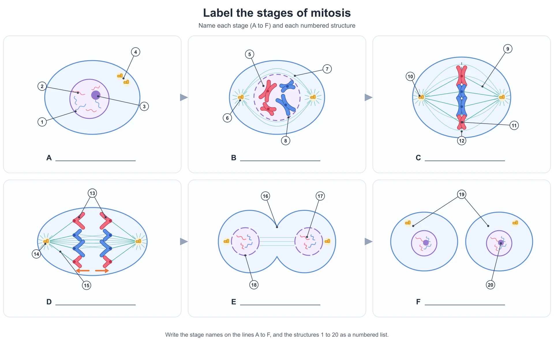
Stages: A interphase, B prophase, C metaphase, D anaphase, E telophase, F cytokinesis.
Structures:
- Nucleus
- Chromatin
- Nucleolus
- Centrosomes (two, after the centrosome has been copied)
- Chromosome, made of two sister chromatids
- Centrosome
- Spindle fibers (the spindle forming)
- Nuclear envelope (breaking down)
- Spindle fiber
- Spindle pole (centrosome)
- Centromere
- Metaphase plate (equator)
- Sister chromatids, separating
- Spindle pole
- Spindle fiber (kinetochore fiber, shortening)
- Cleavage furrow
- Chromosomes (uncoiling)
- Nuclear envelope (re-forming)
- Daughter cells (genetically identical)
- Nucleolus (reappearing)
For a worksheet that matches your own course, such as a plant root tip version, a single stage in close-up, or one with prometaphase added, describe it on the mitosis diagram generator and ask for both a labeled and a blank version. The cell cycle diagram page draws the whole cycle with G1, S and G2 when the question is about interphase too.
Mitosis vs meiosis at a glance
| Mitosis | Meiosis | |
|---|---|---|
| Divisions | One | Two |
| Daughter cells | Two, diploid, genetically identical | Four, haploid, genetically different |
| Homologous chromosomes | Behave independently | Pair up and cross over in prophase I |
| Metaphase | Single chromosomes on the plate | Pairs of homologues on the plate (metaphase I) |
| Used for | Growth, repair, asexual reproduction | Making gametes |
If your diagram needs the second division too, the meiosis diagram page draws meiosis I and II with crossing over.
Frequently asked questions
What are the stages of mitosis in order?
Prophase, metaphase, anaphase and telophase (PMAT). Many courses add prometaphase between prophase and metaphase. Interphase comes before mitosis and cytokinesis follows it; neither is a stage of mitosis itself.
What happens in each stage of mitosis?
Prophase: chromosomes condense and the spindle forms. Metaphase: chromosomes line up on the metaphase plate. Anaphase: sister chromatids are pulled to opposite poles. Telophase: nuclear envelopes re-form around the two sets of chromosomes. Cytokinesis then divides the cytoplasm.
How do you draw prophase?
Draw a round nucleus whose envelope is breaking up (a dashed circle), with no nucleolus and condensed X-shaped chromosomes inside, each made of two sister chromatids joined at a centromere. In an animal cell, draw the two centrosomes on opposite sides of the nucleus with spindle fibers starting to form between them.
How do you draw metaphase?
Draw the chromosomes in one straight row across the middle of the cell, at right angles to the spindle. Draw a spindle fiber from each pole to each centromere. Leave out the nuclear envelope.
How do you draw telophase?
Draw a complete set of chromosomes at each end of the cell, uncoiling, with a new nuclear envelope forming around each set. In an animal cell show the cleavage furrow pinching the middle; in a plant cell, a cell plate.
What is the difference between a chromosome and a chromatid?
A chromatid is one of the two identical copies of a replicated chromosome. Before anaphase, the two sister chromatids are joined at the centromere and together count as one chromosome. Once they separate in anaphase, each is counted as a chromosome.
Why is it called the metaphase plate if there is no plate?
The name describes where the chromosomes line up, the plane halfway between the two spindle poles, not a physical structure. On a diagram it is shown as a dashed line through the centromeres.
What is the longest stage of the cell cycle?
Interphase. Mitosis is a short part of the cycle, which is why most cells on a root tip slide are in interphase.
Do plant cells have centrioles in mitosis?
Flowering plant cells do not. Their spindle forms without centrioles, and they divide their cytoplasm with a cell plate rather than a cleavage furrow.
What is an easy way to draw mitosis?
Use four chromosomes in two sizes and two colors, keep the cell outline identical in all six boxes, draw chromosomes as Xs until anaphase and as Vs after it, and check the counts at the end. The labeled figure at the top of this page follows exactly that pattern.
Where can I get a blank mitosis worksheet?
Use the worksheet above, or describe the version your class needs, such as a plant root tip, a single stage in close-up or a sequence with prometaphase, on the mitosis diagram generator, and generate a labeled and a blank version of the same figure.
Related guides and tools
- How to label a plant cell diagram and how to label an animal cell diagram: the cells before they divide.
- How to label a cell membrane diagram: the membrane the cleavage furrow pinches.
- How to draw a DNA diagram and the DNA replication diagram: how the chromatids were copied in the first place.
- Cell cycle diagram and meiosis diagram for the rest of the cycle and the other kind of division.
- More guides in this series: labeling an ear diagram, labeling the digestive system, labeling a brain diagram.
Sources
- OpenStax, Biology 2e, 10.2 The Cell Cycle: interphase, prophase, prometaphase, metaphase, anaphase, telophase and cytokinesis in animal and plant cells.
- OpenStax, Anatomy and Physiology 2e, 3.5 Cell Growth and Division: the cell cycle and mitosis in human cells.
- Rehman I, Farooq M, Simpson B. Genetics, Mitosis. StatPearls, NCBI Bookshelf.
- AQA, AS and A-level Biology (7401/7402) specification, Cells: required practical 2, root tip squashes and the mitotic index.
- AQA, GCSE Biology (8461) specification, Cell biology: the cell cycle at GCSE and the statement that the phases of mitosis are not required.