Tutorials
How to Label a Cell Membrane Diagram: The Fluid Mosaic Model, Part by Part
How to label a cell membrane diagram: every part of the fluid mosaic model, what it does, the orientation rules, transport, and a worksheet with answers.
 SciFigure AI
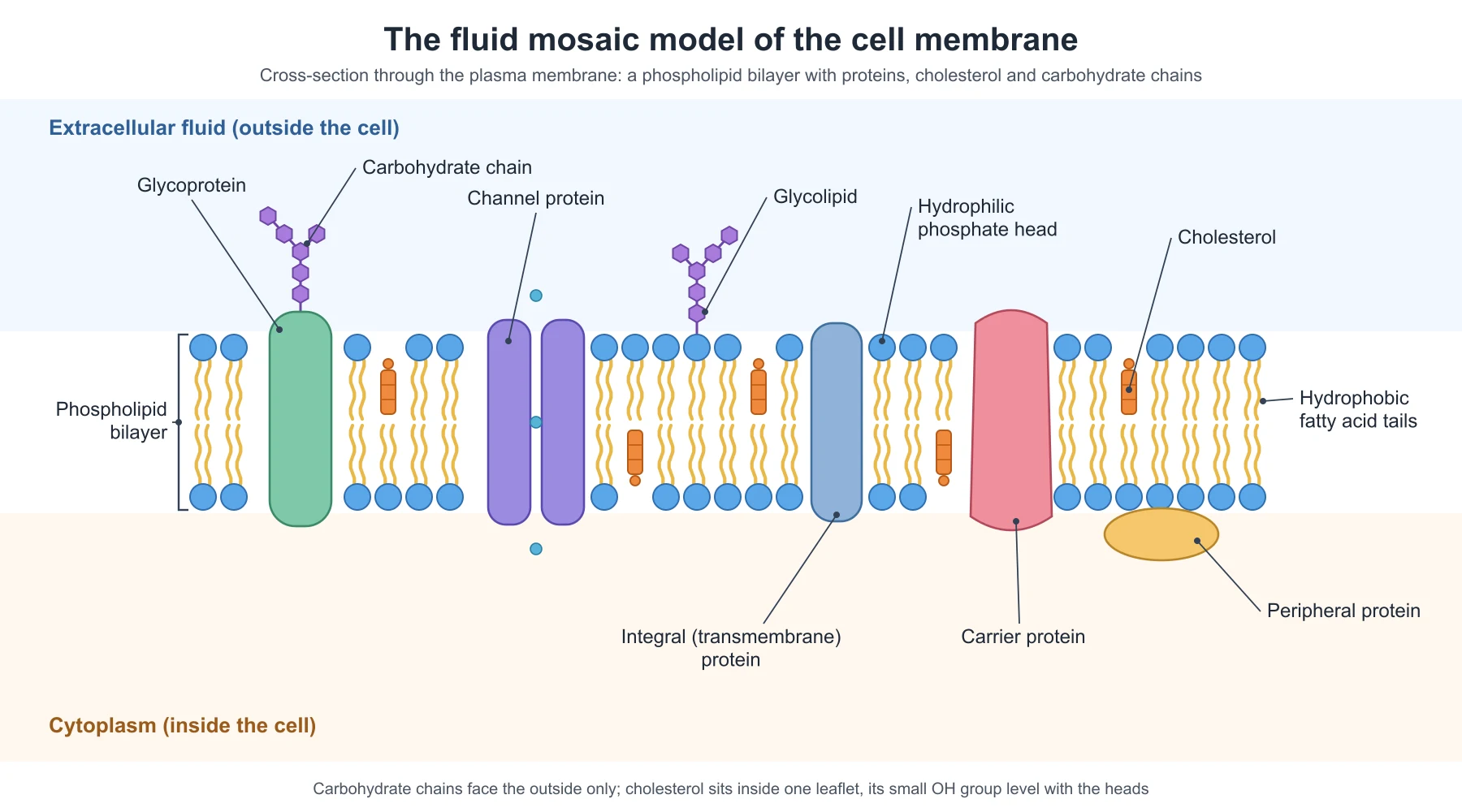
A cell membrane diagram has no awkward anatomy to learn and no organelles to place. It is a strip of circles and tails with a few proteins in it. That simplicity is exactly why examiners mark it so hard: there are only a handful of things to get right, and each one is either right or wrong. Tails pointing outward, carbohydrate chains hanging into the cytoplasm, cholesterol drawn sitting on the surface, a peripheral protein drawn straight through the bilayer: each one is a lost mark, and each one is common.
This guide covers what to label at each level, what every part of the fluid mosaic model looks like and does, the orientation rules that make or break the diagram, how to draw transport across the membrane, the mistakes that cost marks, and a numbered worksheet with an answer key. The labeled figure above was drawn for this guide; to make your own version, with the parts and the level of detail your course needs, use the cell membrane diagram generator.
The short answer: what to label on a cell membrane
A complete labeled cell membrane (plasma membrane) diagram names:
- the phospholipid bilayer, with its hydrophilic phosphate heads and hydrophobic fatty acid tails;
- integral (transmembrane) proteins, including a channel protein and a carrier protein;
- peripheral proteins on one surface;
- cholesterol between the phospholipids;
- glycoproteins and glycolipids, with their carbohydrate chains on the outer surface;
- the two sides: extracellular fluid (outside) and cytoplasm (inside).
Labeling both sides is not optional decoration. Several parts of the diagram are only right or wrong relative to the outside of the cell, and a membrane drawn with no sides marked cannot show that you know which is which.
What to label at each level
| Level | What the diagram shows | Labels expected |
|---|---|---|
| Middle school, KS3 | The membrane as the boundary of a cell | Cell membrane, and that it controls what enters and leaves |
| GCSE, high school biology | A simple bilayer with a protein, often alongside diffusion, osmosis and active transport | Phospholipid bilayer, protein, partially permeable; transport proteins in active transport |
| A-level, AP Biology, IB | The full fluid mosaic model | Phospholipid, hydrophilic head, hydrophobic tails, integral and peripheral proteins, channel and carrier proteins, cholesterol, glycoprotein, glycolipid, carbohydrate chain |
| College cell biology | The fluid mosaic model with function and asymmetry | The above plus glycocalyx, receptor proteins, pumps such as the sodium-potassium pump, aquaporins, lipid rafts, and the different lipid makeup of the two leaflets |
AQA's A-level specification is a good benchmark for the third row: it asks for the arrangement and movement of phospholipids, proteins, glycoproteins and glycolipids in the fluid-mosaic model, and notes that cholesterol may be present, where it restricts the movement of other molecules in the membrane.
Why it is called the fluid mosaic model
The model was proposed by S. J. Singer and Garth Nicolson in 1972, and both words in its name describe something you should be able to see in your diagram.
- Fluid: the phospholipids are not fixed. They move sideways within their own layer, and many proteins drift through the bilayer the same way. Moving from one layer to the other is rare.
- Mosaic: the proteins are scattered through the lipid like tiles in a mosaic, not arranged in a pattern and not forming a continuous coat.
The whole structure is only about 5 to 10 nm thick, far too thin to see with a light microscope, which is why the membrane is always taught as a model diagram rather than a drawing from a slide. By mass, a typical human cell membrane is roughly half protein, about 40% lipid and about 10% carbohydrate.
Every part of the cell membrane and what it does
| Part | How to draw it | What it does |
|---|---|---|
| Phospholipid bilayer | Two rows of phospholipids, heads outward, tails meeting in the middle | The basic barrier: lets small non-polar molecules through and blocks ions and large polar molecules |
| Hydrophilic phosphate head | A circle at the outer end of each phospholipid | Attracted to water, so the heads face the watery fluid on both sides |
| Hydrophobic fatty acid tails | Two wavy lines from each head, pointing into the middle | Repelled by water, so they form the oily core that ions and polar molecules cannot easily cross |
| Integral (transmembrane) protein | A large shape that crosses the whole bilayer | Many transport substances; others act as receptors, enzymes or anchors |
| Peripheral protein | A shape resting on one surface, not entering the core | Signaling, enzyme activity, and linking the membrane to the cytoskeleton |
| Channel protein | A transmembrane protein drawn with a gap (pore) through the middle | A water-filled pore for facilitated diffusion of specific ions and polar molecules |
| Carrier protein | A transmembrane protein that binds a molecule | Changes shape to move a specific molecule across, by facilitated diffusion or active transport |
| Cholesterol | A small, compact shape tucked between the tails of one layer | Buffers membrane fluidity: makes it less fluid when warm and stops it becoming rigid when cold |
| Glycoprotein | A protein with a branched carbohydrate chain on the outer side | Cell recognition and signaling; many receptors and antigens are glycoproteins |
| Glycolipid | A phospholipid-like lipid with a carbohydrate chain on the outer side | Cell recognition; with glycoproteins it forms the glycocalyx on the cell surface |
| Carbohydrate chains (glycocalyx) | Branched chains of small hexagons or beads on the outer surface only | Together form a sugar coat used for recognition, such as telling self from non-self |
How to draw and label a single phospholipid
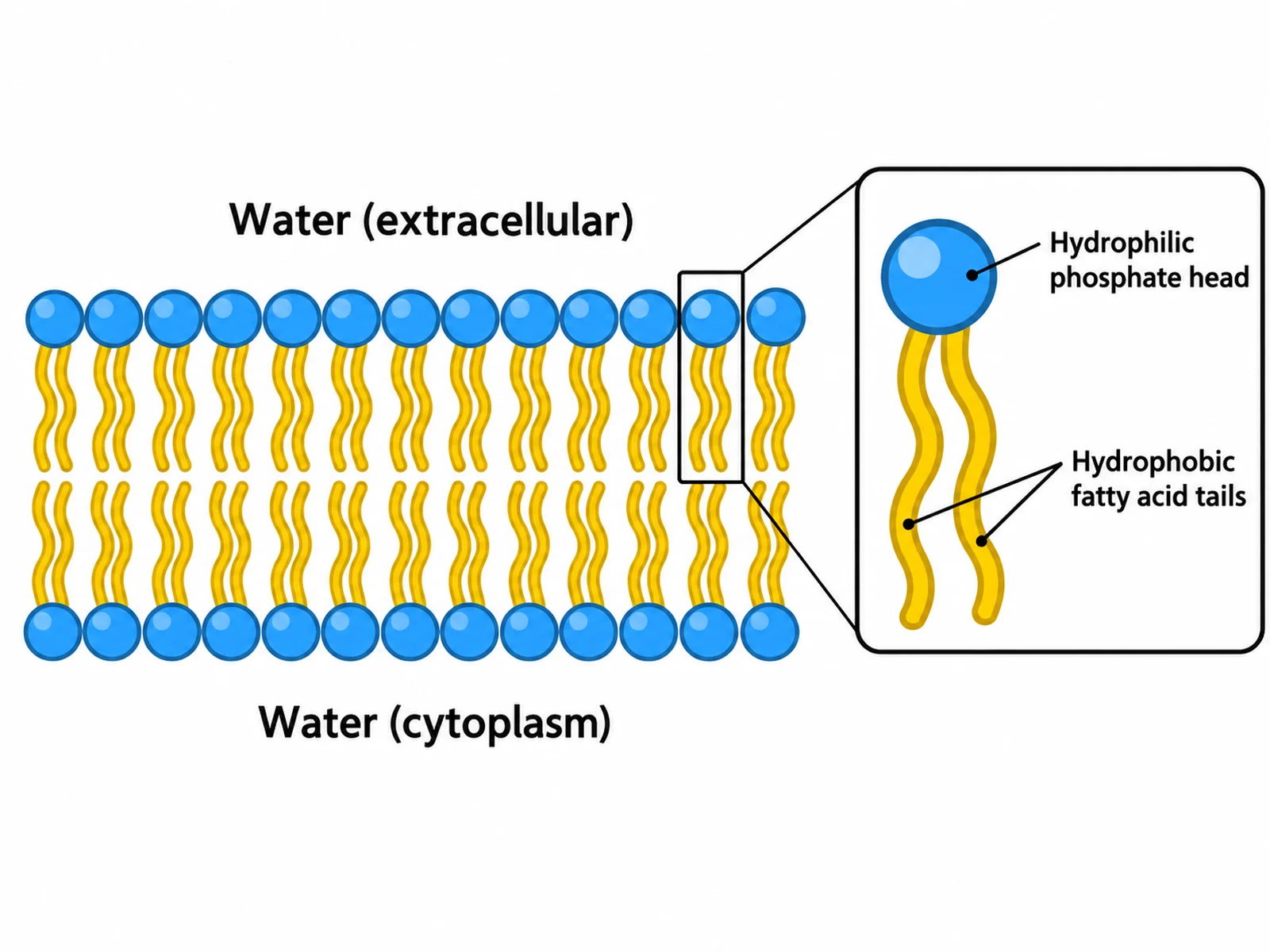
Many questions start smaller, with one phospholipid. Draw a circle for the head and two wavy lines for the tails, and label:
- the phosphate group in the head, which is hydrophilic (water-loving);
- the glycerol that joins the head to the tails (A-level and above);
- the two fatty acid tails, which are hydrophobic (water-fearing).
A molecule with a water-loving end and a water-fearing end is called amphipathic, and that is the whole reason a bilayer forms: in water, the tails hide from water by facing each other while the heads face out. If one of your tails has a kink in it, that kink is a carbon-carbon double bond (an unsaturated fatty acid); kinks stop the phospholipids packing tightly and keep the membrane more fluid.
The orientation rules that make or break the diagram
Almost every lost mark on a membrane diagram breaks one of these six rules.
- Heads face the water on both sides. The outer layer's heads face the extracellular fluid and the inner layer's heads face the cytoplasm. Both sides are watery.
- Tails meet in the middle. Nothing hydrophobic ever faces outward.
- Carbohydrate chains are on the outside only. Glycoproteins and glycolipids carry their sugars on the extracellular surface. A chain hanging into the cytoplasm is wrong, every time.
- Cholesterol sits inside the bilayer. It is wedged between the tails of one layer, with its small polar end level with the heads. It is not a blob on the surface.
- Integral proteins cross the core; peripheral proteins do not. An integral protein spans the hydrophobic middle. A peripheral protein sits on one face, attached to the heads or to an integral protein.
- Label which side is outside. Write "extracellular fluid" (or "outside the cell") above and "cytoplasm" below, so rules 3 and 5 can be checked.
Types of membrane protein
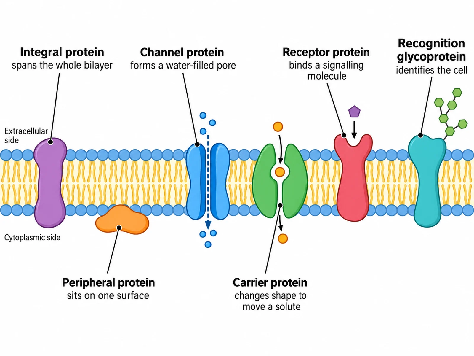
At A-level, AP and IB, you are usually expected to label the protein by what it does, not just "protein":
| Protein | Position | Job |
|---|---|---|
| Channel protein | Integral, spans the bilayer | Water-filled pore for specific ions; often gated (opens and closes) |
| Carrier protein | Integral | Binds a specific molecule and changes shape to move it across |
| Pump | Integral (a carrier that uses ATP) | Moves ions against their concentration gradient, as the sodium-potassium pump does |
| Aquaporin | Integral channel | Lets water cross the membrane quickly, speeding up osmosis |
| Receptor protein | Integral, binding site outside | Binds a signaling molecule such as a hormone and triggers a response inside |
| Recognition glycoprotein | Integral, carbohydrate outside | Identifies the cell to other cells, including the immune system |
| Enzyme | Integral or peripheral | Catalyzes reactions at the membrane surface |
| Peripheral protein | On one surface only | Signaling, enzyme activity, attachment to the cytoskeleton |
Drawing transport across the membrane
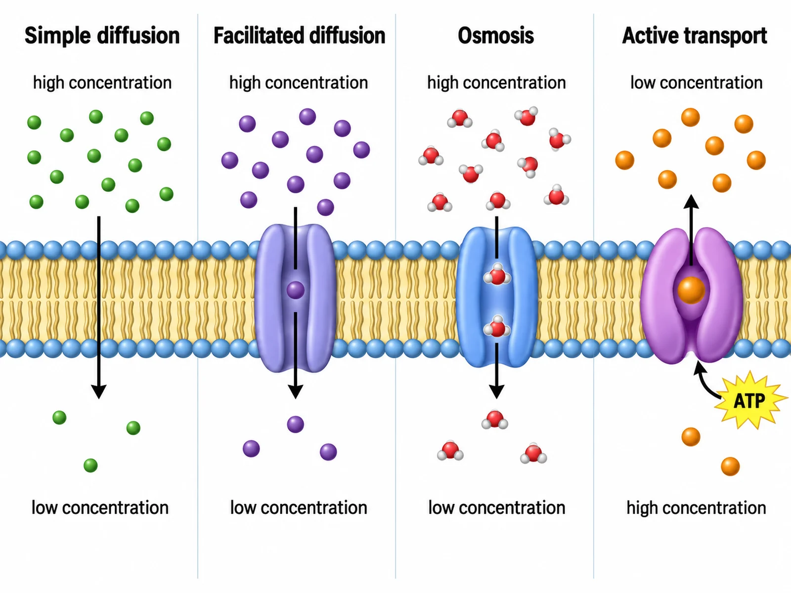
A membrane diagram is often a transport diagram in disguise. The rule of thumb: show which way the gradient runs, and whether a protein or ATP is needed.
| Process | Through | Protein needed? | Energy (ATP)? | Direction |
|---|---|---|---|---|
| Simple diffusion | The bilayer itself | No | No | Down the concentration gradient |
| Facilitated diffusion | Channel or carrier protein | Yes | No | Down the gradient |
| Osmosis | Bilayer and aquaporins | Not always; aquaporins speed it up | No | Water moves from higher to lower water potential (from dilute to concentrated solution) |
| Active transport | Carrier protein (pump) | Yes | Yes | Against the gradient, from low to high concentration |
| Endocytosis and exocytosis | Vesicles formed from or fused with the membrane | No transport protein | Yes | Bulk movement into or out of the cell |
Small, non-polar molecules such as oxygen and carbon dioxide cross the bilayer by simple diffusion. Ions and polar molecules such as glucose need a protein. Draw arrows for direction, mark high and low concentration on the correct sides, and put an ATP symbol beside a pump, not beside a channel.
Labeling conventions that gain marks
- Use straight, ruled label lines with no arrowheads, and keep them from crossing.
- End each line on exactly one thing. A line to "hydrophilic head" must touch one head, not the gap between two; a line to "tails" must end in the tail region, not on a protein.
- Use the precise term. "Phospholipid bilayer", not "lipid layer"; "integral protein" or "transmembrane protein", not just "protein", at A-level; "glycoprotein" for a protein with a carbohydrate chain, and "carbohydrate chain" if the line ends on the sugar itself.
- Label one example of each repeating part. One head, one pair of tails, one cholesterol.
- Bracket the bilayer. A bracket spanning both layers is the clearest way to label "phospholipid bilayer" without pointing at a single molecule.
- Mark both sides of the membrane in words.
- Keep labels outside the strip, above and below, aligned in rows.
How to draw a cell membrane diagram, step by step
- Draw two faint parallel guide lines across the page, about as far apart as you want the membrane to be thick.
- Draw the heads as a row of evenly spaced circles along each guide line.
- Draw two tails from every head, pointing inward so the tails of the two rows meet in the middle.
- Leave gaps and draw proteins into them: at least one integral protein right through both layers, one channel protein with a pore, and one peripheral protein on the inner surface.
- Add cholesterol between the tails in two or three places.
- Add carbohydrate chains on the top surface only: one on a protein (glycoprotein) and one on a lipid (glycolipid).
- Write "extracellular fluid" above and "cytoplasm" below.
- Label from left to right, top labels for the outer-surface parts and bottom labels for the inner-surface parts, and check every line ends on its part.
Mistakes that cost marks
- Fatty acid tails pointing outward, or heads meeting in the middle.
- Carbohydrate chains drawn on the cytoplasmic side.
- Cholesterol drawn on the surface, or as a large block spanning both layers.
- A peripheral protein drawn passing through the hydrophobic core.
- No sides labeled, so the examiner cannot tell inside from outside.
- "Protein" used for every protein when the question asks for types.
- A glycoprotein labeled where the line ends on a phospholipid, or a glycolipid labeled where it ends on a protein.
- A channel protein drawn with no pore, so it looks like any other protein.
- ATP shown beside facilitated diffusion, or active transport drawn down the gradient.
- The phospholipid heads called "hydrophobic" or the tails "hydrophilic". Heads love water; tails avoid it.
- Describing the membrane as fully permeable. It is partially (selectively) permeable; the plant cell wall, by contrast, is fully permeable.
- Drawing the bilayer as a single row of phospholipids.
Printable cell membrane worksheet with answer key
This blank uses exactly the same drawing as the labeled figure at the top of the page, so the answers below line up with it. Save the image and print it across a full page.
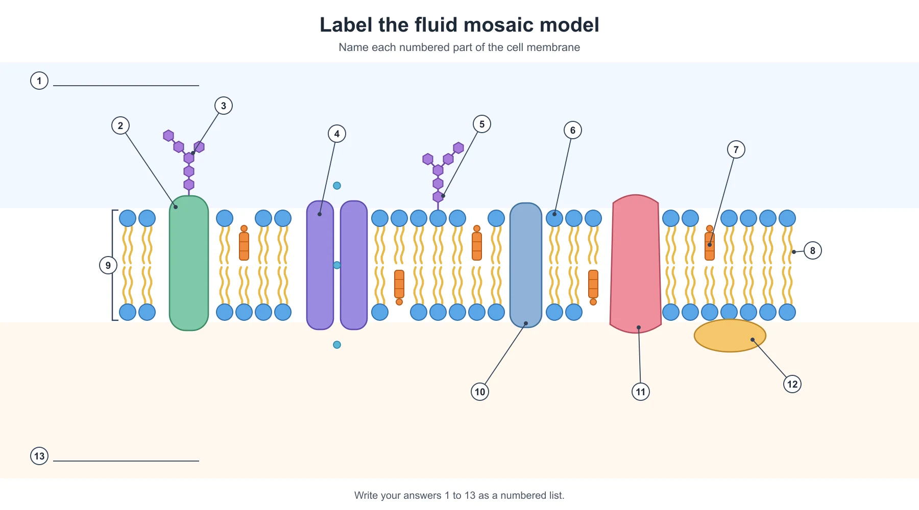
Answer key
- Extracellular fluid (outside the cell)
- Glycoprotein
- Carbohydrate chain
- Channel protein
- Glycolipid
- Hydrophilic phosphate head
- Cholesterol
- Hydrophobic fatty acid tails
- Phospholipid bilayer
- Integral (transmembrane) protein
- Carrier protein
- Peripheral protein
- Cytoplasm (inside the cell)
To make a worksheet for your own course, such as a simpler GCSE version with only the bilayer and one protein, or a transport diagram with blanks, describe it on the cell membrane diagram generator and ask for a labeled and a blank version of the same figure.
Frequently asked questions
What are the main parts of the cell membrane?
A phospholipid bilayer (hydrophilic heads outward, hydrophobic tails inward), integral and peripheral proteins, cholesterol, and glycoproteins and glycolipids carrying carbohydrate chains on the outer surface.
Why is it called the fluid mosaic model?
"Fluid" because the phospholipids and many of the proteins move sideways within the membrane. "Mosaic" because the proteins are scattered through the lipid bilayer like tiles in a mosaic. The model was proposed by Singer and Nicolson in 1972.
Which way do the phospholipid heads face?
Outward, toward the watery fluid on both sides: the extracellular fluid above and the cytoplasm below. The tails point inward and meet in the middle.
What is the difference between integral and peripheral proteins?
Integral proteins are embedded in the bilayer, and many span it completely (transmembrane proteins). Peripheral proteins sit on one surface only, attached to lipid heads or to integral proteins, and do not enter the hydrophobic core.
What does cholesterol do in the cell membrane?
It sits between the phospholipid tails and buffers the membrane's fluidity: it restrains the phospholipids when it is warm and stops them packing too tightly when it is cold. Animal cell membranes contain it; plant membranes rely mainly on related plant sterols.
Why are glycoproteins and glycolipids only on the outside?
Their carbohydrate chains work as identification tags, read by other cells, hormones and the immune system, so they face the extracellular fluid. Together they form the glycocalyx, the sugar coat on the cell's surface.
What is the difference between a channel protein and a carrier protein?
A channel protein forms a water-filled pore that specific ions pass through. A carrier protein binds the molecule and changes shape to move it across. Channels only allow facilitated diffusion; carriers can do facilitated diffusion or, using ATP, active transport.
Is the cell membrane the same as the plasma membrane?
Yes. Cell membrane, plasma membrane and cell surface membrane all mean the membrane around the cell. The organelle membranes inside a eukaryotic cell have the same basic bilayer structure.
How thick is the cell membrane?
About 5 to 10 nanometers. That is why it cannot be seen with a light microscope and is taught as a model.
What is the difference between the cell membrane and the cell wall?
The membrane is a thin, partially permeable phospholipid bilayer around every cell. The cell wall is a thick, fully permeable layer outside the membrane in plant, fungal and bacterial cells. The plant cell labeling guide shows both.
Where can I get a blank cell membrane diagram to label?
Use the worksheet above, or make one that matches your course on the cell membrane diagram generator, which draws the fluid mosaic model labeled or blank, a simple phospholipid bilayer, membrane protein types and transport diagrams.
Related guides and tools
- How to label a plant cell diagram and how to label an animal cell diagram: the membrane in the context of the whole cell.
- How to draw the stages of mitosis: the membrane is pinched in two at cytokinesis.
- Action potential diagram, synapse diagram and neuron diagram: channel proteins and pumps at work in nerve cells.
- Biological pathway diagram generator: signaling that starts at a membrane receptor.
- More guides in this series: labeling an ear diagram, labeling the digestive system, labeling a brain diagram.
Sources
- OpenStax, Biology 2e, 5.1 Components and Structure: the fluid mosaic model, membrane thickness and composition, cholesterol, integral and peripheral proteins, and the glycocalyx.
- OpenStax, Biology 2e, 5.2 Passive Transport and 5.3 Active Transport: diffusion, facilitated diffusion, osmosis, channel and carrier proteins, and pumps.
- OpenStax, Anatomy and Physiology 2e, 3.1 The Cell Membrane: membrane structure and transport in human cells.
- AQA, AS and A-level Biology (7401/7402) specification, Cells: the fluid-mosaic model as examined at A-level.