Tutorials
Ear Diagram Labeled: How to Label the Outer, Middle and Inner Ear
How to label an ear diagram: every part of the outer, middle and inner ear and its job, what to label at each level, the path of sound, and a worksheet.
 SciFigure AI
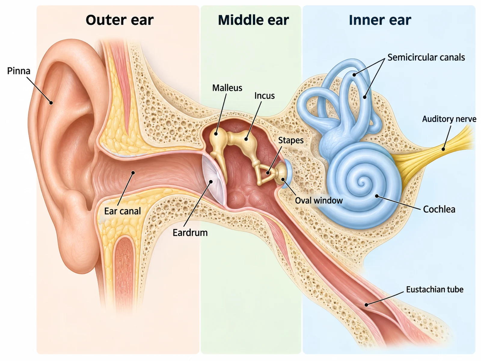
The ear is three organs in a row. The outer ear collects sound, the middle ear turns air vibrations into a stronger push, and the inner ear turns that push into nerve impulses, while also doing a second, unrelated job: balance. A labeled ear diagram is easy to get right once you see it that way, because every structure belongs to exactly one of those three regions and does one job inside it.
This guide covers what to label at each level, every structure of the outer, middle and inner ear with what it looks like and what it does, the path sound takes, the labeling conventions that gain marks, the mistakes that lose them, and a numbered worksheet with an answer key. If you need a figure to work from, the ear diagram generator draws a labeled ear, a blank ear diagram to label, and close-ups of the ossicles or inner ear.
The short answer: what to label on an ear diagram
Label the ear region by region, from the outside in:
- Outer ear: pinna (auricle), ear canal (external auditory canal), eardrum (tympanic membrane).
- Middle ear: malleus, incus and stapes (the ossicles), oval window, Eustachian tube.
- Inner ear: cochlea, semicircular canals, vestibule, and the auditory (vestibulocochlear) nerve.
Those eleven or twelve labels are what a complete school-level ear diagram expects. Add the three region headings, outer, middle and inner ear, as well: many worksheets give a mark for them.
What to label at each level
| Level | Labels expected | Usually also asked |
|---|---|---|
| Middle school, KS3, grades 6–8 | Outer ear (pinna), ear canal, eardrum, the three small bones (hammer, anvil, stirrup), cochlea, auditory nerve | The order sound travels in |
| High school biology, anatomy and physics | Pinna, ear canal, eardrum, malleus, incus, stapes, oval window, Eustachian tube, cochlea, semicircular canals, auditory nerve | Which parts are for hearing and which for balance; the ossicles in order |
| A-level, AP, IB (biology, psychology or physics options that teach hearing) | The above plus round window, vestibule, basilar membrane, hair cells, and the three regions named | How vibration becomes a nerve impulse; how pitch is detected along the cochlea |
| College anatomy and physiology | The above plus external acoustic meatus, tympanic cavity, tensor tympani and stapedius muscles, bony and membranous labyrinth, perilymph and endolymph, scala vestibuli, scala media and scala tympani, organ of Corti, utricle, saccule, ampullae, cochlear and vestibular nerves | Tonotopy, the vestibular organs, and conductive versus sensorineural hearing loss |
The ear is not on every syllabus: AQA's GCSE Biology, for example, examines the eye as its sense organ and does not include the ear. Where the ear does appear, in many US middle and high school courses, physics units on sound, and psychology courses on sensation, the list in the second row is the standard set.
Every part of the ear and what it does
Outer ear
| Part | What it looks like on a diagram | What it does |
|---|---|---|
| Pinna (auricle) | The visible curved flap of skin-covered cartilage on the outside of the head | Collects sound waves and funnels them into the ear canal; its folds help you tell where a sound comes from |
| Ear canal (external auditory canal, external acoustic meatus) | A tube about 2.5 cm long in adults, running inward from the pinna | Carries sound to the eardrum; glands in its lining make earwax, which traps dust and microbes |
| Eardrum (tympanic membrane) | A thin, slightly cone-shaped membrane across the inner end of the canal | Vibrates when sound waves hit it and passes the vibration to the malleus. It is the boundary between the outer and middle ear |
Middle ear
| Part | What it looks like on a diagram | What it does |
|---|---|---|
| Middle ear cavity (tympanic cavity) | An air-filled space behind the eardrum | Holds the ossicles; kept at the same pressure as the outside air |
| Malleus (hammer) | The first ossicle, its handle attached to the inside of the eardrum | Picks up the eardrum's vibration |
| Incus (anvil) | The middle ossicle, joined to the malleus and the stapes | Passes the vibration on |
| Stapes (stirrup) | A stirrup-shaped bone whose flat footplate sits in the oval window; the smallest bone in the body | Pushes on the oval window, moving the fluid of the inner ear |
| Oval window | A membrane-covered opening into the inner ear, filled by the stapes footplate | Where vibration enters the cochlea's fluid |
| Round window | A second membrane-covered opening just below the oval window | Bulges outward as the stapes pushes inward, so the cochlear fluid can move |
| Eustachian tube (auditory tube) | A tube running down and forward from the middle ear to the back of the throat | Equalizes air pressure on both sides of the eardrum; this is what opens when your ears "pop" |
Inner ear
| Part | What it looks like on a diagram | What it does |
|---|---|---|
| Cochlea | A snail-shaped, fluid-filled spiral of about two and a half turns | Hearing: hair cells on its basilar membrane turn fluid vibrations into nerve impulses |
| Semicircular canals | Three fluid-filled loops, each at right angles to the other two | Balance: detect rotation of the head in any direction |
| Vestibule | The central chamber between the cochlea and the canals, containing the utricle and saccule | Balance: detects gravity, head tilt and straight-line acceleration |
| Auditory (vestibulocochlear) nerve | A bundle of fibers leaving the cochlea and vestibule toward the brain | Carries hearing signals (cochlear branch) and balance signals (vestibular branch) to the brain |
Why the middle ear exists: the 20-fold push
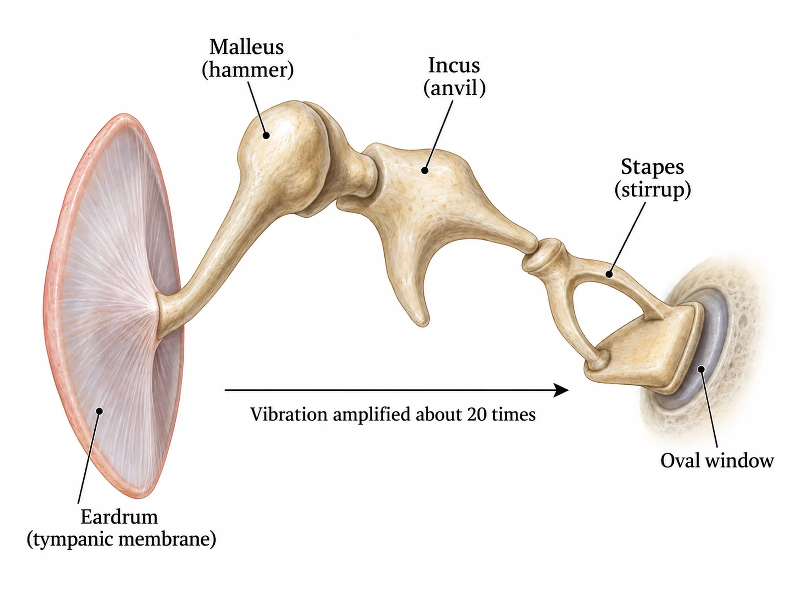
Sound arrives as vibrations in air, but the cochlea is full of fluid, and fluid is far harder to set moving than air. Without help, most of the sound energy would simply bounce off. The middle ear solves this. The eardrum has roughly 17 times the area of the stapes footplate, so the force collected over the whole eardrum is concentrated onto the much smaller oval window, and the lever action of the ossicles adds a little more. The result is a pressure at the oval window about 20 times greater than at the eardrum.
That is why "the ossicles amplify the sound" is accepted at school level, and why the more precise A-level answer is that the middle ear increases the pressure of the vibrations so they can move the cochlear fluid. Two tiny muscles, the tensor tympani on the malleus and the stapedius on the stapes, tighten in response to very loud sounds and damp the chain down.
Inside the inner ear: hearing and balance
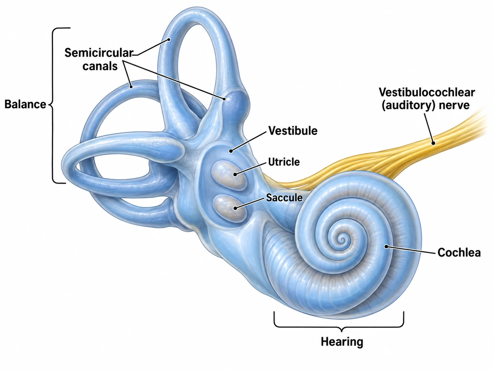
The inner ear is a set of fluid-filled tunnels in the temporal bone (the bony labyrinth, filled with perilymph) with a matching set of membranous tubes inside them (the membranous labyrinth, filled with endolymph). It contains five balance organs and one hearing organ.
Hearing. Cut across, the cochlea has three fluid-filled channels: the scala vestibuli, the scala media (cochlear duct) and the scala tympani. On the floor of the scala media is the basilar membrane, carrying the organ of Corti and its hair cells. When the stapes pushes on the oval window, a pressure wave runs through the fluid and makes the basilar membrane vibrate. High-pitched sounds vibrate it most near the base of the cochlea, by the oval window; low-pitched sounds travel further toward the tip (the apex). Bending the hair cells' stereocilia opens ion channels and starts the nerve impulse.
Balance. The three semicircular canals each end in a swelling, the ampulla, with sensory hair cells that respond when the fluid lags behind as the head turns. Because the three canals lie at right angles to each other, every rotation stimulates at least one of them. The utricle and saccule in the vestibule detect gravity and straight-line movement, such as tilting your head or accelerating in a car.
The path of sound through the ear
If the question asks you to label the ear and explain hearing, number the structures in the order sound reaches them. This sequence is also the best order to label in.
- The pinna collects sound waves and directs them into the ear canal.
- The sound waves make the eardrum vibrate.
- The malleus, incus and stapes pass the vibration across the middle ear, increasing its pressure.
- The stapes pushes on the oval window, setting the fluid in the cochlea moving; the round window bulges out to make room.
- The moving fluid vibrates the basilar membrane, bending the hair cells, which generate nerve impulses.
- The auditory nerve carries the impulses to the brain, where the auditory cortex in the temporal lobe interprets them as sound. The brain diagram labeling guide shows where that is.
Labeling conventions that gain marks
- Group the labels by region. Put a heading over each of the three regions, or a bracket under each, and keep each region's labels on its own side of the drawing where possible.
- Use ruled, straight label lines that never cross. The ossicles are tightly packed, so give each its own line at a different angle.
- End each line on the right bone. The malleus line should end on the bone attached to the eardrum, the stapes line on the stirrup-shaped bone at the oval window.
- Name the bones in full. "Malleus, incus, stapes" scores more reliably than "hammer, anvil, stirrup"; writing both, as in "malleus (hammer)", is safest at school level.
- Use singular and plural correctly. "Semicircular canals" for the set of three; "ossicles" for the bones as a group; "cochlea", not "cochlear", which is the adjective.
- Write "eardrum (tympanic membrane)" and "ear canal", not "drum" or "tube": the Eustachian tube is also a tube.
- Keep text horizontal and outside the drawing.
How to label an ear diagram, step by step
- Find the three regions first. The eardrum marks the end of the outer ear; the oval window marks the start of the inner ear. Everything between them is middle ear.
- Label the outer ear: pinna, then ear canal, then eardrum. These lines are long and run to the left of the drawing.
- Label the ossicles in order: malleus, incus, stapes, from the eardrum inward.
- Label the openings: the oval window where the stapes sits, and, at higher levels, the round window just below it.
- Label the Eustachian tube, running down from the middle ear toward the throat.
- Label the inner ear: the cochlea (the spiral), the semicircular canals (the three loops), the vestibule between them, and the nerve leaving toward the brain.
- Add the region headings and, if asked, mark which structures are for hearing and which for balance.
- Check that the ossicles are in order, the eardrum is at the end of the canal, and the Eustachian tube does not connect to the inner ear.
Mistakes that cost marks
- Ossicles in the wrong order. From the eardrum inward it is always malleus, incus, stapes.
- The eardrum placed in the middle or inner ear. It closes off the end of the ear canal and separates the outer ear from the middle ear.
- "Outer ear" written on the pinna's line. The outer ear is a region; the pinna is the structure.
- The stapes drawn floating instead of sitting in the oval window.
- Oval and round windows swapped. The stapes sits in the oval window; the round window is the separate opening below it.
- The Eustachian tube connected to the ear canal or the inner ear. It connects the middle ear to the throat.
- The semicircular canals labeled as a hearing organ, or the cochlea as the balance organ.
- A cochlea drawn as a flat circle or with a single turn. It is a spiral of about two and a half turns.
- The three semicircular canals drawn in one flat plane. They sit at right angles to each other.
- The auditory nerve leaving from the eardrum or the ossicles. It leaves from the cochlea and vestibule.
- "Cochlear" used as the noun. The structure is the cochlea.
- Labels written inside the drawing. The middle ear is tiny on most diagrams, and text placed in it hides the very bones you are naming.
Printable ear diagram worksheet with answer key
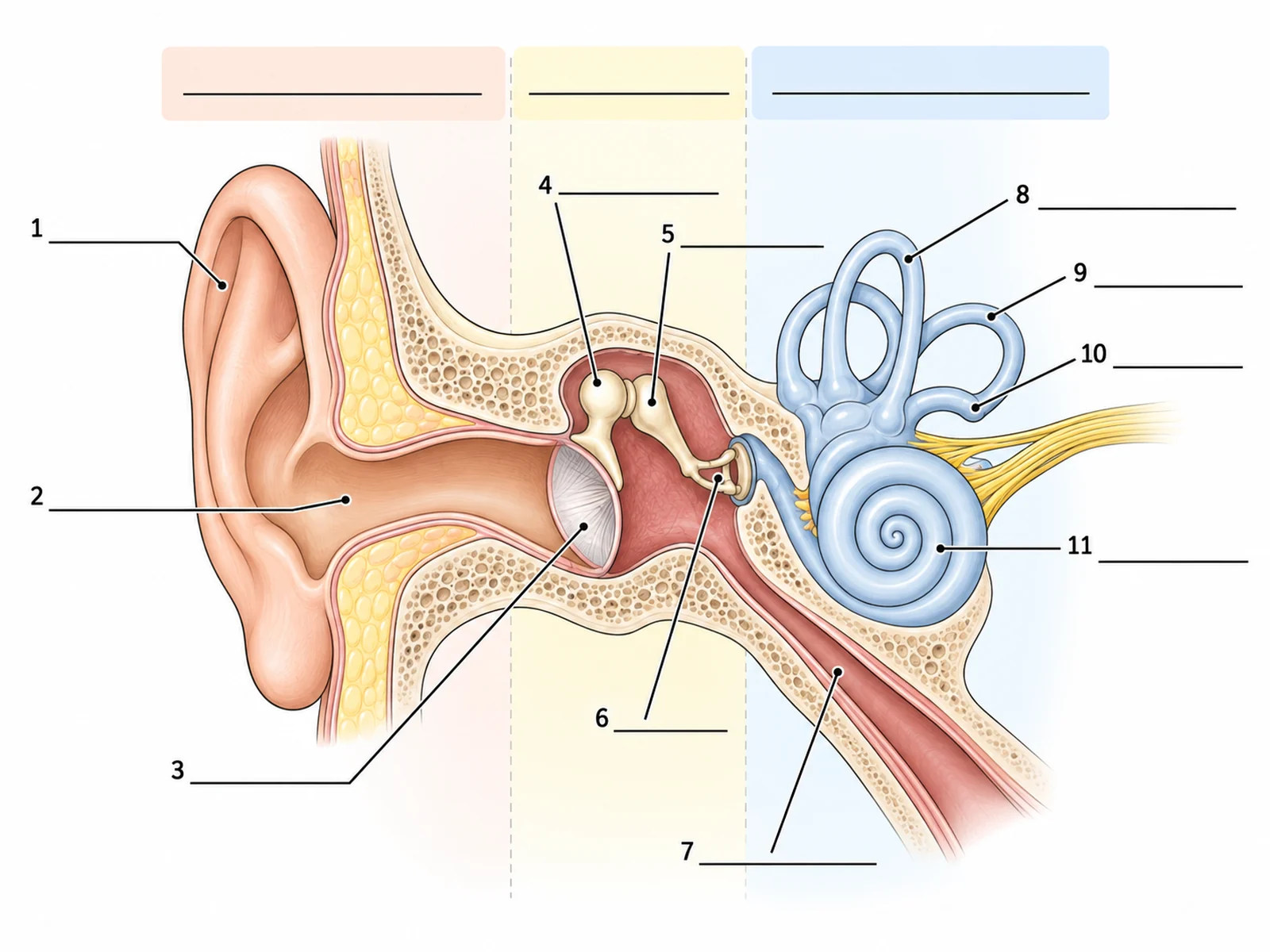
Label the eleven numbered structures and write the three region names in the colored boxes along the top. The answers follow.
Answer key
- Region headings, left to right: outer ear, middle ear, inner ear.
- Pinna (auricle)
- Ear canal (external auditory canal)
- Eardrum (tympanic membrane)
- Malleus (hammer)
- Incus (anvil)
- Stapes (stirrup)
- Eustachian tube
- Semicircular canal
- Semicircular canal
- Semicircular canal (numbers 8 to 10 are the three canals; at college level, name them the anterior, posterior and lateral canals)
- Cochlea
The yellow fibers leaving the cochlea are the auditory (vestibulocochlear) nerve, a good extra label to add. To make a version for your own class, such as a simple five-label ear for younger students or an inner-ear close-up with the utricle and saccule, use the ear diagram generator and ask for a labeled version and a blank version of the same figure.
Frequently asked questions
What are the three main parts of the ear?
The outer ear (pinna, ear canal and eardrum), the middle ear (the three ossicles, the oval window and the Eustachian tube) and the inner ear (the cochlea, the vestibule and the semicircular canals).
What are the three bones in the ear called?
The malleus (hammer), incus (anvil) and stapes (stirrup), together called the ossicles. They are the smallest bones in the body, and the stapes is the smallest of all.
What does the cochlea do?
It turns sound into nerve impulses. Vibrations of the fluid inside it move the basilar membrane, which bends hair cells, and the hair cells trigger impulses in the auditory nerve. Different pitches are detected at different points along the spiral.
Which part of the ear controls balance?
The semicircular canals, which detect rotation of the head, and the vestibule (the utricle and saccule), which detects gravity, head tilt and straight-line acceleration. The cochlea is for hearing only.
What is the Eustachian tube for?
It connects the middle ear to the back of the throat and keeps the air pressure in the middle ear equal to the outside pressure, so the eardrum can vibrate freely. When it opens on a plane or a mountain road, your ears "pop".
Is the eardrum part of the outer ear or the middle ear?
It is the boundary between them. School diagrams usually include it as the last structure of the outer ear; it closes off the ear canal, and the middle ear lies behind it.
Where are the oval window and the round window?
Both are membrane-covered openings between the middle ear and the inner ear. The oval window is filled by the footplate of the stapes; the round window sits just below it and bulges outward as the fluid in the cochlea moves.
Why does the ear need the middle ear bones?
To push sound energy from air into the fluid of the cochlea. Concentrating the vibration of the large eardrum onto the small oval window raises the pressure about 20 times, enough to move the fluid.
How many turns does the cochlea have?
About two and a half turns in humans. Drawing it as a spiral of roughly that many turns is one of the details markers check.
Where can I get an ear diagram to label?
Use the worksheet above, or make one for your own syllabus with the ear diagram generator, which draws labeled and blank versions of the same ear, simple versions for younger students, and close-ups of the ossicles and inner ear.
Related guides and tools
- How to label a brain diagram: where the auditory nerve's signals end up.
- How to label the digestive system: another body system labeled region by region.
- Eye anatomy diagram: the other sense organ that comes up on every anatomy worksheet.
- Neuron diagram and anatomy diagram generator for the nerve pathway and the rest of the body.
- Medical illustration generator for clinical figures such as the middle ear in otitis media.
- More guides in this series: labeling a plant cell, drawing the stages of mitosis, labeling a cell membrane.
Sources
- OpenStax, Biology 2e, 36.4 Hearing and Vestibular Sensation: the path of sound, the ossicles, the cochlea and basilar membrane, and the vestibular organs.
- OpenStax, Anatomy and Physiology 2e, 14.1 Sensory Perception: structure of the outer, middle and inner ear.
- Sutton AE, Geiger Z. External Ear Anatomy. StatPearls, NCBI Bookshelf.
- Peterson DC, Sutton AE. Middle Ear Anatomy and Physiology. StatPearls, NCBI Bookshelf.
- Sutton AE, Peterson DC. Inner Ear Anatomy and Cochlear Physiology. StatPearls, NCBI Bookshelf.