Tutorials
How to Label an Animal Cell Diagram (With Every Part Explained)
A complete guide to labelling an animal cell diagram: every organelle and its function, the labelling conventions markers look for, and the ten mistakes that lose marks.
 SciFigure AI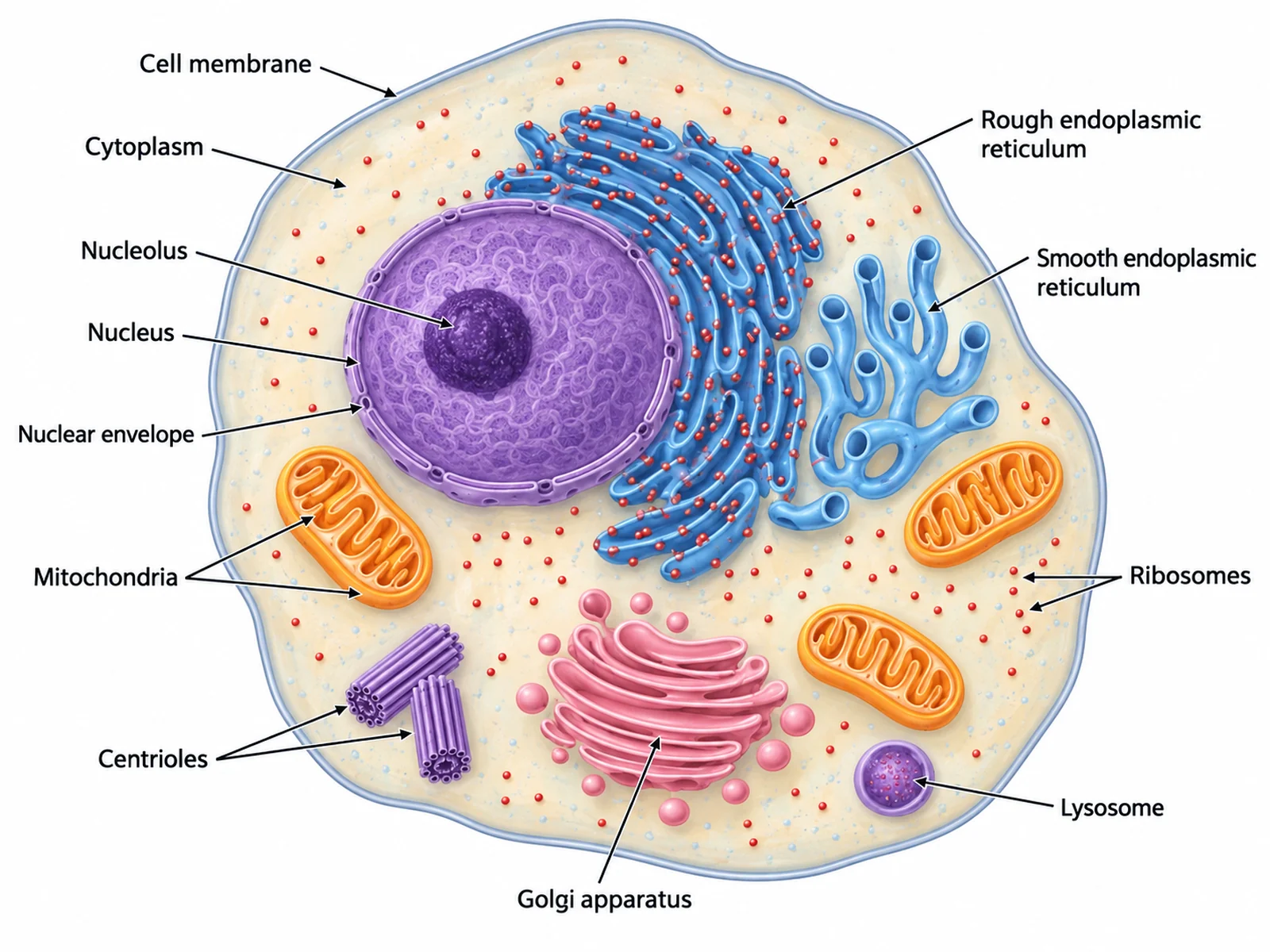
A labelled animal cell diagram is one of the first things asked for in biology and one of the last things done well. The drawing is rarely the problem. Marks are lost on which structures were named, whether the label lines land on the right thing, and whether the cell has parts on it that animal cells do not have.
This guide covers all of it: every organelle you may be asked to label with what it actually does, the labelling conventions that make a diagram readable, the order to work in, and the specific errors that come up again and again. If you want a diagram to start from, the animal cell diagram generator draws both a labelled version and a blank one to fill in.
What to label on an animal cell
The list depends on the level you are working at. Labelling every organelle on an introductory diagram makes it unreadable; leaving half of them off an A-level or AP diagram loses marks. Work out which of these three lists applies before you start.
Introductory (middle school, KS3): cell membrane, cytoplasm, nucleus, mitochondria.
GCSE / IGCSE: the four above plus ribosomes, and often the cell membrane described as a partially permeable membrane.
A-level, IB, AP Biology: all of the above plus the nuclear envelope, nucleolus, rough and smooth endoplasmic reticulum, Golgi apparatus, lysosomes, vesicles, centrioles, and the cytoskeleton.
Every part and what it does
| Part | What it looks like | What it does |
|---|---|---|
| Cell membrane | A thin double line around the whole cell | Controls what enters and leaves. Partially permeable: small molecules pass, large ones do not |
| Cytoplasm | The jelly-like material filling the cell | Where most of the cell's chemical reactions happen |
| Nucleus | A large round structure, usually off-centre | Holds the DNA as chromosomes and controls the cell's activities |
| Nuclear envelope | The double membrane around the nucleus, with pores | Separates the DNA from the cytoplasm; pores let mRNA out |
| Nucleolus | A dense spot inside the nucleus | Makes ribosomes |
| Mitochondrion | Oval, with the inner membrane folded into cristae | Aerobic respiration: releases energy from glucose as ATP |
| Ribosome | Very small dots, free in the cytoplasm or on the rough ER | Protein synthesis |
| Rough endoplasmic reticulum | Folded membrane sheets studded with ribosomes, continuous with the nuclear envelope | Folds and transports the proteins made on its ribosomes |
| Smooth endoplasmic reticulum | Tubular membrane, no ribosomes on it | Makes lipids and steroids; stores calcium |
| Golgi apparatus | A stack of curved flattened sacs with vesicles pinching off | Modifies, sorts and packages proteins for transport |
| Vesicle | Small circles, often near the Golgi | Carries substances around the cell and to the membrane |
| Lysosome | A small round sac with a single membrane | Contains digestive enzymes; breaks down worn-out organelles and pathogens |
| Centrioles | A pair of short cylinders at right angles, near the nucleus | Organise the spindle fibres during cell division |
| Cytoskeleton | Fine threads through the cytoplasm | Holds the cell's shape and moves organelles around |
Two of these are worth extra attention, because they are the two most often drawn wrong.
The mitochondrion. The cristae are folds of the inner membrane, not separate shelves floating in the middle. If your diagram shows loose lines inside the oval that do not join the inner membrane at either end, the label line may be correct and the structure still wrong. The space inside is the matrix; the gap between the two membranes is the intermembrane space.
The rough ER. What makes it rough is the ribosomes on its surface, so rough ER drawn as bare membrane is not rough ER — it is smooth ER with the wrong label. It is also continuous with the nuclear envelope, which is worth showing: the proteins it handles come straight from instructions leaving the nucleus.
What an animal cell does not have
Half the marks available on this question are for knowing what to leave off. An animal cell has no:
- Cell wall. Animal cells have a membrane and nothing outside it. A rigid outer box is a plant, fungal, or bacterial cell.
- Chloroplasts. Animals do not photosynthesise. Drawing green ovals in an animal cell is the single most common way to lose the mark.
- Large permanent vacuole. Animal cells can contain small temporary vacuoles, but not the single big central one that pushes a plant cell's contents to the edge.
The reverse is also examinable: animal cells have centrioles, which most plant cells lack, and their small vacuoles are temporary rather than permanent. A side-by-side comparison is easier to revise from than two separate diagrams, and the plant cell diagram generator will draw the other half of the pair.
Labelling conventions that gain marks
These are not style preferences. Most mark schemes and every journal figure guideline apply some version of them.
- Use a ruler, or draw straight lines. A label line should be a single straight line, not a curve or a freehand scribble.
- Never let label lines cross. Crossing lines make it ambiguous which label belongs to which structure, and an ambiguous label scores nothing. If two lines want to cross, move one label to the other side of the cell.
- The line must touch the structure it names. Stopping short of the membrane, or overshooting into the cytoplasm, both count as pointing at the wrong thing.
- Put the labels outside the cell and line them up in a column on each side. The interior is crowded; text inside it covers the structures.
- Write labels horizontally, even where the line arrives at an angle. Rotated text is harder to read and looks like a different label on a photocopy.
- Do not use arrowheads unless the whole diagram uses them. A plain line is the convention for a label; arrows suggest movement or a process.
- Get the singular right. One is a mitochondrion; several are mitochondria. A single line pointing at one oval labelled "mitochondria" is wrong. The same applies to nucleus/nuclei and vesicle/vesicles.
- Capitalise Golgi, because it is named after Camillo Golgi. Everything else on the list is lower case unless it starts a line.
- Do not shade or colour for decoration. If you use colour, use it to mean something, and say what in a key.
- Add a title and a magnification or scale if the diagram is from a microscope. A biological drawing without a scale cannot be interpreted.
The order to work in
Labelling in a sensible order is what keeps the lines from crossing, and it is much easier than fixing a tangle afterwards.
- Draw or choose the cell first, and leave a wide margin all the way round for the labels.
- Label the outermost structures first — cell membrane, then cytoplasm. Their lines are the shortest and will not need to cross anything.
- Label the nucleus next, then the structures inside it (nuclear envelope, nucleolus) with short lines that stay in that region.
- Work through the remaining organelles by position, not by importance: everything on the left of the cell labelled to the left, everything on the right labelled to the right. This one habit prevents most crossings.
- Label any repeated structure once, pointing at one clear example. A diagram with eight lines all saying "ribosome" is harder to read, not more thorough.
- Check every line ends on the structure and not just near it.
Ten mistakes that lose marks
- A cell wall drawn round an animal cell.
- Chloroplasts in an animal cell.
- One large central vacuole instead of small temporary ones.
- "Mitochondria" used for a single mitochondrion.
- The nucleolus labelled as the nucleus, or the nucleolus omitted at A-level.
- Cristae drawn as free-floating lines rather than folds of the inner membrane.
- Rough ER drawn without ribosomes on it.
- The Golgi apparatus drawn as a single sac instead of a stack.
- Label lines crossing each other.
- Labels written inside the cell, on top of the structures they name.
Getting a diagram to label
You need two versions of the same cell: a labelled one to learn from, and a blank one to test yourself on. Drawing both by hand takes longer than it is worth.
The animal cell diagram generator produces both from a description, along with a simplified version for younger students and a cutaway if you need to show the interior more clearly. For other cell types, the biology diagram maker covers plant, bacterial, and specialised cells, and the mitochondria diagram page goes into the organelle on its own when the cell diagram does not have room for the detail.
Once you have the blank, the fastest way to revise is to label it from memory, check it against the list above, and then label it again the next day. The parts you miss twice are the ones to write out.