Generate an apoptosis pathway diagram that gets the signaling right: death receptors, FADD and caspase-8 on the extrinsic side; DNA damage, p53, the BCL-2 family, BAX and BAK, cytochrome c and the apoptosome on the intrinsic side; both meeting at caspases-3 and -7 and ending in blebbing, apoptotic bodies and phagocytosis. Pick a textbook illustration or a publication schematic, a level, and a labeled diagram or a blank worksheet with its answer key.
Extrinsic + intrinsic routesBCL-2, MOMP, apoptosomeLabeled or blank worksheet
Checked against NCBI Bookshelf and review papers
Activation arrows, inhibition bars
Worksheet + answer key pairs
Print-ready PNG
AI Apoptosis Pathway Diagram Generator
Describe the pathway and the proteins to show, or pick a pathway, level, and style from the menus.
621 / 2000 Zeichen
Generate a publication-ready PNG from your description.
Preview
Apoptosis Pathway Diagram Example Previews
Ziel-Seitenverhältnis: 16:9
Sample previews. Describe your pathway to generate your own apoptosis figure.
Apoptosis Pathway Diagram Examples
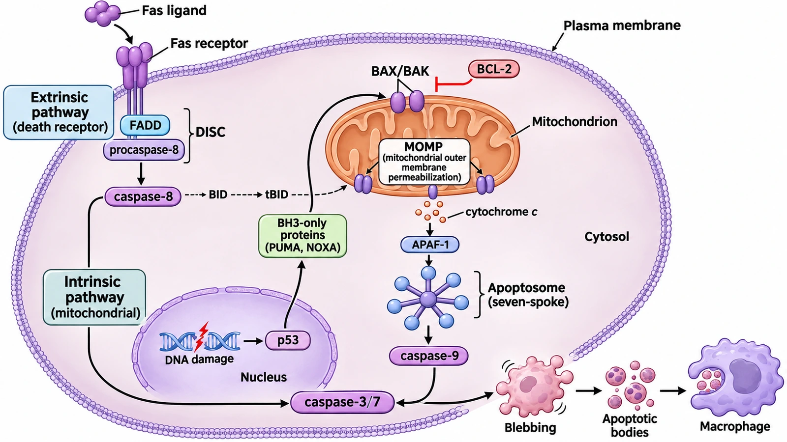
A labeled textbook diagram of both pathways with its blank worksheet, apoptosis vs necrosis, a publication-style overview, a mitochondrial close-up and the stages of a dying cell. Load any of them into the generator, then switch the pathway, level, or labels to make your own version.
Prompt
Create a labeled apoptosis pathway diagram as a clean textbook illustration on a white background, wide layout, inside one large cell labeled "Plasma membrane" and "Cytosol". Extrinsic pathway (box "Extrinsic pathway (death receptor)"): a "Fas ligand" trimer outside the cell binds a "Fas receptor" spanning the membrane; "FADD" and "procaspase-8" under it, bracketed as "DISC"; then "caspase-8", with a dashed arrow through "BID" to "tBID" and on to the mitochondrion. Intrinsic pathway (box "Intrinsic pathway (mitochondrial)"): inside the "Nucleus", "DNA damage" leads to "p53"; an arrow into the cytosol to "BH3-only proteins (PUMA, NOXA)", which act on "BAX/BAK" in the outer membrane of the "Mitochondrion"; "BCL-2" has a flat inhibition bar on BAX/BAK; a box "MOMP (mitochondrial outer membrane permeabilization)"; "cytochrome c" leaves the mitochondrion, joins "APAF-1" and forms the "Apoptosome (seven-spoke)" in the cytosol, which activates "caspase-9". Caspase-8 and caspase-9 both activate "caspase-3/7". Outside the cell at the bottom right: "Blebbing", then "Apoptotic bodies", then a "Macrophage" engulfing them. Keep DNA damage and p53 inside the nucleus, the apoptosome in the cytosol (never inside the mitochondrion), and BCL-2 with an inhibition bar only.
Dieses Beispiel verwenden
Prompt
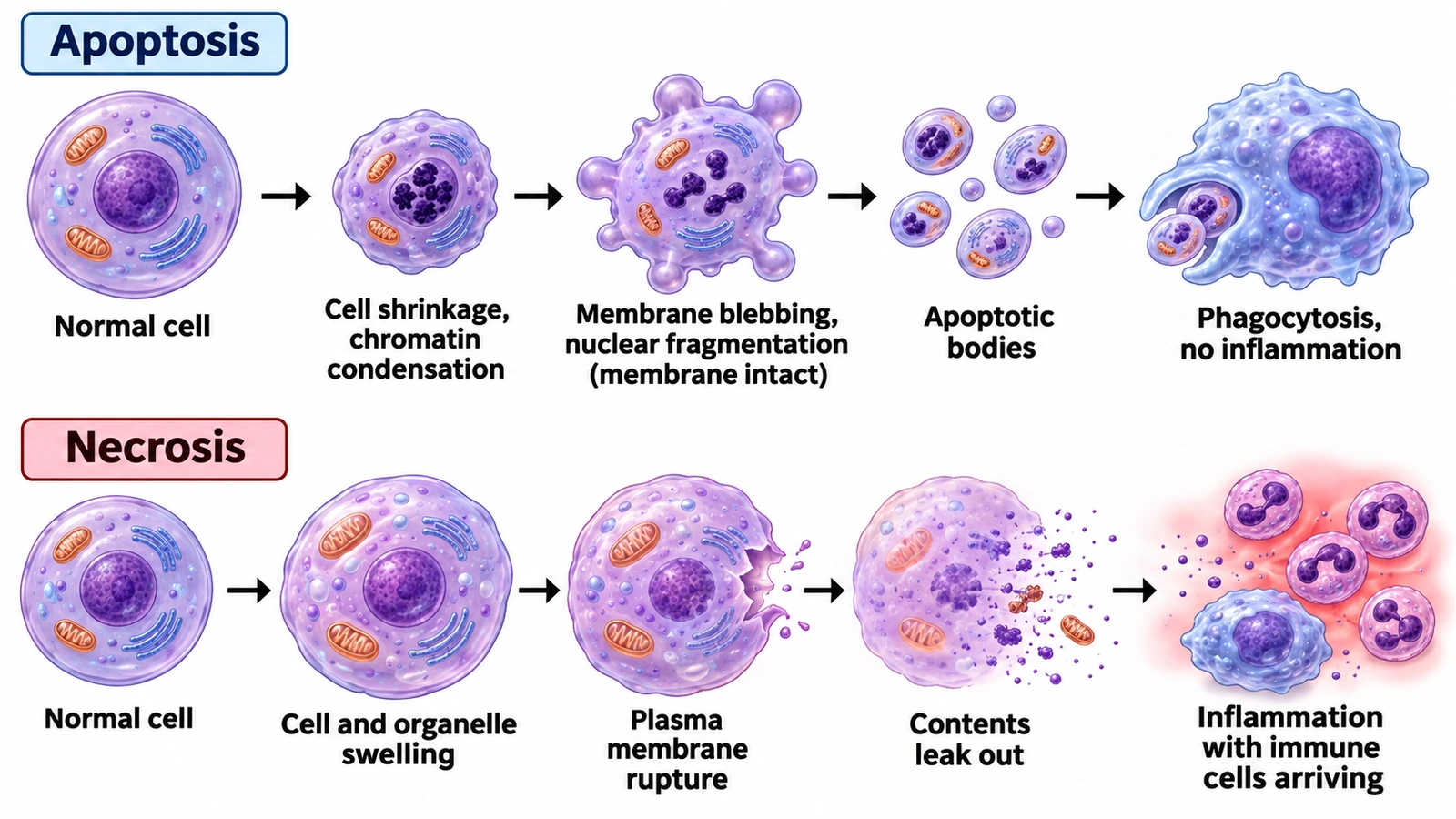
Create an apoptosis vs necrosis comparison diagram on a white background in two rows of five cells joined by arrows. Top row, headed "Apoptosis": "Normal cell"; "Cell shrinkage, chromatin condensation"; "Membrane blebbing, nuclear fragmentation (membrane intact)"; "Apoptotic bodies"; "Phagocytosis, no inflammation", with a macrophage engulfing the bodies. Bottom row, headed "Necrosis": "Normal cell"; "Cell and organelle swelling"; "Plasma membrane rupture"; "Contents leak out"; "Inflammation with immune cells arriving", with neutrophils gathering in a reddish zone. No blebs in the necrosis row and no rupture anywhere in the apoptosis row.
Apoptosis vs necrosis
Dieses Beispiel verwenden
Prompt
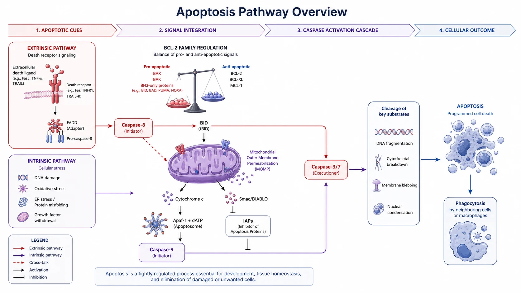
Create a publication-style apoptosis pathway overview titled "Apoptosis Pathway Overview" in a wide layout on a white background, with four chevron headers: "1. Apoptotic cues", "2. Signal integration", "3. Caspase activation cascade", "4. Cellular outcome". On the left, an "Extrinsic pathway" box (death receptor signaling): an extracellular death ligand (FasL, TNF-α, TRAIL) binding a death receptor (Fas, TNFR1, TRAIL-R) in the membrane, FADD (adapter) and pro-caspase-8; below it an "Intrinsic pathway" box (cellular stress): DNA damage, oxidative stress, ER stress / protein misfolding, growth factor withdrawal. In the middle, "BCL-2 family regulation" as a balance: pro-apoptotic BAX, BAK and BH3-only proteins (BID, BAD, PUMA, NOXA) against anti-apoptotic BCL-2, BCL-XL and MCL-1. Caspase-8 (initiator) activates BID (tBID), which acts on a mitochondrion labeled "Mitochondrial outer membrane permeabilization (MOMP)"; a dashed crosstalk arrow runs from caspase-8 to the mitochondrion. Cytochrome c goes to "APAF-1 + dATP (Apoptosome)" and then "Caspase-9 (initiator)"; Smac/DIABLO blocks "IAPs (inhibitor of apoptosis proteins)", which block the caspases. Caspase-8 and caspase-9 both activate "Caspase-3/7 (executioner)", leading to "Cleavage of key substrates": DNA fragmentation, cytoskeletal breakdown, membrane blebbing, nuclear condensation; then "Apoptosis" and "Phagocytosis by neighbouring cells or macrophages". Legend: red extrinsic arrows, purple intrinsic arrows, dashed crosstalk, arrows for activation, bars for inhibition.
Graduate / researchPublication schematic
Dieses Beispiel verwenden
Prompt
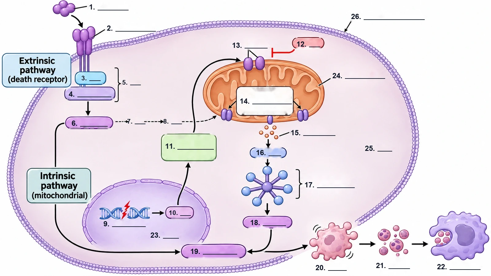
Create a blank apoptosis pathway worksheet from the labeled two-pathway cell drawing: keep every shape, arrow, inhibition bar and the boxes "Extrinsic pathway (death receptor)" and "Intrinsic pathway (mitochondrial)", but replace every other name with a numbered empty answer line, in this order: 1 Fas ligand, 2 Fas receptor, 3 FADD, 4 procaspase-8, 5 DISC, 6 caspase-8, 7 BID, 8 tBID, 9 DNA damage, 10 p53, 11 BH3-only proteins, 12 BCL-2, 13 BAX/BAK, 14 MOMP, 15 cytochrome c, 16 APAF-1, 17 apoptosome, 18 caspase-9, 19 caspase-3/7, 20 blebbing, 21 apoptotic bodies, 22 macrophage, 23 nucleus, 24 mitochondrion, 25 cytosol, 26 plasma membrane. No word bank; the labeled diagram is the answer key.
Blank worksheet
Dieses Beispiel verwenden
Prompt
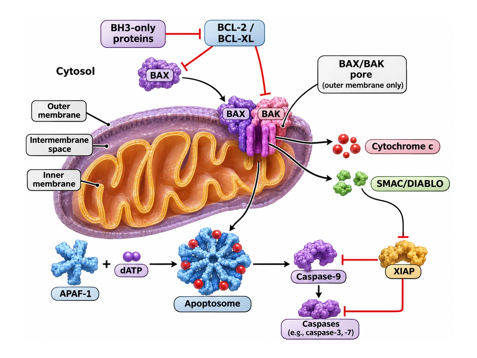
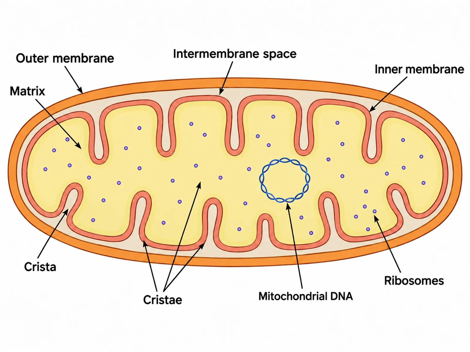
Create a close-up of the mitochondrial apoptosis pathway as a textbook illustration on a white background. One large mitochondrion with "Outer membrane", "Inner membrane" (folded cristae) and "Intermembrane space" labeled, and "Cytosol" written outside it. "BH3-only proteins" have an inhibition bar on "BCL-2 / BCL-XL", which has inhibition bars on "BAX", moving in from the cytosol, and on "BAK", already in the outer membrane. BAX and BAK form a "BAX/BAK pore (outer membrane only)" through which "Cytochrome c" and "SMAC/DIABLO" leave. In the cytosol: "APAF-1" + "dATP" form the "Apoptosome", which activates "Caspase-9", which activates "Caspases (e.g., caspase-3, -7)". "XIAP" has inhibition bars on caspase-9 and on the executioner caspases, and SMAC/DIABLO has an inhibition bar on XIAP. No pores in the inner membrane and no apoptosome inside the mitochondrion.
Mitochondria close-upGraduate / research
Dieses Beispiel verwenden
Prompt
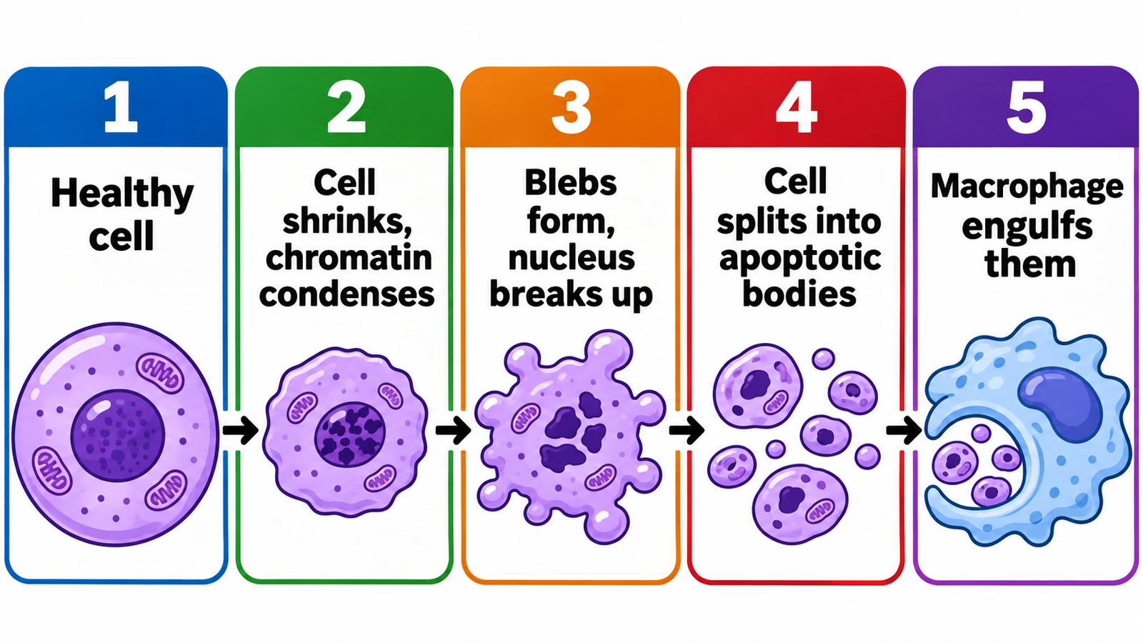
Create a simple stages-of-apoptosis diagram in a bold, flat, colourful style with large text on a white background: five numbered panels in a row joined by arrows, each with a coloured number header. 1 "Healthy cell"; 2 "Cell shrinks, chromatin condenses"; 3 "Blebs form, nucleus breaks up"; 4 "Cell splits into apoptotic bodies"; 5 "Macrophage engulfs them". Show only the cell shapes, no pathway proteins.
Stages of a dying cellHigh school
Dieses Beispiel verwenden
How to make an apoptosis pathway diagram
1
Choose the pathway, level, and style
Both pathways together, the extrinsic or intrinsic route alone, a mitochondria close-up, the stages of a dying cell, or apoptosis vs necrosis. Set the level from high school to research, then a textbook illustration or a publication schematic.
2
Generate the figure
The generator places the ligand outside the cell, FADD and caspase-8 at the membrane, the BCL-2 family on the mitochondrion, the apoptosome in the cytosol, and both routes meeting at caspases-3 and -7, with arrows for activation and bars for inhibition.
3
Check every arrow, then export
Confirm BCL-2 blocks rather than promotes death, the apoptosome sits outside the mitochondrion, and arrows run from initiator to executioner caspases. For a worksheet, make the answer key from the same result, then download the PNG.
Apoptosis key facts
Term coined
1972, by Kerr, Wyllie and Currie
Initiator caspases
Caspase-8 and -10 (extrinsic), caspase-9 (intrinsic), caspase-2
Executioner caspases
Caspase-3, -6 and -7
Extrinsic platform
DISC: death receptor + FADD + procaspase-8
Intrinsic platform
Apoptosome: 7 APAF-1 + cytochrome c + procaspase-9
Point of commitment
MOMP by BAX and BAK
Anti-apoptotic BCL-2 proteins
BCL-2, BCL-XL, MCL-1
Link between pathways
Caspase-8 cuts BID to tBID
Eat-me signal
Phosphatidylserine on the outer membrane leaflet
C. elegans development
131 of 1,090 cells die by programmed death
What an apoptosis pathway diagram shows
Apoptosis is programmed cell death: a cell that is damaged, infected, or simply no longer needed dismantles itself in an orderly way, without spilling its contents. The word was coined in 1972 by Kerr, Wyllie, and Currie, from the Greek for leaves falling from a tree. An apoptosis pathway diagram turns that process into a map: which signal starts it, which proteins pass the signal on, where in the cell each step happens, and what the dying cell looks like at the end.
Every complete diagram of apoptosis has the same skeleton. Two entry routes, the extrinsic pathway at the plasma membrane and the intrinsic pathway at the mitochondrion, each switch on an initiator caspase. Both initiators activate the same executioner caspases, caspase-3 and caspase-7. The executioners cut the cell’s proteins and DNA, and the cell shrinks, blebs, and breaks into apoptotic bodies for phagocytes to eat. Draw that skeleton correctly and the details fit around it.
The extrinsic pathway, step by step
A death ligand on another cell or in the fluid around the cell, such as Fas ligand on a cytotoxic T cell, TNF-α, or TRAIL, binds its death receptor: Fas (also called CD95), TNFR1, or TRAIL-R1/R2. Ligands and receptors work as trimers.
Ligand binding brings the receptors’ intracellular death domains together, and the adaptor protein FADD binds to them (TNFR1 first recruits TRADD, which then binds FADD).
FADD recruits several procaspase-8 molecules (and procaspase-10) through its death effector domain, forming the death-inducing signaling complex, or DISC.
Packed together in the DISC, procaspase-8 molecules activate one another. Active caspase-8 is released into the cytosol.
Caspase-8 cuts and activates the executioner caspases-3 and -7, and it also cuts BID into tBID, which carries the signal to the mitochondria.
The brake: c-FLIP, a caspase-8 look-alike without enzyme activity, can occupy the DISC and stop caspase-8 activation.
The intrinsic (mitochondrial) pathway, step by step
Stress builds up inside the cell: DNA damage, loss of growth factors, misfolded proteins in the endoplasmic reticulum, oxidative damage, or viral infection.
The stress produces or activates BH3-only proteins such as BIM, BID, PUMA, NOXA, and BAD. After DNA damage, the transcription factor p53 switches on the genes for PUMA and NOXA.
BH3-only proteins neutralize the protective proteins BCL-2, BCL-XL, and MCL-1, and some of them also activate BAX and BAK directly.
BAX moves from the cytosol to the mitochondrion and, with BAK, which already sits in the outer membrane, forms oligomers that make the outer membrane leaky. This is mitochondrial outer membrane permeabilization, MOMP, usually treated as the point of no return.
Cytochrome c and SMAC/DIABLO escape from the intermembrane space into the cytosol.
Cytochrome c binds APAF-1, the apoptosome forms, and it activates caspase-9, which activates caspases-3 and -7.
The BCL-2 family: who protects and who kills
Whether a stressed cell survives is decided at the mitochondrion by the balance of BCL-2 family proteins, which Czabotar and colleagues divide into three groups. Drawing them as a balance, or as an inhibition network, makes the logic of the intrinsic pathway visible.
Anti-apoptotic guardians: BCL-2, BCL-XL, and MCL-1. They bind and hold back the other two groups. BCL-2 is named after B-cell lymphoma, in which a chromosome translocation makes too much of it.
Pro-apoptotic effectors: BAX and BAK. Once released and activated, they oligomerize in the outer mitochondrial membrane and cause MOMP.
BH3-only sensors: BIM, BID (and its cut form tBID), PUMA, NOXA, BAD, and others. They respond to specific stresses and tip the balance by blocking the guardians or activating the effectors.
Drugs that mimic BH3-only proteins, such as venetoclax, which blocks BCL-2, are used to treat some leukaemias, a direct clinical use of this diagram.
Cytochrome c, MOMP and the apoptosome
In a healthy cell, cytochrome c is a small protein in the space between the two mitochondrial membranes, where it shuttles electrons in the respiratory chain. MOMP lets it out. In the cytosol, cytochrome c binds the protein APAF-1 and, with dATP or ATP, seven APAF-1 molecules assemble into a wheel-shaped platform, the apoptosome. Its hub recruits procaspase-9 and activates it.
On a diagram, the details that matter most are positions. BAX and BAK work in the outer membrane, not the inner one; cytochrome c comes from the intermembrane space; and the apoptosome forms in the cytosol, outside the mitochondrion. SMAC/DIABLO, released at the same time, binds XIAP and stops it from inhibiting caspases, which is why it is often drawn as a second arrow leaving the mitochondrion.
Caspases: initiators and executioners
Caspases are cysteine proteases that cut their targets just after an aspartate residue. Every one is made as an inactive procaspase, which is why a cell can keep them on hand without harm. Initiator caspases are activated by being brought together in a platform; executioner caspases are activated when an initiator cuts them.
Initiators: caspase-8 and caspase-10 (extrinsic, at the DISC), caspase-9 (intrinsic, at the apoptosome), and caspase-2.
Executioners: caspase-3, caspase-6, and caspase-7, which cut hundreds of proteins.
ICAD/CAD: cutting ICAD frees the nuclease CAD, which cuts DNA between nucleosomes into fragments of about 180 to 200 base pairs and multiples of it, the “DNA ladder” seen on a gel.
Lamins: cutting the nuclear lamina lets the nucleus collapse and fragment.
PARP: cleavage of this DNA-repair enzyme is a standard laboratory marker of apoptosis.
Cytoskeleton and membrane: cutting cytoskeletal regulators produces blebbing, and changes to membrane lipid transporters expose phosphatidylserine on the outer surface.
Where the two pathways meet
The extrinsic and intrinsic routes are drawn as two columns, but they are connected. Caspase-8 cuts the BH3-only protein BID into tBID, which travels to the mitochondrion and activates BAX and BAK. In type I cells, such as thymocytes, the DISC makes enough caspase-8 to activate caspase-3 directly, and the mitochondria are not needed. In type II cells, such as hepatocytes, the extrinsic signal is too weak on its own, and XIAP keeps caspase-3 in check until the mitochondria release cytochrome c and SMAC/DIABLO. Draw the crosstalk as a single arrow from caspase-8 to BID to the mitochondrion, often dashed so that it reads as a link rather than a third pathway.
The brakes: IAPs, XIAP, SMAC/DIABLO and c-FLIP
XIAP, the best-studied inhibitor of apoptosis protein (IAP), binds and blocks caspase-9, caspase-3, and caspase-7. Draw it with flat-ended bars on those three.
SMAC/DIABLO, released from the mitochondria during MOMP, binds XIAP and removes the block. Draw it with a bar on XIAP: an inhibitor of an inhibitor.
c-FLIP resembles caspase-8 but cannot cut, and it blocks caspase-8 activation at the DISC.
The anti-apoptotic BCL-2 proteins are the brake on the intrinsic pathway, upstream of all of these.
Apoptosis vs necrosis
Apoptosis and necrosis are the two classic ways cells die, and a side-by-side diagram is one of the most common exam figures. The Nomenclature Committee on Cell Death now also names regulated forms of necrosis, such as necroptosis, which uses the kinases RIPK1 and RIPK3 and the protein MLKL, and pyroptosis, which uses inflammatory caspases and gasdermins, but the basic contrast below is what most diagrams show.
Cell size: apoptotic cells shrink; necrotic cells and their organelles swell.
Nucleus: apoptosis condenses the chromatin and fragments the nucleus in an orderly way; necrosis lets the nucleus fade and break down.
Membrane: in apoptosis the plasma membrane blebs but stays sealed until the cell is eaten; in necrosis it ruptures early and the contents leak out.
Energy: apoptosis is an active program that needs ATP; classic necrosis follows a collapse in energy supply.
Tissue response: apoptotic bodies are removed quietly, without inflammation; necrosis triggers inflammation in the surrounding tissue.
Extent: apoptosis usually removes single cells; necrosis often kills patches of cells together, as in an infarct.
The perforin and granzyme route
Cytotoxic T cells and natural killer cells have a third way to trigger apoptosis in an infected or cancerous cell, which Elmore describes as the perforin/granzyme pathway. The killer cell releases perforin, which lets granzymes into the target cell. Granzyme B cuts and activates caspases, including caspase-3, and also cuts BID, engaging the mitochondria. A full immunology figure may add this route beside the Fas ligand that the same killer cells use for the extrinsic pathway; ask for it in your description if you need it.
Why apoptosis matters
Development: OpenStax describes how apoptosis removes the web of tissue between developing fingers and toes. In the nematode C. elegans, 131 of the 1,090 cells formed during development die by programmed cell death, the finding behind the 2002 Nobel Prize in Physiology or Medicine.
The immune system: apoptosis removes lymphocytes that would attack the body’s own tissues and shuts down an immune response once an infection is over.
Cancer: cancer cells often escape apoptosis, for example by losing p53 or making too much BCL-2, which is why many cancer treatments work by restoring it.
Tissue turnover: every day the body removes huge numbers of old or damaged cells by apoptosis without any inflammation.
Disease: too much apoptosis contributes to the loss of neurons in neurodegenerative disease and of heart cells after a heart attack.
How apoptosis is detected in the lab
Research figures often pair a pathway diagram with the assays used to measure it. If your figure needs a panel on detection, these are the standard methods to name.
Annexin V staining binds phosphatidylserine on the outer surface of early apoptotic cells; combined with a DNA dye such as propidium iodide, it separates early apoptotic, late apoptotic, and necrotic cells by flow cytometry.
TUNEL labels the ends of fragmented DNA in tissue sections.
Antibodies against cleaved caspase-3 or cleaved PARP show caspase activity on a western blot or in tissue.
A DNA ladder on an agarose gel shows internucleosomal cleavage.
Cytochrome c release can be shown by comparing mitochondrial and cytosolic fractions, or by imaging.
How to draw an apoptosis pathway diagram
Start with the compartments. Draw the plasma membrane across the top, a mitochondrion in the middle on the right, and a nucleus at the bottom or to one side. Put the extrinsic pathway down the left: ligand outside, receptor in the membrane, FADD and caspase-8 just inside. Put the intrinsic pathway down the right: the stress signal, p53 and the BH3-only proteins, the BCL-2 family on the mitochondrion, cytochrome c leaving it, and the apoptosome beside it. Bring both columns together at caspases-3 and -7 in the centre, and end with the dying cell at the bottom.
Then add the logic. Use pointed arrows for activation and flat-ended bars for inhibition, and keep them consistent: BCL-2 ⊣ BAX/BAK, XIAP ⊣ caspases, SMAC/DIABLO ⊣ XIAP, c-FLIP ⊣ caspase-8. Give each pathway its own colour, add the tBID crosstalk as a dashed arrow, and finish with a small legend. A figure built in this order reads top to bottom like the process itself.
Most common apoptosis diagram mistakes
These are the errors that appear most often in student drawings, online images, and AI output. Check each one before the figure goes into a paper, a slide, or graded work.
Häufiger FehlerRichtig
Häufiger FehlerBCL-2 drawn with an arrow promoting cell death
RichtigBCL-2 is anti-apoptotic: an inhibition bar on BAX and BAK
Häufiger FehlerThe apoptosome inside the mitochondrion or the nucleus
RichtigCytochrome c leaves the mitochondrion; the apoptosome assembles in the cytosol
Häufiger FehlerCaspase-3 labeled as an initiator, or arrows from caspase-3 to caspase-9
RichtigInitiators 8, 9 (and 2, 10) activate executioners 3, 6 and 7, never the reverse
Häufiger FehlerFADD drawn as the receptor or as a caspase
RichtigFADD is the adaptor linking the death receptor to procaspase-8
Häufiger FehlerBAX and BAK pores in the inner mitochondrial membrane
RichtigBAX and BAK permeabilize the outer membrane (MOMP)
Häufiger FehlerThe death ligand drawn inside the cell
RichtigThe ligand is extracellular; the receptor spans the plasma membrane
Häufiger FehlerThe apoptotic cell swells and bursts
RichtigApoptotic cells shrink and bleb with intact membranes; swelling and rupture are necrosis
Häufiger FehlerCytochrome c written as “Cytochrome C” or shown cutting DNA
RichtigCytochrome c (lowercase c) activates APAF-1; the nuclease CAD cuts the DNA
Intrinsic vs extrinsic apoptosis pathway
Most courses ask students to tell the two routes apart. This is how they differ at every step, and where they meet.
Extrinsic (death receptor) pathway
Intrinsic (mitochondrial) pathway
Trigger
A death ligand from another cell: FasL, TNF-α, TRAIL
Stress inside the cell: DNA damage, growth-factor loss, ER or oxidative stress
Where it starts
Death receptors in the plasma membrane
BCL-2 family proteins at the mitochondrion
Adaptor and platform
FADD; the DISC
APAF-1 and cytochrome c; the apoptosome
Initiator caspase
Caspase-8 (and -10)
Caspase-9
Key regulators
c-FLIP blocks caspase-8 activation
BCL-2, BCL-XL and MCL-1 block BAX and BAK; XIAP blocks caspases
Role of mitochondria
Not needed in type I cells; amplified through tBID in type II cells
Central: MOMP releases cytochrome c and SMAC/DIABLO
Executioners
Caspase-3 and -7
Caspase-3 and -7
Typical example
A cytotoxic T cell’s Fas ligand killing an infected cell
A cell with unrepairable DNA damage after p53 activation
Apoptosis Pathway Diagram FAQ
It is a figure that shows how a cell switches on its own death program, from the signal that starts it to the enzymes that dismantle the cell. A complete diagram of apoptosis has two entry routes, the extrinsic (death receptor) pathway at the cell surface and the intrinsic (mitochondrial) pathway inside the cell, which meet at the executioner caspases-3 and -7, and it ends with the shrinking, blebbing cell breaking into apoptotic bodies that a phagocyte engulfs.
Generate signaling pathways, cell cycle and mitochondria diagrams, blank labeling worksheets with matching answer keys, and publication-ready molecular biology figures with the same SciFigure AI workflow.